THE LIVING WORLD
Unit two. The Living Cell
4.4. Eukaryotic Cells
For the first 1 billion years of life on earth, all organisms were prokaryotes, cells with very simple interiors. Then, about 1.5 billion years ago, a new kind of cell appeared for the first time, the eukaryotic cell. Eukaryotic cells are much larger and profoundly different from prokaryotic cells, with a complex interior organization. All cells alive today except bacteria and archaea are of this new kind.
Figures 4.6 and 4.7 present cross-sectional diagrams of idealized animal and plant cells. As you can see, the interior of a eukaryotic cell is much more complex than the prokaryotic cell you encountered in figure 4.4. The plasma membrane 1 encases a semifluid matrix called the cytoplasm 2, which contains within it the nucleus and various cell structures called organelles. An organelle is a specialized structure within which particular cell processes occur. Each organelle, such as a mitochondrion 3, has a specific function in the eukaryotic cell. The organelles are anchored at specific locations in the cytoplasm by an interior scaffold of protein fibers, the cytoskeleton 4.
One of the organelles is very visible when these cells are examined with a microscope, filling the center of the cell like the pit of a peach. Seeing it, the English botanist Robert Brown in 1831 called it the nucleus 5 (plural, nuclei), from the Latin word for “kernel.” Inside the nucleus, the DNA is wound tightly around proteins and packaged into compact units called chromosomes. It is the nucleus that gives eukaryotes their name, from the Greek words eu, true, and karyon, nut; by way of contrast, the earlier-evolving bacteria and archaea are called prokaryotes (“before the nut”).
If you examine the organelles in figures 4.6 and 4.7, you can see that most of them form separate compartments within the cytoplasm, bounded by their own membranes. The hallmark of the eukaryotic cell is this compartmentalization. This internal compartmentalization is achieved by an extensive endomembrane system 6 that weaves through the cell interior, providing extensive surface area for many membrane-associated cell processes to occur.
Vesicles 7 (small membrane-bounded sacs that store and transport materials) form in the cell either by budding off of the endomembrane system or by the incorporation of lipids and protein in the cytoplasm. These many closed-off compartments allow different processes to proceed simultaneously without interfering with one another, just as rooms do in a house. Thus the organelles called lysosomes are recycling centers. Their very acid interiors break down old organelles, and the component molecules are recycled. This acid would be very destructive if released into the cytoplasm. Similarly, chemical isolation is essential to the function of the organelles called peroxisomes. Toxic chemicals are degraded and food molecules are processed within peroxisomes by enzymes that act by removing electrons and associated hydrogen atoms. If not isolated within the peroxisomes, these enzymes would tend to short-circuit chemical reactions occurring in the cytoplasm, which often involves adding hydrogen atoms to molecules.
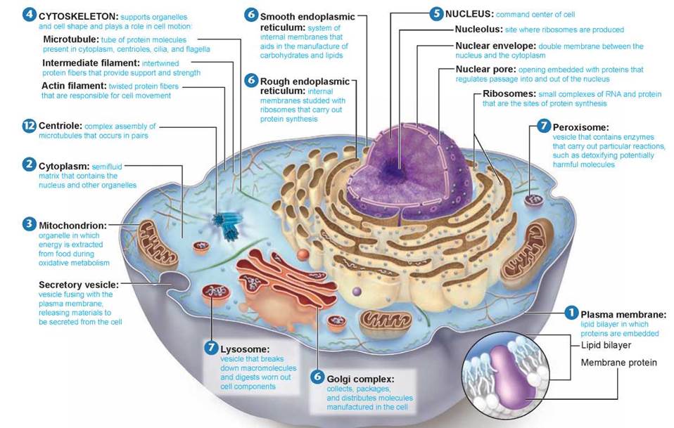
Figure 4.6. Structure of an animal cell.
In this generalized diagram of an animal cell, the plasma membrane encases the cell, which contains the cytoskeleton and various cell organelles and interior structures suspended in a semifluid matrix called the cytoplasm. Some kinds of animal cells possess fingerlike projections called microvilli. Other types of eukaryotic cells, for example many protist cells, may possess flagella, which aid in movement, or cilia, which can have many different functions.
If you compare figure 4.6 with figure 4.7, you will see the same set of organelles, with a few interesting exceptions. For example, the cells of plants, fungi, and many protists have strong thick exterior cell walls 8 composed of cellulose or chitin fibers, while the cells of animals lack cell walls. All plants and many kinds of protists have chloroplasts 9, within which photosynthesis occurs. No animal or fungal cells contain chloroplasts. Plant cells also contain a large central vacuole 10 that stores water, and cytoplasmic connections through openings in the cell wall called plasmodesmata 11. Centrioles 12 are present in animal cells but absent in plant and fungal cells. Some kinds of animal cells possess fingerlike projections called microvilli. Many animal and protist cells possess flagella, which aid in movement, or cilia, which have many different functions. Flagella occur in sperm of a few plant species, but are otherwise absent in plant and fungal cells.
We will now journey into the interior of a typical eukaryotic cell and explore it in more detail, using diagrams with the particular organelle you are examining highlighted. While the various organelles are color-coded for easier identification, remember that most are actually colorless.
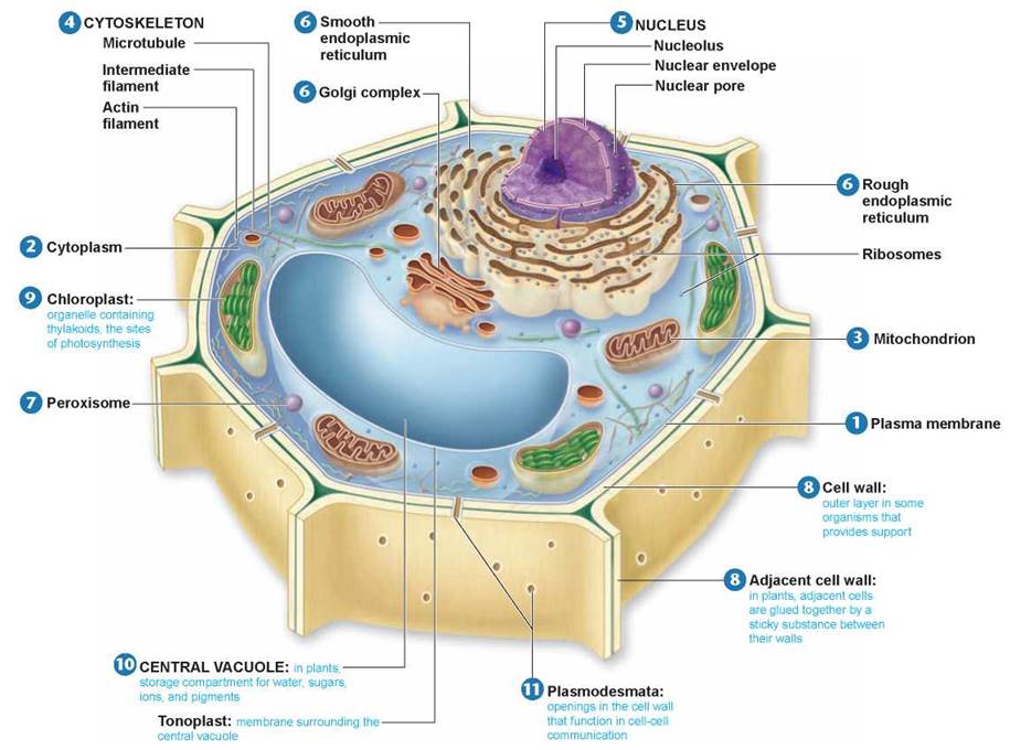
Figure 4.7. Structure of a plant cell.
Most mature plant cells contain large central vacuoles that occupy a major portion of the internal volume of the cell and organelles called chloroplasts, within which photosynthesis takes place. The cells of plants, fungi, and some protists have cell walls, although the composition of the walls varies among the groups. Plant cells have cytoplasmic connections through openings in the cell wall called plasmodesmata. Flagella occur in sperm of a few plant species, but are otherwise absent in plant and fungal cells. Centrioles are also absent in plant and fungal cells.
Key Learning Outcome 4.4. Eukaryotic cells have a system of interior membranes and organelles that subdivide the interior into functional compartments.
Today’s Biology
Membrane Defects Can Cause Disease
The year 1993 marked an important milestone in the treatment of human disease. That year the first attempt was made to cure cystic fibrosis (CF), a deadly genetic disorder, by transferring healthy genes into sick individuals. Cystic fibrosis is a fatal disease in which the body cells of affected individuals secrete a thick mucus that clogs the airways of the lungs. The cystic fibrosis patient in the photograph is breathing into a Vitalograph, a device that measures lung function. These same secretions block the ducts of the pancreas and liver so that the few patients who do not die of lung disease die of liver failure. Cystic fibrosis is usually thought of as a children's disease because until recently few affected individuals lived long enough to become adults. Even today half die before their midtwenties. There is no known cure.
Cystic fibrosis results from a defect in a single gene that is passed down from parent to child. It is the most common fatal genetic disease of Caucasians. One in 20 individuals possesses at least one copy of the defective gene. Most of these individuals are not afflicted with the disease; only those children who inherit a copy of the defective gene from each parent succumb to cystic fibrosis—about 1 in 2,500 infants.
Cystic fibrosis has proven difficult to study. Many organs are affected, and until recently it was impossible to identify the nature of the defective gene responsible for the disease. In 1985 the first clear clue was obtained.
An investigator, Paul Quinton, seized on a commonly observed characteristic of cystic fibrosis patients, that their sweat is abnormally salty, and performed the following experiment. He isolated a sweat duct from a small piece of skin and placed it in a solution of salt (NaCl) that was three times as concentrated as the NaCl inside the duct. He then monitored the movement of ions. Diffusion tends to drive both the sodium (Na+) and the chloride (Cl-) ions into the duct because of the higher outer ion concentrations.
In skin isolated from normal individuals, Na+ and Cl- both entered the duct, as expected. In skin isolated from cystic fibrosis individuals, however, only Na+ entered the duct—no Cl- entered. For the first time, the molecular nature of cystic fibrosis became clear. Water accompanies chloride, and was not entering the ducts because chloride was not, creating thick mucus. Cystic fibrosis is a defect in a plasma membrane protein called CFTR (cystic fibrosis fransmembrane conductance regulator) that normally regulates passage of Cl- into and out of the body's cells.
The defective cf gene was isolated in 1987, and its position on a particular human chromosome (chromosome 7) was pinpointed in 1989. Interestingly, many cystic fibrosis patients produce a CFTR protein with a normal amino acid sequence. The cf mutation in these cases appears to interfere with how the CFTR protein folds, preventing it from folding into a functional shape.
Soon after the cf gene was isolated, experiments were begun to see if it would be possible to cure cystic fibrosis by gene therapy—that is, by transferring healthy cf genes into the cells with defective ones. In 1990 a working cf gene was successfully transferred into human lung cells growing in tissue culture, using adenovirus, a cold virus, to carry the gene into the cells. The CFTR-defective cells were "cured,” becoming able to transport chloride ions across their plasma membranes. Then in 1991 a team of researchers successfully transferred a normal human cf gene into the lung cells of a living animal—a rat. The cf gene was first inserted into the adenovirus genome because adenovirus is a cold virus and easily infects lung cells. The treated virus was then inhaled by the rat. Carried piggyback, the cf gene entered the rat lung cells and began producing the normal human CFTR protein within these cells!
These results were very encouraging, and at first the future for all cystic fibrosis patients seemed bright. Clinical tests using adenovirus to introduce healthy cf genes into cystic fibrosis patients were begun with much fanfare in 1993.
They were not successful. As described further in chapter 13, there were insurmountable problems with the adenovirus being used to transport the cf gene into cystic fibrosis patients. The difficult and frustrating challenge that cystic fibrosis researchers had faced was not over. Research into clinical problems is often a time-consuming and frustrating enterprise, never more so than in this case. Recently, as chapter 13 recounts, new ways of introducing the healthy cf gene have been tried with better results. The long, slow journey toward a cure has taught us not to leap to the assumption that a cure is now at hand, but the steady persistence of researchers has taken us a long way, and again the future for cystic fibrosis patients seems bright.