THE LIVING WORLD
Unit two. The Living Cell
4.13. Bulk Passage into and out of Cells
Endocytosis and Exocytosis
The cells of many eukaryotes take in food and liquids by extending their plasma membranes outward toward food particles. The membrane engulfs the particle and forms a vesicle—a membrane-bounded sac—around it. This process is called endocytosis (figure 4.22).
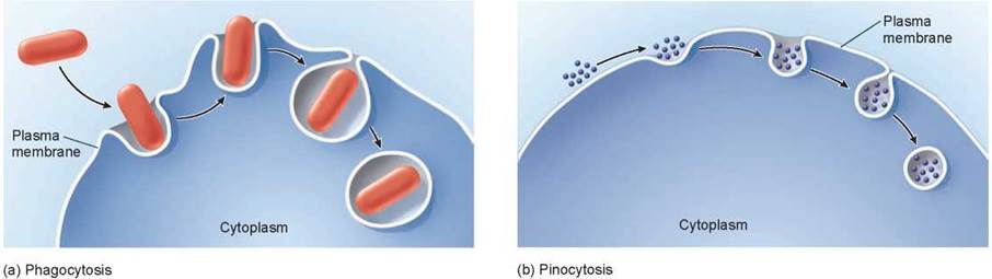
Figure 4.22. Endocytosis.
Endocytosis is the process of engulfing material by folding the plasma membrane around it, forming a vesicle. (a) When the material is an organism or some other relatively large fragment of organic matter, the process is called phagocytosis. (b) When the material is a liquid, the process is called pinocytosis.
The reverse of endocytosis is exocytosis, the discharge of material from vesicles at the cell surface. The vesicle in figure 4.23 contains a substance to be discharged, or released, from the cell. The purple particles remain suspended in the vesicle as it fuses with the plasma membrane. The membrane that forms the vesicle is made of phospholipids, and as it comes in contact with the plasma membrane, the phospholipids of both membranes interact, forming a pore through which the contents leave the vesicle to the outside. In plant cells, exocytosis is an important means of exporting the materials needed to construct the cell wall through the plasma membrane. Among protists, the discharge of a contractile vacuole is a form of exocytosis. In animal cells, exocytosis provides a mechanism for secreting many hormones, neurotransmitters, digestive enzymes, and other substances.
Phagocytosis and Pinocytosis
If the material the cell takes in is particulate (made up of discrete particles), such as an organism, like the red bacterium in figure 4.22a, or some other fragment of organic matter, the process is called phagocytosis (Greek phagein, to eat, and cytos, cell). If the material the cell takes in is liquid or substances dissolved in a liquid like the small particles in figure 4.22b, it is called pinocytosis (Greek pinein, to drink). Pinocytosis is common among animal cells. Mammalian egg cells, for example, “nurse” from surrounding cells; the nearby cells secrete nutrients that the maturing egg cell takes up by pinocytosis. Virtually all eukaryotic cells constantly carry out these kinds of endocytosis, trapping particles and extracellular fluid in vesicles and ingesting them. Endocytosis rates vary from one cell type to another. They can be surprisingly high: Some types of white blood cells ingest 25% of their cell volume each hour!
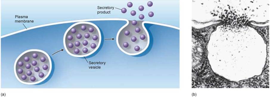
Figure 4.23. Exocytosis.
Exocytosis is the discharge of material from vesicles at the cell surface. (a) Proteins and other molecules are secreted from cells in small pockets called secretory vesicles, whose membranes fuse with the plasma membrane, thereby allowing the secretory vesicles to release their contents to the cell surface. (b) In the photomicrograph, you can see exocytosis taking place explosively.
Receptor-Mediated Endocytosis
Specific molecules are often transported into eukaryotic cells through receptor-mediated endocytosis, illustrated in figure 4.24. Molecules to be transported into the cell, the red balls in the figure, first bind to specific receptors in the plasma membrane. The transport process is specific to only molecules that have a shape that fits snugly into the receptor. The plasma membrane of a particular kind of cell contains a characteristic battery of receptor types, each for a different kind of molecule.
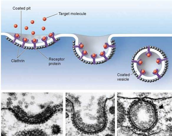
Figure 4.24. Receptor-mediated endocytosis.
Cells that undergo receptor-mediated endocytosis have pits coated with the protein clathrin that initiate endocytosis when target molecules bind to receptor proteins in the plasma membrane. In the photomicrographs, a coated pit appears in the plasma membrane of a developing egg cell, covered with a layer of proteins (80,000x). When an appropriate collection of molecules gathers in the coated pit, the pit deepens, and eventually seals off to form a vesicle.
The portion of the receptor molecule inside the membrane is trapped in an indented pit coated with the protein clathrin, visible in the photos as well as in the drawing above. The pits act like molecular mousetraps, closing over to form an internal vesicle when the right molecule enters the pit. The trigger that releases the trap is the binding of the properly fitted target molecule to a receptor embedded in the membrane of the pit. When binding occurs, the cell reacts by initiating endocytosis. The process is highly specific and very fast.
One type of molecule that is taken up by receptor-mediated endocytosis is called low-density lipoprotein (LDL). The LDL molecules bring cholesterol into the cell where it can be incorporated into membranes. Cholesterol plays a key role in determining the stiffness of the cell’s membrane. In the human genetic disease called hypercholesterolemia, the receptors lack tails and so are never caught in the clathrin-coated pits and, thus, are never taken up by the cells. The cholesterol stays in the bloodstream of affected individuals, coating their arteries and leading to heart attacks.
It is important to understand that receptor-mediated endocytosis in itself does not bring substances directly into the cytoplasm of a cell. The material taken in is still separated from the cytoplasm by the membrane of the vesicle. Other processes break down or release the contents.
Key Learning Outcome 4.13. The plasma membrane can engulf materials by endocytosis, folding the membrane around the material to encase it within a vesicle. Exocytosis is essentially this process in reverse, expelling substances using vesicles. Receptor-mediated endocytosis brings in only selected substances.