THE LIVING WORLD
Unit Three. The Continuity of Life
8.3. Chromosomes
Chromosomes were first observed by the German embryologist Walther Fleming in 1879 while he was examining the rapidly dividing cells of salamander larvae. When Fleming looked at the cells through what would now be a rather primitive light microscope, he saw minute threads within their nuclei that appeared to be dividing lengthwise. Fleming called their division mitosis, based on the Greek word mitos, meaning “thread.”
Chromosome Number
Since their initial discovery, chromosomes have been found in the cells of all eukaryotes examined. Their number may vary enormously from one species to another. A few kinds of organisms—such as the Australian ant Myrmecia spp.; the plant Haplopappus gracilis, a relative of the sunflower that grows in North American deserts; and the fungus Penicillium— have only 1 pair of chromosomes, while some ferns have more than 500 pairs. Most eukaryotes have between 10 and 50 chromosomes in their body cells.
Homologous Chromosomes
Chromosomes exist in somatic cells as pairs, called homologous chromosomes, or homologues. Homologues carry information about the same traits at the same locations on each chromosome but the information can vary between homologues, which will be discussed in chapter 10. Cells that have two of each type of chromosome are called diploid cells. One chromosome of each pair is inherited from the mother (colored green in figure 8.2) and the other from the father (colored purple). Before cell division, each homologous chromosome replicates, resulting in two identical copies, called sister chromatids. You see in figure 8.2 that the sister chromatids remain joined together after replication at a special linkage site called the centromere, the knoblike structure in the middle of each chromosome. Human body cells have a total of 46 chromosomes, which are actually 23 pairs of homologous chromosomes. In their duplicated state, before mitosis, there are still only 23 pairs of chromosomes, but each chromosome has duplicated and consists of two sister chromatids, for a total of 92 chromatids. The duplicated sister chromatids can make it confusing to count the number of chromosomes in an organism, but keep in mind that the number of centromeres doesn’t increase with replication, and so you can always determine the number of chromosomes simply by counting the centromeres.
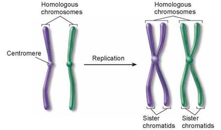
Figure 8.2. The difference between homologous chromosomes and sister chromatids.
Homologous chromosomes are a pair of the same chromosome— say, chromosome number 16. Sister chromatids are the two replicas of a single chromosome held together by the centromere after DNA replication. A duplicated chromosome looks somewhat like an X.
The Human Karyotype
The 46 human chromosomes can be paired as homologues by comparing size, shape, location of centromeres, and so on. This arrangement of chromosomes is called a karyotype. An example of a human karyotype is shown in figure 8.3. You can see how the different sizes and shapes of chromosomes allow scientists to pair together the ones that are homologous.
For example, chromosome 1 is much larger than chromosome 14, and its centromere is more centrally located on the chromosome. Each chromosome contains thousands of genes that play important roles in determining how a person’s body develops and functions. For this reason, possession of all the chromosomes is essential to survival. Humans missing even one chromosome, a condition called monosomy, do not usually survive embryonic development. Nor does the human embryo develop properly with an extra copy of any one chromosome, a condition called trisomy. For all but a few of the smallest chromosomes, trisomy is fatal; even in those cases, serious problems result. We will revisit this issue of differences in chromosome number in chapter 10.
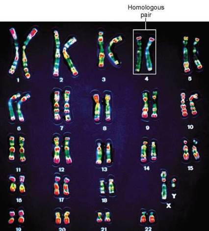
Figure 8.3. The 46 chromosomes of a human.
In this presentation, photographs of the individual chromosomes of a human male have been cut out and paired with their homologues, creating an organized display called a karyotype. The chromosomes are in a duplicated state, and the sister chromatids can actually be seen in many of the homologous pairs.
Chromosome Structure
Chromosomes are composed of chromatin, a complex of DNA and protein; most are about 40% DNA and 60% protein. A significant amount of RNA is also associated with chromosomes because chromosomes are the sites of RNA synthesis. The DNA of a chromosome is one very long, double-stranded fiber that extends unbroken through the entire length of the chromosome. A typical human chromosome contains about 140 million (1.4 x 10⁸) nucleotides in its DNA. Furthermore, if the strand of DNA from a single chromosome were laid out in a straight line, it would be about 5 centimeters (2 inches) long. The amount of information in one human chromosome would fill about 2,000 printed books of 1,000 pages each! Fitting such a strand into a nucleus is like cramming a string the length of a football field into a baseball—and that’s only 1 of 46 chromosomes! In the cell, however, the DNA is coiled, allowing it to fit into a much smaller space than would otherwise be possible.
Chromosome Coiling
The DNA of eukaryotes is divided into several chromosomes, although the chromosomes you see in figure 8.3 hardly look like long, double-stranded molecules of DNA. These chromosomes, duplicated as sister chromatids, are formed by winding and twisting the long DNA strands into a much more compact structure. Winding up DNA presents an interesting challenge. Because the phosphate groups of DNA molecules have negative charges, it is impossible to just tightly wind up DNA because all the negative charges would simply repel one another. As you can see in figure 8.4, the DNA helix wraps around proteins with positive charges called histones. The positive charges of the histones counteract the negative charges of the DNA, so that the complex has no net charge. Every 200 nucleotides, the DNA duplex is coiled around a core of eight histone proteins, forming a complex known as a nucleosome. The nucleosomes, which resemble beads on a string in figure 8.4, are further coiled into a solenoid. This solenoid is then organized into looped domains. The final organization of the chromosome is not known, but it appears to involve further radial looping into rosettes around a preexisting scaffolding of protein. This complex of DNA and histone proteins, coiled tightly, forms a compact chromosome.
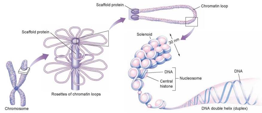
Figure 8.4. Levels of eukaryotic chromosomal organization.
Compact, rod-shaped chromosomes are in fact highly wound-up molecules of DNA. The arrangement illustrated here is one of many possibilities.

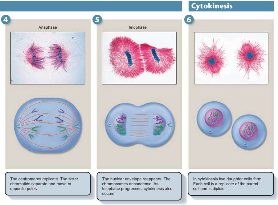
Figure 8.5. How cell division works.
Cell division in eukaryotes begins in interphase, carries through the four stages of mitosis, and ends with cytokinesis. Several features of the spindle illustrated in the drawings above appear in dividing animal cells but not in plant cells, and cannot be seen in the photographs, which are of the African blood lily Haemanthus katharinae. (In these exceptional photographs, the chromosomes are stained blue and microtubules stained red)
Key Learning Outcome 8.3. All eukaryotic cells store their hereditary information in chromosomes, but different kinds of organisms use very different numbers of chromosomes to store this information. Coiling of the DNA into chromosomes allows it to fit in the nucleus.