THE LIVING WORLD
Unit Three. The Continuity of Life
10. Foundations of Genetics
The principles of genetics first proposed by Mendel apply not only to pea plants and fruit flies, but also to you. How closely you resemble your father or mother was largely established before your birth by the chromosomes you received from them, just as meiosis in pea plants determined the segregation of Mendel’s traits. But many of the alleles found in human populations demand more serious concern than the color of a pea. Some of the most devastating human disorders result from alleles specifying defective forms of proteins that have important functions in our bodies. By studying human heredity, scientists are more able to predict which disorders parents might expect to pass on to their children, and with what probability.
Although humans pass genes on to the next generation in much the same way that other organisms do, we naturally have a special curiosity about ourselves. Because we know that some illnesses are hereditary and others are not, we cannot escape concern when a member of our family becomes ill. If a family member has had a stroke, we tend to worry about our own future health because we know that the propensity to suffer strokes can be hereditary. Few parents have babies without worrying about the possibility of birth defects. Genes are also clearly involved in such conditions as diabetes, depression, and alcoholism. The way in which genes interact with the environment to produce individuals with differing personalities is the subject of continuing intensive study. Because of the importance of genes in determining the course of our lives, we are all human geneticists, interested in what the laws of genetics reveal about ourselves and our families.
Human Chromosomes
Although chromosomes were discovered more than a century ago, the exact number of chromosomes that humans possess (46) was not established until 1956, when new techniques for accurately determining the number and form of human and other mammalian chromosomes were developed.
Biologists examine human chromosomes by collecting a blood sample, adding chemicals that induce the white blood cells in the sample to divide (red blood cells have lost their nuclei and cannot divide), and then adding other chemicals that arrest cell division at metaphase. Metaphase is the stage of mitosis when the chromosomes are most condensed and thus most easily distinguished from one another. The cells are then flattened, spreading out their contents, and the individual chromosomes are separated for examination. The chromosomes are stained and photographed, and a chromosomal “portrait” called a karyotype is prepared with the photographs of the individual chromosomes. A human karyotype is presented in figure 10.23. By convention, the chromosomes in a karyotype are presented with homologues together, and chromosomes arranged in order of descending size.
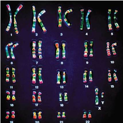
Figure 10.23. A human karyotype.
Photographs of each of the 46 chromosomes have been arranged in descending order of size. The banding patterns revealed by staining allow the investigator to identify homologues and pair them together.
Of the 23 pairs of human chromosomes you see in figure 10.23, 22 consist of members that are similar in size and morphology in both males and females. These chromosomes are called autosomes. In many plants and animals, including peas, fruit flies, and humans, the two members of the remaining pair—the so-called sex chromosomes—are unlike each other in males and similar in females. In humans, females are designated XX and males are designated XY. The Y chromosome is much smaller than the X chromosome and carries only a tenth the number of genes. Among the genes present on the Y chromosome are those that determine “maleness,” and, therefore, humans who inherit the Y chromosome develop into males.
The karyotypes of individuals are often examined to detect genetic abnormalities arising from extra or lost chromosomes. For example, the human birth defect Down syndrome (discussed on the facing page) is associated with the presence of an extra copy of chromosome 21, which can be recognized easily in karyotypes, as there are 47 chromosomes rather than 46, and the extra chromosome can be identified by its banding pattern as a third copy of chromosome 21. Karyotypes of fetal cells taken before birth can reveal genetic abnormalities of this sort.
Nondisjunction
Some of the most significant human hereditary disorders arise as a result of problems with how human chromosomes sort during meiosis.
Sometimes during meiosis, sister chromatids or homologous chromosomes that paired up during metaphase remain stuck together instead of separating. The failure of chromosomes to separate correctly during either meiosis I or II is called nondisjunction. Nondisjunction leads to aneuploidy, an abnormal number of chromosomes. The nondisjunction you see in figure 10.24 occurs because the homologous pair of larger chromosomes failed to separate in anaphase I. The gametes that result from this division have unequal numbers of chromosomes. Under normal meiosis, all gametes would be expected to have two chromosomes, but as you can see, two of these gametes have three chromosomes, while two others have just one.
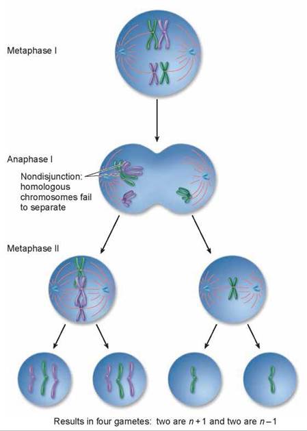
Figure 10.24. Nondisjunction in anaphase I.
In nondisjunction that occurs during meiosis I, one pair of homologous chromosomes fails to separate in anaphase I, and the gametes that result have one too many or one too few chromosomes. Nondisjunction can also occur in meiosis II, when sister chromatids fail to separate during anaphase II.
Almost all humans of the same sex have the same karyotype simply because other arrangements don’t work well. Humans who have lost even one copy of an autosome (called monosomics) do not survive development. In all but a few cases, humans who have gained an extra autosome (called trisomics) also do not survive. However, five of the smallest chromosomes—those numbered 13, 15, 18, 21, and 22—can be present in humans as three copies and still allow the individual to survive for a time. The presence of an extra chromosome 13, 15, or 18 causes severe developmental defects, and infants with such a genetic makeup die within a few months. In contrast, individuals who have an extra copy of chromosome 21 or, more rarely, chromosome 22, usually survive to adulthood. In such individuals, the maturation of the skeletal system is delayed, so they generally are short and have poor muscle tone. Their mental development is also affected.
Down Syndrome The developmental defect produced by the trisomy 21 seen in figure 10.25 was first described in 1866 by J. Langdon Down; for this reason, it is called Down syndrome. About 1 in every 750 children exhibits Down syndrome, and the frequency is similar in all racial groups. It is much more common in children of older mothers. The graph in figure 10.26 (see next page) shows the increasing incidence in older mothers. In mothers under 30 years old, the incidence is only about 0.6 per 1,000 (or 1 in 1,500 births), while in mothers 30 to 35 years old, the incidence doubles to about 1.3 per 1,000 births (or 1 in 750 births). In mothers over 45, the risk is as high as 63 per 1,000 births (or 1 in 16 births). The reason that older mothers are more prone to Down syndrome babies is that all the eggs that a woman will ever produce are present in her ovaries by the time she is born, and as she gets older they may accumulate damage that can result in nondisjunction.
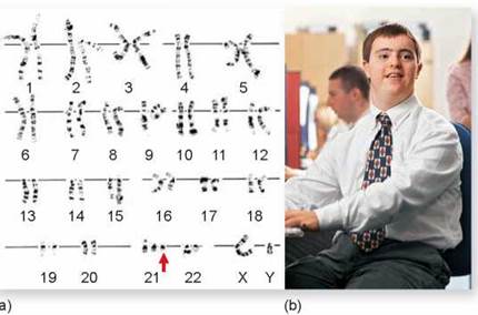
Figure 10.25. Down syndrome.
(a) In this karyotype of a male individual with Down syndrome, the trisomy at position 21 can be clearly seen. (b) A person with Down syndrome.
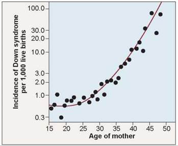
Figure 10.26. Correlation between maternal age and the incidence of Down syndrome.
As women age, the chances they will bear a child with Down syndrome increase. After a woman reaches age 35, the frequency of Down syndrome increases rapidly.
Nondisjunction Involving the Sex Chromosomes
As noted, 22 of the 23 pairs of human chromosomes are perfectly matched in both males and females and are called autosomes. The remaining pair are the sex chromosomes, X and Y. In humans, as in Drosophila (but by no means in all diploid species), females are XX and males XY; any individual with at least one Y chromosome is male. The Y chromosome is highly condensed and bears few functional genes in most organisms. Some of the active genes the Y chromosome does possess are responsible for the features associated with “maleness.” Individuals who gain or lose a sex chromosome do not generally experience the severe developmental abnormalities caused by changes in autosome numbers. Such individuals may reach maturity, but with somewhat abnormal features.
Nondisjunction of the X Chromosome. When X chromosomes fail to separate during meiosis, some of the gametes that are produced possess both X chromosomes and so are XX gametes; the other gametes that result from such an event have no sex chromosome and are designated “O.”
Figure 10.27 shows what happens if gametes from X chromosome nondisjunction combine with sperm. If an XX gamete combines with an X gamete, the resulting XXX zygote (in the upper left of the Punnett square) develops into a female who is taller than average but other symptoms can vary greatly. Some are normal in most respects, others may have lower reading and verbal skills, and still others are mentally retarded. If an XX gamete combines with a Y gamete (in the lower left), the XXY zygote develops into a sterile male who has many female body characteristics and, in some cases, diminished mental capacity. This condition, called Klinefelter syndrome, occurs in about 1 in 500 male births.
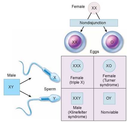
Figure 10.27. Nondisjunction of the X chromosome.
Nondisjunction of the X chromosome can produce sex chromosome aneuploidy—that is, abnormalities in the number of sex chromosomes.
If an O gamete fuses with a Y gamete (in the lower right), the OY zygote is nonviable and fails to develop further because humans cannot survive when they lack the genes on the X chromosome. If an O gamete fuses with an X gamete (in the upper right), the XO zygote develops into a sterile female of short stature, with a webbed neck and immature sex organs that do not undergo changes during puberty. The mental abilities of XO individuals are normal in verbal learning but lower in nonverbal/math-based problem solving. This condition, called Turner syndrome, occurs roughly once in every 5,000 female births.
Nondisjunction of the Y Chromosome. The Y chromosome can also fail to separate in meiosis, leading to the formation of YY gametes. When these gametes combine with X gametes, the XYY zygotes develop into fertile males of normal appearance. The frequency of the XYY genotype is about 1 per 1,000 newborn males.
Key Learning Outcome 10.8. The particular array of chromosomes that an individual possesses is called the karyotype. The human karyotype usually contains 23 pairs of chromosomes. Autosome loss is always lethal, and an extra autosome is with few exceptions lethal too.