MCAT Biology Review
Chapter 4: The Nervous System
4.3 Organization of the Human Nervous System
The nervous system is a remarkable collection of cells that governs both involuntary and voluntary behavior, while also maintaining homeostasis. Functions of the nervous system include:
· Sensation and perception
· Motor function
· Cognition (thinking) and problem-solving
· Executive function and planning
· Language comprehension and creation
· Memory
· Emotion and emotional expression
· Balance and coordination
· Regulation of endocrine organs
· Regulation of heart rate, breathing rate, vascular resistance, temperature, and exocrine glands
The human nervous system is a complex web of over 100 billion cells that communicate, coordinate, and regulate signals for the rest of the body. Mental and physical action occurs when the body can react to external stimuli using the nervous system. In this section, we will look at the nervous system and its basic organization.
Note: Much of the information contained in this section is also discussed in Chapter 1 of MCAT Behavioral Sciences Review.
CENTRAL AND PERIPHERAL NERVOUS SYSTEMS
Generally speaking, there are three kinds of nerve cells in the nervous system: sensory neurons, motor neurons, and interneurons. Sensory neurons (also known as afferent neurons) transmit sensory information from receptors to the spinal cord and brain. Motor neurons (also known asefferent neurons) transmit motor information from the brain and spinal cord to muscles and glands. Interneurons are found between other neurons and are the most numerous of the three types. Interneurons are located predominantly in the brain and spinal cord and are often linked to reflexive behavior.
MNEMONIC
Afferent neurons ascend in the spinal cord toward the brain; efferent neurons exit the spinal cord on their way to the rest of the body.
Different types of information require different types of processing. Processing of stimuli and response generation may happen at the level of the spinal cord, or may require input from the brainstem or cerebral cortex. Reflexes, discussed later in this section, only require processing at the level of the spinal cord. For example, when a reflex hammer hits the patellar tendon, the sensory information goes to the spinal cord, where a motor signal is sent to the quadriceps muscle, causing the leg to jerk forward at the knee. No input from the brain is required. However, some scenarios require input from the brain or brainstem. When this happens, supraspinal circuits are used.
Let’s turn to the overall structure of the human nervous system, which is diagrammed in Figure 4.9.
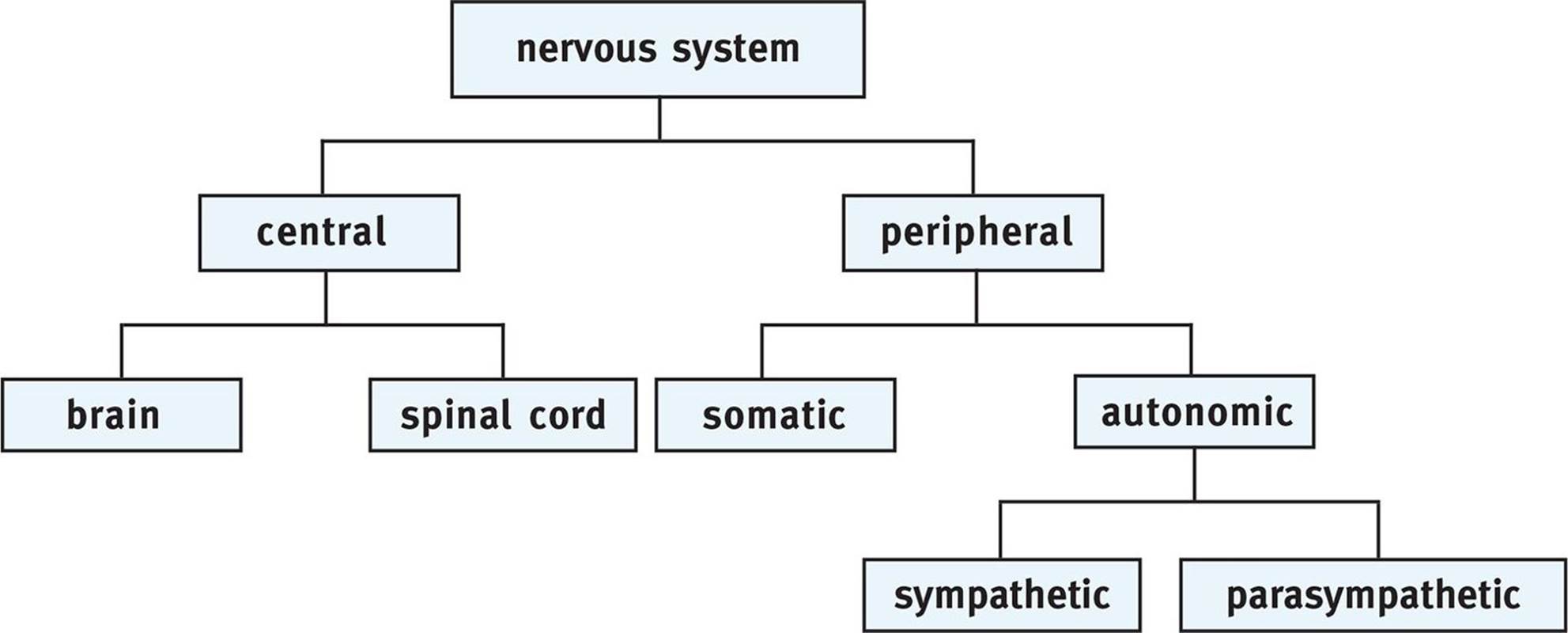 Figure 4.9. Major Divisions of the Nervous System
The nervous system can be broadly divided into two primary components: the central and peripheral nervous systems. The central nervous system (CNS) is composed of the brain and spinal cord. The brain consists of white matter and grey matter. The white matter consists of axons encased in myelin sheaths. The grey matter consists of unmyelinated cell bodies and dendrites. In the brain, the white matter lies deeper than the grey matter. At the base of the brain is the brainstem, which is largely responsible for basic life functions such as breathing. Note that the lobes of the brain and major brain structures are discussed in Chapter 1 of MCAT Behavioral Sciences Review.
The spinal cord extends downward from the brainstem and can be divided into four divisions: cervical, thoracic, lumbar, and sacral. Almost all of the structures below the neck receive sensory and motor innervation from the spinal cord. The spinal cord is protected by the vertebral column, which transmits nerves at the space between adjacent vertebrae. Like the brain, the spinal cord also consists of white and grey matter. The white matter lies on the outside of the cord, and the grey matter is deep within it. The axons of motor and sensory neurons are in the spinal cord. The sensory neurons bring information in from the periphery and enter on the dorsal (back) side of the spinal cord. The cell bodies of these sensory neurons are found in the dorsal root ganglia. Motor neurons exit the spinal cord ventrally, or on the side closest to the front of the body. The structure of the spinal cord can be seen in Figure 4.10.
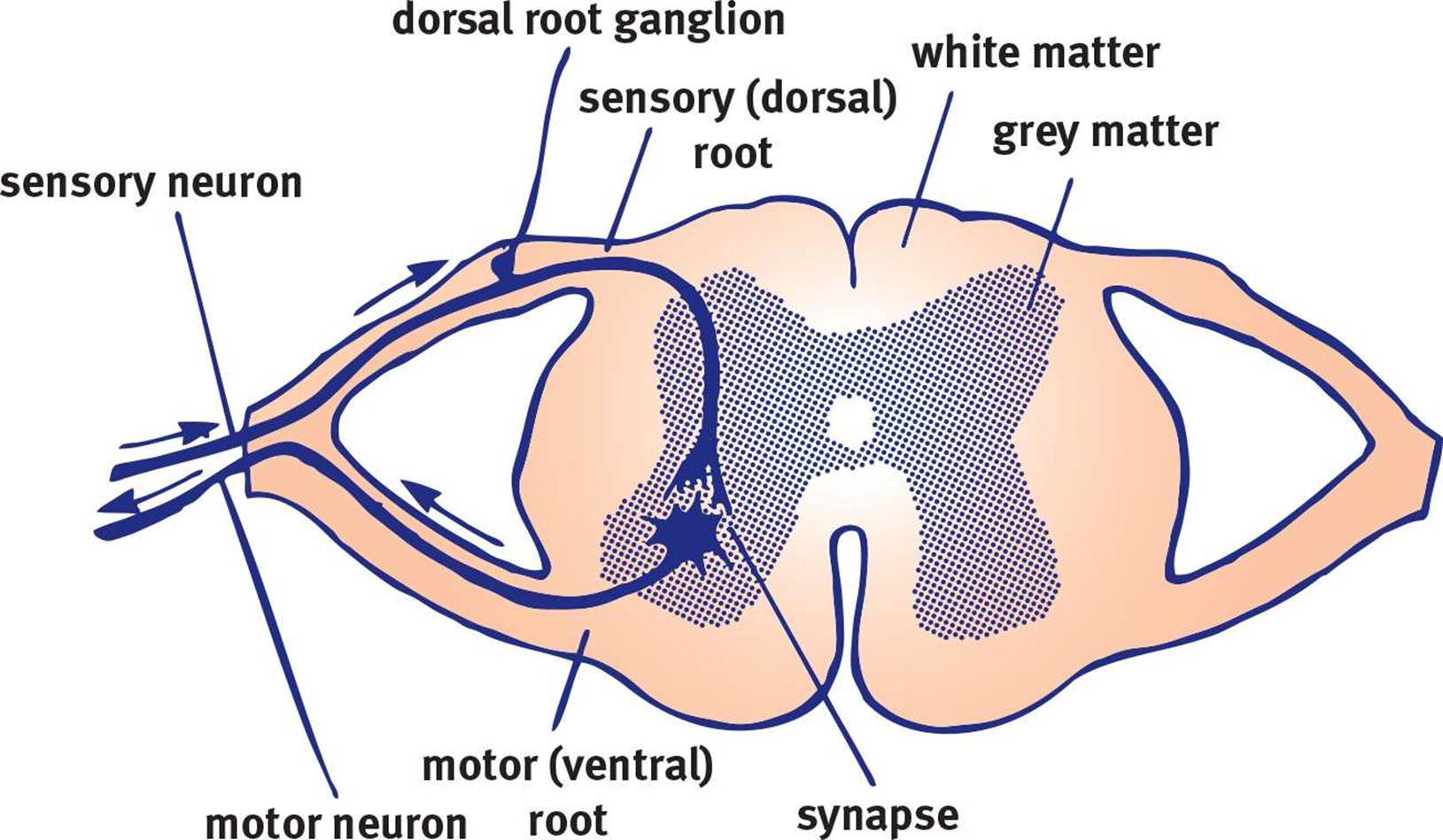 Figure 4.10. The Spinal Cord Sensory neurons transmit information about pain, temperature, and vibration up to the brain and have cell bodies in the dorsal root ganglia toward the back of the spinal cord; the motor neurons run from the brain along the opposite side of the spinal cord in the ventral root and control movements of skeletal muscle and glandular secretions.
The peripheral nervous system (PNS), in contrast, is made up of nerve tissue and fibers outside the brain and spinal cord, such as the 12 pairs of cranial and 31 pairs of spinal nerves. The PNS thus connects the CNS to the rest of the body and can itself be subdivided into the somatic and autonomic nervous systems.
The somatic nervous system consists of sensory and motor neurons distributed throughout the skin, joints, and muscles. Sensory neurons transmit information through afferent fibers. Motor impulses, in contrast, travel along efferent fibers.
The autonomic nervous system (ANS) generally regulates heartbeat, respiration, digestion, and glandular secretions. In other words, the ANS manages the involuntary muscles associated with many internal organs and glands. The ANS also helps regulate body temperature by activating sweating or piloerection, depending on whether we are too hot or too cold. The main thing to understand about these functions is that they are automatic, or independent of conscious control. Note the similarity between the words autonomic and automatic. This association makes it easy to remember that the autonomic nervous system manages automatic functions such as heartbeat, respiration, digestion, and temperature control.
One primary difference between the somatic and autonomic nervous systems is that the peripheral component of the autonomic nervous system contains two neurons. A motor neuron in the somatic nervous system goes directly from the spinal cord to the muscle without synapsing. In the autonomic nervous system, two neurons work in series to transmit messages from the spinal cord. The first neuron is known as the preganglionic neuron, whereas the second is the postganglionic neuron. The soma of the preganglionic neuron is in the CNS, and its axon travels to a ganglion in the PNS. Here it synapses on the cell body of the postganglionic neuron, which then affects the target tissue.
KEY CONCEPT
The first neuron in the autonomic nervous system is called the preganglionic neuron. The second neuron is the postganglionic neuron.
THE AUTONOMIC NERVOUS SYSTEM
The ANS has two subdivisions: the sympathetic nervous system and the parasympathetic nervous system. These two branches often act in opposition to one another, meaning that they are antagonistic. For example, the sympathetic nervous system acts to accelerate heart rate and inhibit digestion, while the parasympathetic nervous system, in contrast, decelerates heart rate and increases digestion.
The main role of the parasympathetic nervous system is to conserve energy. It is associated with resting and sleeping states and acts to reduce heart rate and constrict the bronchi. The parasympathetic nervous system is also responsible for managing digestion by increasing peristalsis and exocrine secretions. Acetylcholine is the neurotransmitter responsible for parasympathetic responses in the body and is released by both preganglionic and postganglionic neurons. The vagus nerve (cranial nerve X), is responsible for much of the parasympathetic innervation of the thoracic and abdominal cavity. The functions of the parasympathetic nervous system are summarized in Figure 4.11.
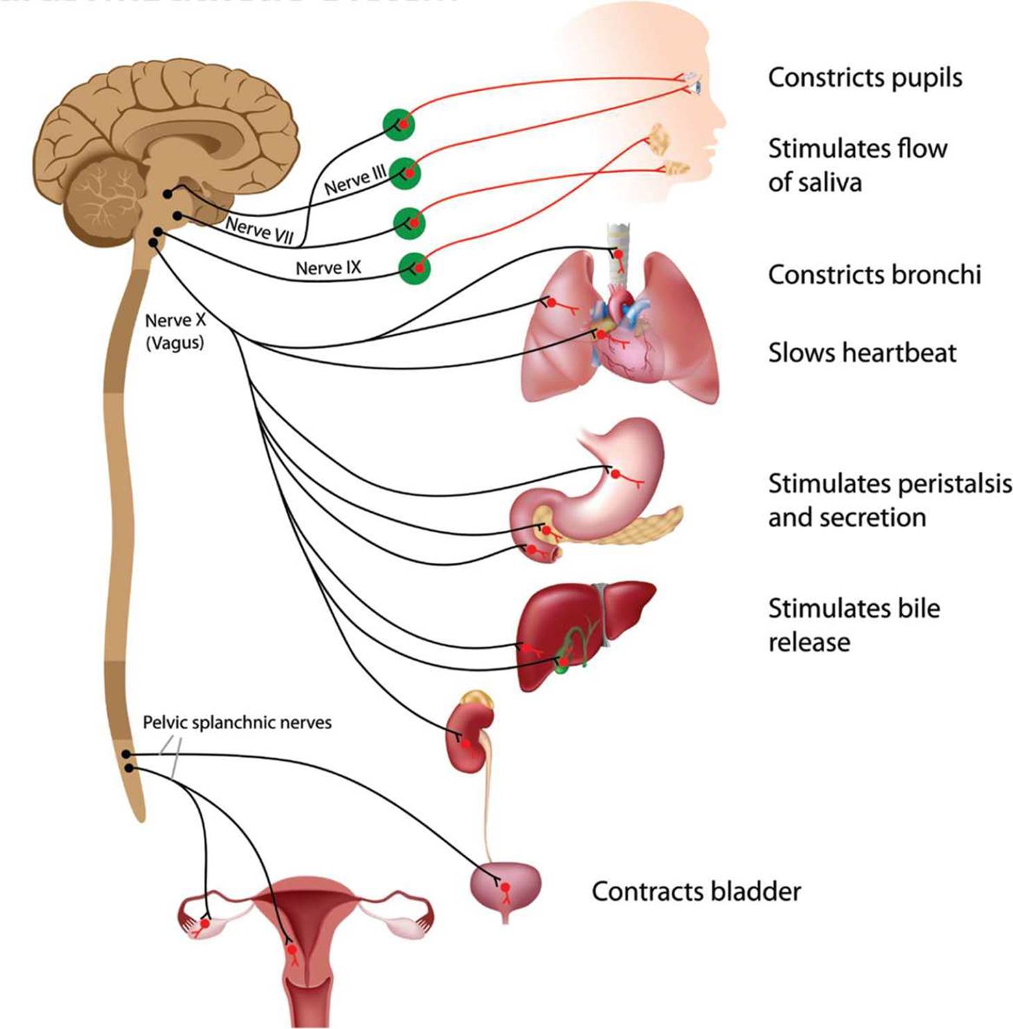 Figure 4.11. Functions of the Parasympathetic Nervous System
In contrast, the sympathetic nervous system is activated by stress. This can include everything from a mild stressor, such as keeping up with schoolwork, to emergencies that mean the difference between life and death. The sympathetic nervous system is closely associated with rage and fear reactions, also known as “fight-or-flight” reactions. When activated, the sympathetic nervous system:
· Increases heart rate
· Redistributes blood to muscles of locomotion
· Increases blood glucose concentration
· Relaxes the bronchi
· Decreases digestion and peristalsis
· Dilates the eyes to maximize light intake
· Releases epinephrine into the bloodstream
MNEMONIC
Sympathetic and parasympathetic nervous systems:
· Sympathetic: “fight-or-flight”
· Parasympathetic: “rest-and-digest”
The functions of the sympathetic nervous system are summarized in Figure 4.12. In the sympathetic nervous system, preganglionic neurons release acetylcholine, while most postganglionic neurons release norepinephrine.
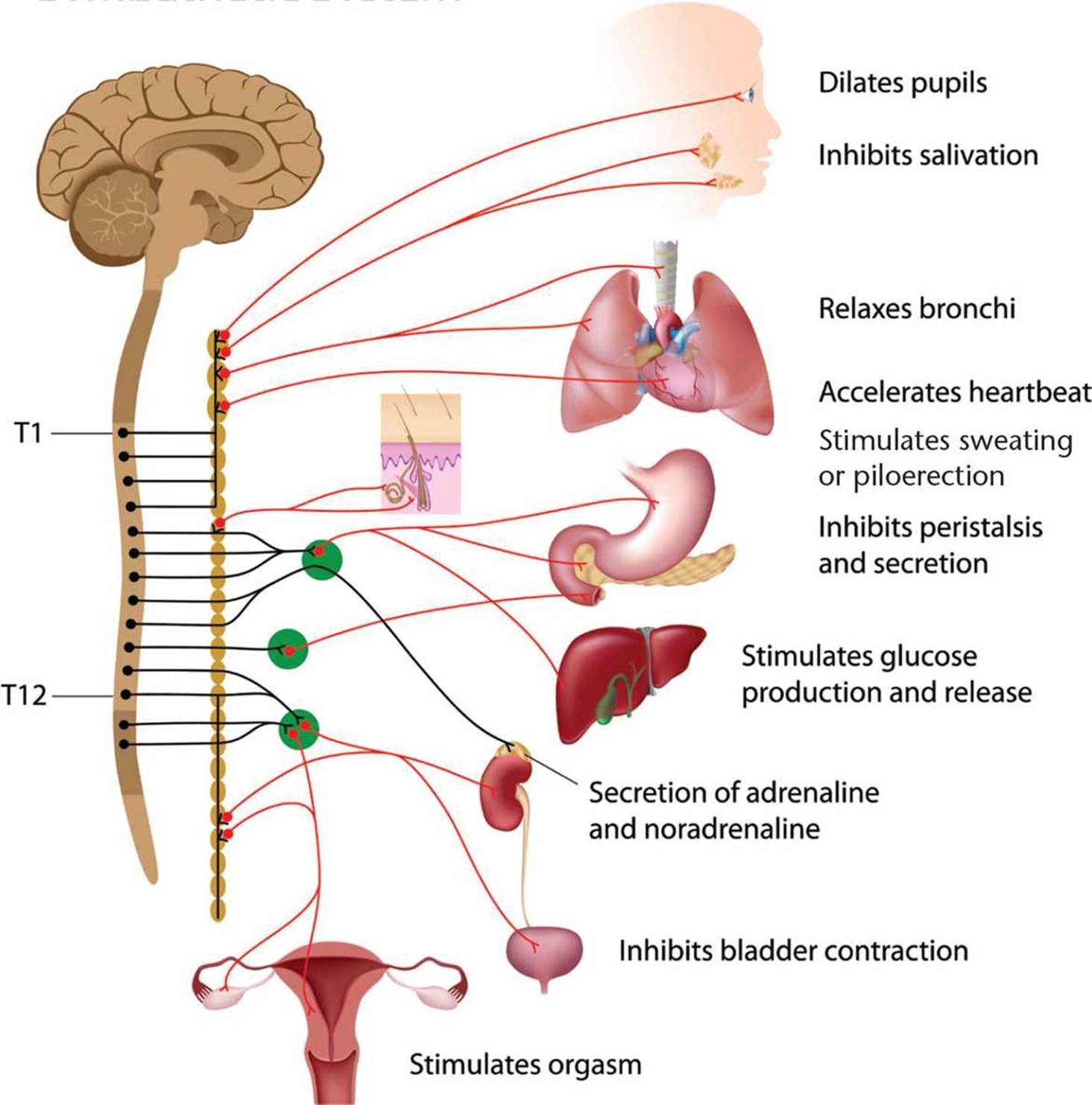 Figure 4.12. Functions of the Sympathetic Nervous System
REFLEXES
Neural circuits called reflex arcs control reflexive behavior. For example, consider what occurs when someone steps on a nail. Receptors in the foot detect pain, and the pain signal is transmitted by sensory neurons up to the spinal cord. At that point, the sensory neurons connect with interneurons, which can then relay pain impulses up to the brain. Rather than wait for the brain to send out a signal, interneurons in the spinal cord can also send signals to the muscles of both legs directly, causing the individual to withdraw the foot with pain while supporting with the other foot. The original sensory information still makes its way up to the brain; however, by the time it arrives there, the muscles have already responded to the pain, thanks to the reflex arc. There are two types of reflex arcs: monosynaptic and polysynaptic.
KEY CONCEPT
Consider the purpose of reflexes. Although it may be amusing to make your friends’ legs jump when you tap them, there is a more functional reason why this response occurs. The stretch on the patellar tendon makes the body think that the muscle may be getting overstretched. In response, the muscle contracts in order to prevent injury.
Monosynaptic
In a monosynaptic reflex arc, there is a single synapse between the sensory neuron that receives the stimulus and the motor neuron that responds to it. A classic example is the knee-jerk reflex, shown in Figure 4.13. When the patellar tendon is stretched, information travels up the sensory (afferent, presynaptic) neuron to the spinal cord, where it interfaces with the motor (efferent, postsynaptic) neuron that contracts the quadriceps muscle. The net result is extension of the leg, which lessens the tension on the patellar tendon. Note that the reflex is simply a feedback loop and a response to potential injury. If the patellar tendon or quadriceps muscles are stretched too far, they may tear, damaging the knee joint. Thus, the reflex serves to protect the muscle.
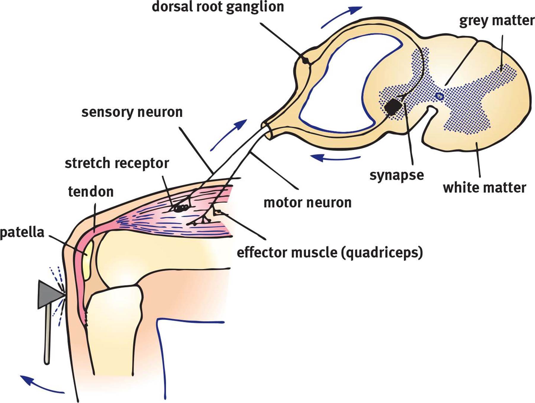 Figure 4.13. The Knee-Jerk Reflex The knee-jerk or knee extension reflex may be elicited by swiftly stretching the patellar tendon with a reflex hammer.
Polysynaptic
In a polysynaptic reflex arc, there is at least one interneuron between the sensory and motor neurons. A real-life example is the reaction to stepping on a nail described earlier, which involves the withdrawal reflex. The leg with which one steps on the nail will be stimulated to flex, using the hip muscles and hamstring muscles, pulling the foot away from the nail. This is a monosynaptic reflex, similar to the knee-jerk reflex described previously. However, if the person is to maintain balance, the other foot must be planted firmly on the ground. For this to occur, the motor neuron that controls the quadriceps muscles in the opposite leg must be stimulated, extending that leg. Interneurons in the spinal cord provide the connections from the incoming sensory information to the motor neurons in the supporting leg.
MCAT Concept Check 4.3:
Before you move on, assess your understanding of the material with these questions.
1. What parts of the nervous system are in the central nervous system (CNS)? Peripheral nervous system (PNS)?
· CNS:
· PNS:
2. What do afferent neurons do? Efferent neurons?
· Afferent:
· Efferent:
3. What functions are accomplished by the somatic nervous system? The autonomic nervous system?
· Somatic:
· Autonomic:
4. What are the effects of the sympathetic nervous system? The parasympathetic nervous system?
· Sympathetic:
· Parasympathetic:
5. What is the pathway of neural impulses in a monosynaptic reflex? In a polysynaptic reflex?
· Monosynaptic reflex:
· Polysynaptic reflex: