MCAT Biology Review
Chapter 1: The Cell
1.3 Classification and Structure of Prokaryotic Cells
Prokaryotes are the simplest of all organisms and include all bacteria. Prokaryotes do not contain any membrane-bound organelles, and their genetic material is organized into a single circular molecule of DNA concentrated in an area of the cell called the nucleoid region. Despite the simplicity of prokaryotes, they are incredibly diverse, and knowledge of this diversity is essential for the study of medicine because many prokaryotes can cause infection. In fact, choosing the appropriate antibiotic to fight an infection requires knowledge about the basic structure of the bacteria causing the infection.
PROKARYOTIC DOMAINS
There are three overarching domains into which all life is classified: Archaea, Bacteria, and Eukarya. Two of these—Archaea and Bacteria—contain prokaryotes. Initially, Archaea and Bacteria were classified together into the kingdom of Monera. However, modern genetics and biochemical techniques have indicated that the differences in the evolutionary pathways between Archaea and Bacteria are at least as significant as between either of these domains and Eukarya.
Archaea
Archaea are single-celled organisms that are visually similar to bacteria, but contain genes and several metabolic pathways that are more similar to eukaryotes than to bacteria. Historically, Archaea were considered extremophiles, in that they were most commonly isolated from harsh environments with extremely high temperatures, high salinity, or no light. More recent research has demonstrated a greater variety of habitats for these organisms, including the human body. Archaea are notable for their ability to use alternative sources of energy. While some are photosynthetic, many are chemosynthetic and are able to generate energy from inorganic compounds, including sulfur- and nitrogen-based compounds, such as ammonia.
Due to the similarities of this domain to eukaryotes, it is hypothesized that eukaryotes and the domain Archaea share a common origin. Both eukaryotes and Archaea start translation with methionine, contain similar RNA polymerases, and associate their DNA with histones. However, Archaea contain a single circular chromosome, divide by binary fission or budding, and overall share a similar structure to bacteria. Interestingly, Archaea are resistant to many antibiotics.
Bacteria
All bacteria contain a cell membrane and cytoplasm, and some have flagella or fimbriae (similar to cilia), as shown in Figure 1.6. Because bacteria and eukaryotes often share analogous structures, it can be difficult to develop medicines that target only bacteria. However, in some cases, even seemingly similar structures have enough biochemical differences to allow the targeting of one organism over the other. For example, bacterial flagella and eukaryotic flagella are distinct enough that scientists are able to develop antibacterial vaccines that specifically target the bacterial flagellum. Also, many antibiotics target the bacterial ribosome, which is significantly smaller than the eukaryotic ribosome.
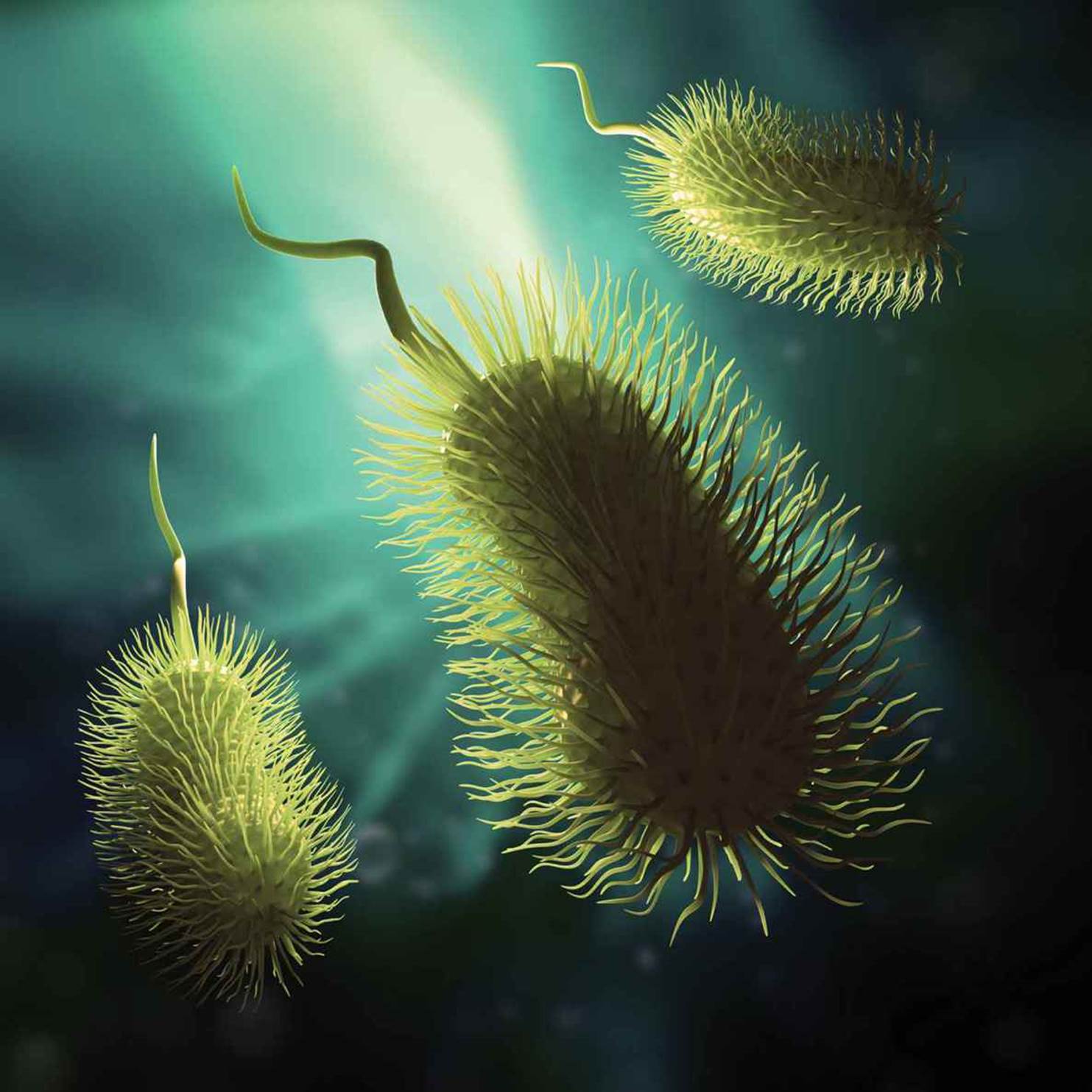 Figure 1.6. Prokaryotic Cell Specializations: Flagella and Fimbriae
REAL WORLD
Bacteria perform essential functions for human beings, including the production of vitamin K in the intestine. Vitamin K is required for production of plasma proteins necessary for blood clotting. Newborn infants are not yet colonized by bacteria and cannot produce clotting factors, putting them at risk for hemorrhage. When babies are born, they are given an injection of vitamin K to aid in the production of clotting factors until they have been colonized with bacteria.
There are approximately 5 × 1030 bacteria on earth, outnumbering all of the plants and animals combined. As mentioned in the introduction to this chapter, bacteria outnumber human cells in the body by 10:1. The relationship between the human body and bacteria is complex. Some bacteria are symbiotes, meaning that both humans and the bacteria benefit from the relationship. Examples include the bacteria in the human gut that produce vitamin K and biotin (vitamin B7), and which also prevent the overgrowth of harmful bacteria. Other bacteria are pathogens, meaning that they provide no advantage or benefit to the host, but rather cause disease. Pathogenic bacteria may live intracellularly or extracellularly. For example, Chlamydia trachomatis, a common sexually transmitted infection, lives inside cells of the reproductive tract; Clostridium tetani, the cause of tetanus, lives outside of cells and produces toxins that enter the bloodstream.
CLASSIFICATION OF BACTERIA BY SHAPE
Classification of bacteria by shape provides scientists and pathologists (physicians who specialize in the identification and characterization of disease) a common language to talk about the bacteria, as well as a way to identify different species of bacteria.
Most bacteria exist in one of three shapes, as shown in Figure 1.7. Spherical bacteria, known as cocci, include common pathogens such as Streptococcus pyogenes.
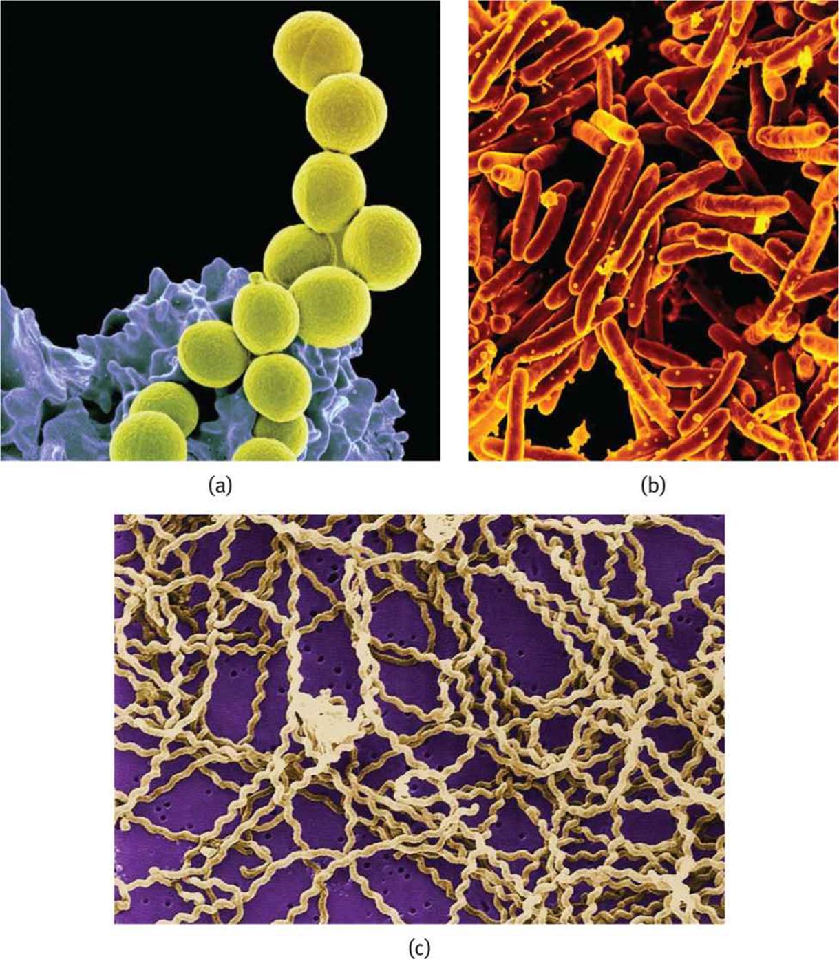 Figure 1.7. Prokaryotic Cell Shapes (a) Cocci (Staphylococcus aureus), (b) Bacilli (Mycobacterium tuberculosis), (c) Spirilli (Leptospira interrogans).
Rod-shaped bacteria, like Escherichia coli, are known as bacilli. Finally, spiral-shaped bacteria, known as spirilli, include such species as Treponema pallidum, which causes syphilis.
REAL WORLD
Very few pathogenic bacteria are spiral shaped. The three most common are:
· Treponema pallidum, the cause of syphilis
· Borrelia burgdorferi, the cause of Lyme disease
· Leptospira interrogans, the cause of Weil’s syndrome
AEROBES AND ANAEROBES
Some bacteria require oxygen for survival, while others do not. Bacteria that require oxygen for metabolism are termed obligate aerobes. Other bacteria that use fermentation, or some other form of cellular metabolism that does not require oxygen, are called anaerobes. There are different types of anaerobes. Anaerobes that cannot survive in an oxygen-containing environment are called obligate anaerobes; the presence of oxygen leads to the production of reactive oxygen-containing radicals in these species, which leads to cell death. Other bacteria can toggle between metabolic processes, using oxygen for aerobic metabolism if it is present, and switching to anaerobic metabolism if it is not. These bacteria are called facultative anaerobes. Finally, aerotolerant anaerobes are unable to use oxygen for metabolism, but are not harmed by its presence in the environment.
PROKARYOTIC CELL STRUCTURE
One of the main differences between prokaryotes and eukaryotes is that prokaryotes lack a nucleus and membrane-bound organelles, as shown in Figure 1.8. Prokaryotes are also single-celled organisms, meaning that each cell must be able to perform all of the functions necessary for life on its own. However, prokaryotes may live in colonies with other cells and may signal these cells to share information about the environment.
 Figure 1.8. Prokaryotic Cell Structure
Cell Wall
Because prokaryotes do not form multicellular organisms, each bacterium is responsible for protecting itself from the environment. The cell wall forms the outer barrier of the cell. The next layer is the cell membrane (plasma membrane), which is composed of phospholipids, similar to that of a eukaryote. Together, the cell wall and the cell membrane are known as the envelope.
The cell wall both provides structure and controls the movement of solutes into and out of the bacterium. This allows the cell to maintain concentration gradients relative to the environment. In bacteria, there are two main types of cell wall: gram positive and gram negative. The type of cell wall is determined by the Gram staining process with a crystal violet stain, followed by a counterstain with a substance called safranin. If the envelope absorbs the crystal violet stain, it will appear deep purple, and the cell is said to be gram positive. If the envelope does not absorb the crystal violet stain, but absorbs the safranin counterstain, then the cell will appear pink-red, and the cell is said to be gram negative.
REAL WORLD
The antibiotic penicillin targets the enzyme that catalyzes the cross-linking of peptidoglycan. If a gram-positive cell cannot cross-link its cell wall, it no longer serves as an effective barrier. The bacterium becomes susceptible to osmotic damage and lyses. Most bacteria have developed resistance mechanisms to penicillin, although a few bacteria—including Streptococcus pyogenes, which causes strep throat and some skin infections, and Treponema pallidum, which causes syphilis—are still very sensitive to this antibiotic.
Gram-positive cell walls consist of a thick layer of peptidoglycan, a polymeric substance made from amino acids and sugars. In addition to its structural and barrier functions, the cell wall may also aid a pathogen by providing protection from a host organism’s immune system. In addition to peptidoglycan, the gram-positive cell wall also contains lipoteichoic acid. The function of this acid is unknown, but the human immune system may be activated by exposure to these chemicals.
Gram-negative cell walls are very thin and also contain peptidoglycan, but in much smaller amounts. The cell walls of these bacteria directly abut the cell membrane. In addition to the cell wall and cell membrane, gram-negative bacteria also have outer membranes containing phospholipids and lipopolysaccharides. Interestingly, lipopolysaccharides are the part of the gram-negative bacteria that triggers an immune response in human beings; the inflammatory response to lipopolysaccharides is much stronger than the response to lipoteichoic acid.
KEY CONCEPT
Bacteria contain a cell wall, the composition of which is different in gram-positive and gram-negative bacteria. The human immune system can respond to the components of the cell wall, inciting an inflammatory response.
Flagella
Flagella are long, whiplike structures that can be used for propulsion; bacteria may have one, two, or many flagella, depending on the species. Flagella can be used to move toward food or away from toxins or immune cells. This ability of a cell to detect chemical stimuli and move toward or away from them is called chemotaxis. The flagella are composed of a filament, a basal body, and a hook, as shown in Figure 1.9. The filament is a hollow, helical structure composed of flagellin. The basal body is a complex structure that anchors the flagellum to the cytoplasmic membrane and is also the motor of the flagellum, rotating at rates up to 300 Hz. The hook connects the filament and the basal body so that, as the basal body rotates, it exerts torque on the filament, which can thereby spin and propel the bacterium forward. The overall structure of flagella is similar in both gram-positive and gram-negative bacteria, but there are slight differences due to the different physical structure and chemical composition of the envelope in gram-positive and gram-negative bacteria. Archaea also contain flagella, but this structure is quite different from that of bacteria and is unlikely to be asked about on Test Day.
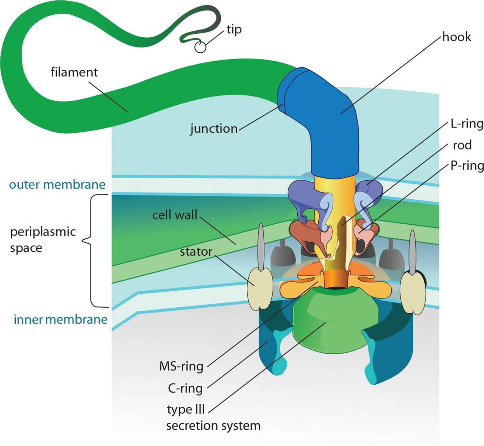 Figure 1.9. Prokaryotic Flagellum Structure The filament connects via the hook to the basal body (the complex structure of which is shown).
Other Organelles
As mentioned earlier, prokaryotes concentrate DNA in a region of the cell known as the nucleoid region, but do not contain a nuclear envelope. Prokaryotic DNA is carried on a single circular chromosome. This DNA is not coiled around histones, as it is in eukaryotes. In addition, DNA acquired from external sources may also be carried on smaller circular structures known as plasmids. Plasmids carry DNA that is not necessary for survival of the prokaryote—and therefore is not considered part of the genome of the bacterium—but may confer an advantage such as antibiotic resistance.
BRIDGE
The fact that prokaryotes and eukaryotes have different-sized ribosomes should imply to you that they carry out protein synthesis in slightly different ways. These differences are highlighted in Chapter 7 of MCAT Biochemistry Review. This difference also allows us to target bacterial ribosomes with a number of antibiotics, including tetracyclines, aminoglycosides, and macrolides, while leaving the eukaryotic ribosome more or less unaffected.
Prokaryotes lack several key organelles, such as mitochondria. Instead, the cell membrane is used for the electron transport chain and generation of ATP. Prokaryotes do contain a primitive cytoskeleton, but it is not nearly as complex as the one found in eukaryotes. Prokaryotes also contain ribosomes, but this ribosome is a different size from that found in eukaryotes: prokaryotic ribosomes contain 30S and 50S subunits, whereas eukaryotic ribosomes contain 40S and 60S ribosomes.
MCAT Concept Check 1.3:
Before you move on, assess your understanding of the material with these questions.
1. In what ways are Archaea similar to bacteria? In what ways are Archaea similar to eukaryotes?
· Similar to bacteria:
· Similar to eukaryotes:
2. What are the three common shapes of bacteria?
·
·
·
3. Compare and contrast the metabolisms of aerobic and anaerobic bacteria: (Note: Put “yes” or “no” in each box.)
|
Oxygen Present |
Oxygen Absent |
|||
|
Type of Bacteria |
Can survive |
Can carry out aerobic metabolism |
Can survive |
Can carry out anaerobic metabolism |
|
Obligate aerobe |
||||
|
Facultative anaerobe |
||||
|
Obligate anaerobe |
||||
|
Aerotolerant anaerobe |
||||
4. How do the envelopes of gram-positive and gram-negative bacteria differ?
· Gram-positive:
· Gram-negative:
5. How do the structures of eukaryotic and prokaryotic flagella differ?
· Eukaryotic:
· Prokaryotic: