Lippincott’s Illustrated Reviews: Biochemistr, Sixth Edition (2014)
UNIT II: Bioenergetics and Carbohydrate Metabolism
Chapter 6. Bioenergetics and Oxidative Phosphorylation
I. OVERVIEW
Bioenergetics describes the transfer and utilization of energy in biologic systems. It makes use of a few basic ideas from the field of thermodynamics, particularly the concept of free energy. Changes in free energy provide a measure of the energetic feasibility of a chemical reaction and can, therefore, allow prediction of whether a reaction or process can take place. Bioenergetics concerns only the initial and final energy states of reaction components, not the mechanism or how much time is needed for the chemical change to take place (the rate). In short, bioenergetics predicts if a process is possible, whereas kinetics measures how fast the reaction occurs (see p. 54).
II. FREE ENERGY
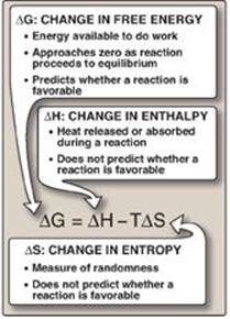
The direction and extent to which a chemical reaction proceeds is determined by the degree to which two factors change during the reaction. These are enthalpy (∆H, a measure of the change in heat content of the reactants and products) and entropy (∆S, a measure of the change in randomness or disorder of reactants and products; Figure 6.1). Neither of these thermodynamic quantities by itself is sufficient to determine whether a chemical reaction will proceed spontaneously in the direction it is written. However, when combined mathematically (see Figure 6.1), enthalpy and entropy can be used to define a third quantity, free energy (G), which predicts the direction in which a reaction will spontaneously proceed.
Figure 6.1 Relationship between changes in free energy (G), enthalpy (H), and entropy (S). T is the absolute temperature in Kelvin (K): K = oC + 273.
III. FREE ENERGY CHANGE
The change in free energy is represented in two ways, ∆G and ∆Go. The first, ∆G (without the superscript “o”), represents the change in free energy and, thus, the direction of a reaction at any specified concentration of products and reactants. DG, then, is a variable. This contrasts with the standard free energy change, ∆Go (with the superscript “o”), which is the energy change when reactants and products are at a concentration of 1 mol/l. [Note: The concentration of protons is assumed to be 10–7 mol/l (that is, pH = 7). This may be shown by a prime sign (ʹ), for example, ΔGoʹ.] Although ∆Go, a constant, represents energy changes at these nonphysiologic concentrations of reactants and products, it is nonetheless useful in comparing the energy changes of different reactions. Furthermore, ∆Go can readily be determined from measurement of the equilibrium constant (see p. 72). This section outlines the uses of ∆G, and ∆Go is described on p. 71.
A. Sign of ∆G and the direction of a reaction
∆G can be used to predict the direction of a reaction at constant temperature and pressure. Consider the reaction:
Figure 6.2 Change in free energy (ΔG) during a reaction. ![]() The product has a lower free energy (G) than the reactant.
The product has a lower free energy (G) than the reactant. ![]() The product has a higher free energy than the reactant.
The product has a higher free energy than the reactant.
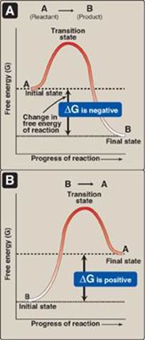
A ![]() B
B
1. Negative ∆G: If ∆G is negative, there is a net loss of energy, and the reaction goes spontaneously as written (that is, A is converted into B) as shown in Figure 6.2A. The reaction is said to be exergonic.
2. Positive ∆G: If ∆G is positive, there is a net gain of energy, and the reaction does not go spontaneously from B to A (see Figure 6.2B). Energy must be added to the system to make the reaction go from B to A. The reaction is said to be endergonic.
3. ∆G is zero: If ∆G = 0, the reactants are in equilibrium. [Note: When a reaction is proceeding spontaneously (that is, free energy is being lost) then the reaction continues until ∆G reaches zero and equilibrium is established.]
B. ∆G of the forward and back reactions
The free energy of the forward reaction (A → B) is equal in magnitude but opposite in sign to that of the back reaction (B → A). For example, if ∆G of the forward reaction is −5 kcal/mol, then that of the back reaction is +5 kcal/mol. [Note: ∆G can also be expressed in kilojoules per mole or kJ/mol (1 kcal = 4.2 kJ).]
C. ∆G and the concentration of reactants and products
The ∆G of the reaction A → B depends on the concentration of the reactant and product. At constant temperature and pressure, the following relationship can be derived:
![]()
|
where |
where ∆Go is the standard free energy change (see below) |
|
|
R is the gas constant (1.987 cal/mol K) |
||
|
T is the absolute temperature (K) |
||
|
[A] and [B] are the actual concentrations of the reactant and product |
||
|
In represents the natural logarithm |
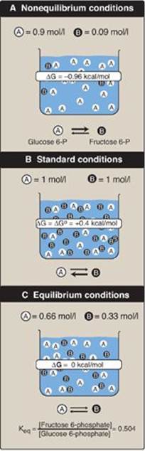
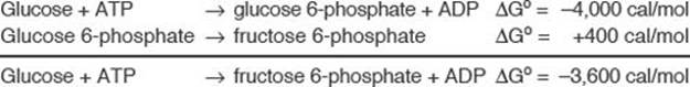
A reaction with a positive ∆Go can proceed in the forward direction (have a negative overall ∆G) if the ratio of products to reactants ([B]/[A]) is sufficiently small (that is, the ratio of reactants to products is large). For example, consider the reaction:
Glucose 6-phosphate ![]() fructose 6-phosphate
fructose 6-phosphate
Figure 6.3A shows reaction conditions in which the concentration of reactant, glucose 6-phosphate, is high compared with the concentration of product, fructose 6-phosphate. This means that the ratio of the product to reactant is small, and RT ln([fructose 6-phosphate]/[glucose 6-phosphate]) is large and negative, causing ∆G to be negative despite ∆Go being positive. Thus, the reaction can proceed in the forward direction.
Figure 6.3 Free energy change (![]() ) of a reaction depends on the concentration of reactant and product
) of a reaction depends on the concentration of reactant and product ![]() . For the conversion of glucose 6-phosphate to fructose 6-phosphate, ΔG is negative when the ratio of reactant
. For the conversion of glucose 6-phosphate to fructose 6-phosphate, ΔG is negative when the ratio of reactant ![]() to product
to product ![]() is large (top, panel A), is positive under standard conditions (middle, panel B), and is zero at equilibrium (bottom, panel C). ΔG0 = standard free energy change.
is large (top, panel A), is positive under standard conditions (middle, panel B), and is zero at equilibrium (bottom, panel C). ΔG0 = standard free energy change.
D. Standard free energy change
Τhe standard free energy change, ∆Go, is so called because it is equal to the free energy change, ∆G, under standard conditions (that is, when reactants and products are at 1 mol/l concentrations; see Figure 6.3B). Under these conditions, the natural logarithm of the ratio of products to reactants is zero (ln1 = 0), and, therefore, the equation shown at the bottom of the previous page becomes:
∆G = ∆Go + 0
1. ∆Go and the direction of a reaction: Under standard conditions, ∆Go can be used to predict the direction a reaction proceeds because, under these conditions, ∆Go is equal to ∆G. However, ∆Go cannot predict the direction of a reaction under physiologic conditions, because it is composed solely of constants (R, T, and Keq [see below]) and is not, therefore, altered by changes in product or substrate concentrations.
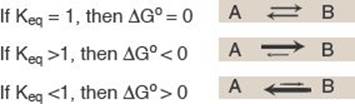
2. Relationship between ∆Go and Keq: In a reaction A ![]() B, a point of equilibrium is reached at which no further net chemical change takes place (that is, when A is being converted to B as fast as B is being converted to A). In this state, the ratio of [B] to [A] is constant, regardless of the actual concentrations of the two compounds:
B, a point of equilibrium is reached at which no further net chemical change takes place (that is, when A is being converted to B as fast as B is being converted to A). In this state, the ratio of [B] to [A] is constant, regardless of the actual concentrations of the two compounds:
![]()
where Keq is the equilibrium constant, and [A]eq and [B]eq are the concentrations of A and B at equilibrium. If the reaction A ![]() B is allowed to go to equilibrium at constant temperature and pressure, then, at equilibrium, the overall ∆G is zero. Therefore,
B is allowed to go to equilibrium at constant temperature and pressure, then, at equilibrium, the overall ∆G is zero. Therefore,
![]()
where the actual concentrations of A and B are equal to the equilibrium concentrations of reactant and product [A]eq and [B]eq, and their ratio is equal to the Keq. Thus,
![]()
This equation allows some simple predictions:
3. ∆Go of two consecutive reactions: The ∆Gos are additive in any sequence of consecutive reactions, as are the ∆Gs. For example:
4. ∆Gs of a pathway: The additive property of free energy changes is very important in biochemical pathways through which substrates must pass in a particular direction (for example, A → B → C → D →…). As long as the sum of the ∆Gs of the individual reactions is negative, the pathway can potentially proceed as written, even if some of the individual reactions of the pathway have a positive ∆G. The actual rate of the reactions does, of course, depend on the lowering of activation energies by the enzymes that catalyze the reactions (see p. 55).
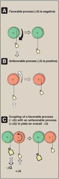
Figure 6.4 Mechanical model of coupling of favorable and unfavorable processes. ![]() Gear with weight attached spontaneously turns in the direction that achieves the lowest energy state.
Gear with weight attached spontaneously turns in the direction that achieves the lowest energy state. ![]() The reverse movement is energetically unfavorable (not spontaneous).
The reverse movement is energetically unfavorable (not spontaneous). ![]() The energetically favorable movement can drive the unfavorable one.
The energetically favorable movement can drive the unfavorable one.
IV. ADENOSINE TRIPHOSPHATE AS AN ENERGY CARRIER
Reactions or processes that have a large positive ∆G, such as moving ions against a concentration gradient across a cell membrane, are made possible by coupling the endergonic movement of ions with a second, spontaneous process with a large negative ∆G such as the exergonic hydrolysis of adenosine triphosphate (ATP). [Note: In the absence of enzymes, ATP is a stable molecule because its hydrolysis has a high activation energy (see p. 55).] Figure 6.4 shows a mechanical model of energy coupling. The simplest example of energy coupling in biologic reactions occurs when the energy-requiring and the energy-yielding reactions share a common intermediate.
A. Common intermediates
Two chemical reactions have a common intermediate when they occur sequentially so that the product of the first reaction is a substrate for the second. For example, given the reactions
A + B → C + D
D + X → Y + Z
D is the common intermediate and can serve as a carrier of chemical energy between the two reactions. Many coupled reactions use ATP to generate a common intermediate. These reactions may involve the transfer of a phosphate group from ATP to another molecule. Other reactions involve the transfer of phosphate from an energy-rich intermediate to adenosine diphosphate (ADP), forming ATP.
Figure 6.5 Adenosine triphosphate.
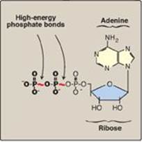
B. Energy carried by adenosine triphosphate
ATP consists of a molecule of adenosine (adenine + ribose) to which three phosphate groups are attached (Figure 6.5). If one phosphate is removed, ADP is produced. If two phosphates are removed, adenosine monophosphate (AMP) results. The standard free energy of hydrolysis of ATP, ∆Go, is approximately –7.3 kcal/mol for each of the two terminal phosphate groups. Because of this large negative ∆Go, ATP is called a high-energy phosphate compound.
V. ELECTRON TRANSPORT CHAIN
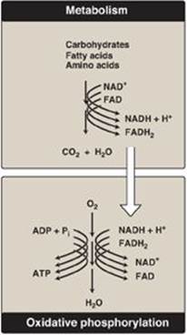
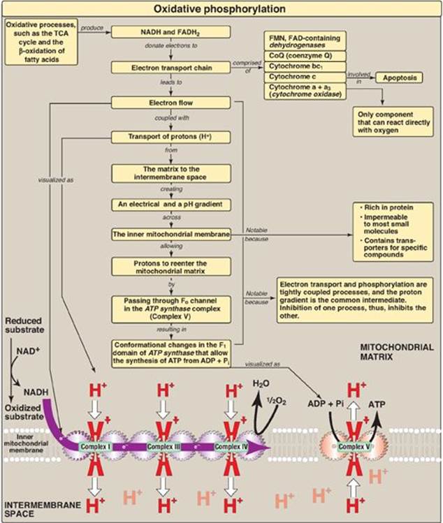
Energy-rich molecules, such as glucose, are metabolized by a series of oxidation reactions ultimately yielding CO2 and water (Figure 6.6). The metabolic intermediates of these reactions donate electrons to specific coenzymes, nicotinamide adenine dinucleotide (NAD+) and flavin adenine dinucleotide (FAD), to form the energy-rich reduced forms, NADH and FADH2. These reduced coenzymes can, in turn, each donate a pair of electrons to a specialized set of electron carriers, collectively called the electron transport chain (ETC), described in this section. As electrons are passed down the ETC, they lose much of their free energy. This energy is used to move protons across the inner mitochondrial membrane, creating a proton gradient that drives the production of ATP from ADP and inorganic phosphate (Pi), described on p. 77. The coupling of electron transport with ATP synthesis is called oxidative phosphorylation, often denoted as OXPHOS. It proceeds continuously in all tissues that contain mitochondria. [Note: The remainder of the free energy not trapped as ATP is used to drive ancillary reactions such as calcium transport into mitochondria (see p. 133) and to generate heat.]
A. The electron transport chain of the mitochondrion
The ETC (except for cytochrome c; see p. 75) is located in the inner mitochondrial membrane and is the final common pathway by which electrons derived from different fuels of the body flow to oxygen (O2).
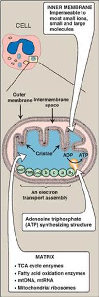
1. Membranes of the mitochondrion: The mitochondrion contains an outer and an inner membrane separated by the intermembrane space. Although the outer membrane contains special channels (formed by the protein porin), making it freely permeable to most ions and small molecules, the inner membrane is a specialized structure that is impermeable to most small ions, including protons and small molecules such as ATP, ADP, pyruvate, and other metabolites important to mitochondrial function (Figure 6.7). Specialized carriers or transport systems are required to move ions or molecules across this membrane. The inner mitochondrial membrane is unusually rich in protein, over half of which is directly involved in oxidative phosphorylation. It also is highly convoluted. The convolutions, called cristae, serve to greatly increase the surface area of the inner membrane.
Figure 6.6 The metabolic breakdown of energyyielding molecules. NAD(H) = nicotinamide adenine dinucleotide; FAD(H2)= flavin adenine dinucleotide; ADP = adenosine diphosphate; ATP = adenosine triphosphate; Pi = inorganic phosphate.
2. Matrix of the mitochondrion: This gel-like solution in the interior of mitochondria is also rich in protein. These molecules include the enzymes responsible for the oxidation of pyruvate, amino acids, and fatty acids (by β-oxidation) as well as those of the tricarboxylic acid (TCA) cycle. The synthesis of glucose, urea, and heme occurs partially in the matrix of mitochondria. In addition, the matrix contains NAD+ and FAD (the oxidized forms of the two coenzymes that are required as hydrogen acceptors), and ADP and Pi, which are used to produce ATP. [Note: The matrix also contains mitochondrial DNA (mtDNA) and RNA (mtRNA) and ribosomes.]
Figure 6.7 Structure of a mitochondrion showing schematic representation of the electron transport chain and the ATP synthesizing structure on the inner membrane. [Note: In contrast to the inner membrane, the outer membrane is highly permeable, and the milieu of the intermembrane space is like that of the cytosol.] mtDNA = mitochondrial DNA; mtRNA = mitochondrial RNA; TCA = tricarboxylic acid.
B. Organization of the electron transport chain
The inner mitochondrial membrane contains five separate protein complexes, called Complexes I, II, III, IV, and V. Complexes I–IV each contain part of the ETC (Figure 6.8). These complexes accept or donate electrons to the relatively mobile electron carriers, coenzyme Q and cytochrome c. Each carrier in the ETC can receive electrons from an electron donor and can subsequently donate electrons to the next acceptor in the chain. The electrons ultimately combine with O2 and protons to form water. This requirement for O2 makes the electron transport process the respiratory chain, which accounts for the greatest portion of the body’s use of O2. Complex V is described on p. 78.
C. Reactions of the electron transport chain
With the exception of coenzyme Q, which is a lipid-soluble quinone, all members of this chain are proteins. These may function as enzymes as is the case with the flavin-containing dehydrogenases, may contain iron as part of an iron-sulfur center, may contain iron as part of the porphyrin prosthetic group of heme as in the cytochromes, or may contain copper as does the cytochrome a + a3 complex.
1. Formation of NADH: NAD+ is reduced to NADH by dehydrogenases that remove two hydrogen atoms from their substrate. (For examples of these reactions, see the discussion of the dehydrogenases of the TCA cycle, p. 112.) Both electrons but only one proton (that is, a hydride ion [:H–]) are transferred to the NAD+, forming NADH plus a free proton.
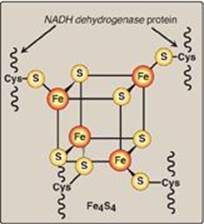
2. NADH dehydrogenase: The free proton plus the hydride ion carried by NADH are transferred to NADH dehydrogenase, a protein complex (Complex I) embedded in the inner mitochondrial membrane. Complex I has a tightly bound molecule of flavin mononucleotide (FMN), a coenzyme structurally related to FAD (see Figure 28.15, p. 380) that accepts the two hydrogen atoms (2e– + 2H+), becoming FMNH2. NADH dehydrogenase also contains peptide subunits with iron-sulfur centers (Figure 6.9). At Complex I, electrons move from NADH to FMN to the iron of the iron-sulfur centers and then to coenzyme Q. As electrons flow, they lose energy. This energy is used to pump protons across the inner mitochondrial membrane, from the matrix to the intermembrane space.
3. Succinate dehydrogenase: At Complex II, electrons from the succinate dehydrogenase–catalyzed oxidation of succinate to fumarate move from the coenzyme, FADH2, to an iron-sulfur protein, and then to coenzyme Q. [Note: No energy is lost in this process, and, therefore, no protons are pumped at Complex II.]
Figure 6.8 Electron transport chain. The flow of electrons is shown by the magenta arrows. NAD(H) = nicotinamide adenine dinucleotide; FMN = flavin mononucleotide; FAD = flavin adenine dinucleotide; Fe-S = iron-sulfur center; CoQ = coenzyme Q.
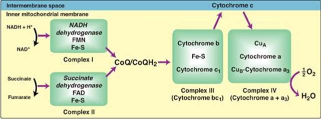
4. Coenzyme Q: Coenzyme Q (CoQ) is a quinone derivative with a long, hydrophobic isoprenoid tail. It is also called ubiquinone because it is ubiquitous in biologic systems. CoQ is a mobile electron carrier and can accept hydrogen atoms from NADH dehydrogenase (Complex I), from succinate dehydrogenase (Complex II), and from other mitochondrial dehydrogenases: glycerophosphate dehydrogenase (see p. 79) and acyl CoA dehydrogenase(see p. 192). CoQ transfers electrons to Complex III (cytochrome bc1). CoQ, then, links the flavoprotein dehydrogenases to the cytochromes.
5. Cytochromes: The remaining members of the ETC are cytochrome proteins. Each contains a heme group (a porphyrin ring plus iron). Unlike the heme groups of hemoglobin, the cytochrome iron is reversibly converted from its ferric (Fe3+) to its ferrous (Fe2+) form as a normal part of its function as an acceptor and donor of electrons. Electrons are passed along the chain from cytochrome bc1 (Complex III), to cytochrome c, and then to cytochromes a + a3 (Complex IV; see Figure 6.8). As electrons flow, protons are pumped across the inner mitochondrial membrane at Complexes III and IV. [Note: Cytochrome c is located in the intermembrane space, loosely associated with the outer face of the inner membrane. As seen with CoQ, cytochrome c is a mobile carrier of electrons.]
6. Cytochrome a + a3: This cytochrome complex (Complex IV) is the only electron carrier in which the heme iron has an available coordination site that can react directly with O2 and so also is called cytochrome oxidase. At Complex IV, the transported electrons, O2, and free protons are brought together, and O2 is reduced to water (see Figure 6.8). [Note: Four electrons are required to reduce one molecule of O2 to two molecules of water.] Cytochrome oxidase contains copper (Cu) atoms that are required for this complicated reaction to occur. Electrons move from CuA to cytochrome a to cytochrome a3 (in association with CuB) to O2.
Figure 6.9 Iron-sulfur (Fe-S) center of Complex I. [Note: Complexes II and III also contain iron-sulfur centers.] NADH = nicotinamide adenine dinucleotide;Cys = cysteine.
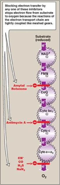
7. Site-specific inhibitors: Site-specific inhibitors of electron transport have been identified and are illustrated in Figure 6.10. These compounds prevent the passage of electrons by binding to a component of the chain, blocking the oxidation-reduction reaction. Therefore, all electron carriers before the block are fully reduced, whereas those located after the block are oxidized. [Note: Inhibition of electron transport inhibits ATP synthesis because these processes are tightly coupled (see p. 77).]
Incomplete reduction of oxygen to water produces reactive oxygen species (ROS), such as superoxide (O2–•), hydrogen peroxide (H2O2), and hydroxyl radicals (OH•). ROS damage DNA and proteins and cause lipid peroxidation. Enzymes such as superoxide dismutase (SOD), catalase, and glutathione peroxidase are cellular defenses against ROS.
Figure 6.10 Site-specific inhibitors of electron transport shown using a mechanical model for the coupling of oxidationreduction reactions. [Note: Figure illustrates normal direction of electron flow.] CN- = cyanide; CO = carbon monoxide; H2S = hydrogen sulfide; NaN3 = sodium azide; FMN = flavin mononucleotide; FAD = flavin adenine dinucleotide; CoQ = coenzyme Q; Cyto = cytochrome.
D. Release of free energy during electron transport
The free energy released as electrons are transferred along the ETC from an electron donor (reducing agent or reductant) to an electron acceptor (oxidizing agent or oxidant) is used to pump protons at Complexes I, III, and IV. [Note: The electrons can be transferred as hydride ions (:H–) to NAD+; as hydrogen atoms (•H) to FMN, CoQ, and FAD; or as electrons (e–) to cytochromes.]
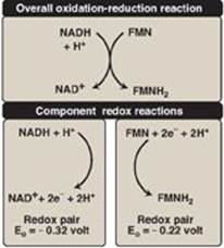
1. Redox pairs: Oxidation (loss of electrons) of one substance is always accompanied by reduction (gain of electrons) of a second. For example, Figure 6.11 shows the oxidation of NADH to NAD+ by NADH dehydrogenase at Complex I, accompanied by the reduction of FMN, the prosthetic group, to FMNH2. Such oxidation-reduction reactions can be written as the sum of two separate half-reactions, one an oxidation and the other a reduction (see Figure 6.11). NAD+ and NADH form a redox pair, as do FMN and FMNH2. Redox pairs differ in their tendency to lose electrons. This tendency is a characteristic of a particular redox pair and can be quantitatively specified by a constant, Eo (the standard reduction potential), with units in volts.
2. Standard reduction potential: The Eo of various redox pairs can be ordered from the most negative Eo to the most positive. The more negative the Eo of a redox pair, the greater the tendency of the reductant member of that pair to lose electrons. The more positive the Eo, the greater the tendency of the oxidant member of that pair to accept electrons. Therefore, electrons flow from the pair with the more negative Eo to that with the more positive Eo. The Eo values for some members of the ETC are shown in Figure 6.12. [Note: The components of the chain are arranged in order of increasingly positive Eo values.]
3. Relationship of ∆Go to ∆Eο : The ∆Go is related directly to the magnitude of the change in Eo:
∆Go = – n F ∆Eo
|
where |
n = number of electrons transferred (1 for a cytochrome, 2 for NADH, FADH2, and coenzyme Q) |
|
|
F = Faraday constant (23.1 kcal/volt mol) |
||
|
∆Eo = Eo of the electron-accepting pair minus the Eo of the electron-donating pair |
||
|
∆Go = change in the standard free energy |
4. ∆Go of ATP: The ∆Go for the phosphorylation of ADP to ATP is +7.3 kcal/mol. The transport of a pair of electrons from NADH to O2 through the ETC releases 52.58 kcal. Therefore, more than sufficient energy is available to produce 3 ATP from 3 ADP and 3 Pi (3 × 7.3 = 21.9 kcal/mol), sometimes expressed as a P:O ratio (ATP made per O atom reduced) of 3:1. The remaining calories are used for ancillary reactions or released as heat. [Note: The P:O for FADH2 is 2:1 because Complex I is bypassed.]
Figure 6.11 Oxidation of NADH by FMN, separated into two component half-reactions. NAD(H) = nicotinamide adenine dinucleotide; FMN(H2) = flavin mononucleotide.
VI. PHOSPHORYLATION OF ADP TO ATP
The transfer of electrons down the ETC is energetically favored because NADH is a strong electron donor and O2 is an avid electron acceptor. However, the flow of electrons does not directly result in ATP synthesis.
A. Chemiosmotic hypothesis
The chemiosmotic hypothesis (also known as the Mitchell hypothesis) explains how the free energy generated by the transport of electrons by the ETC is used to produce ATP from ADP + Pi.
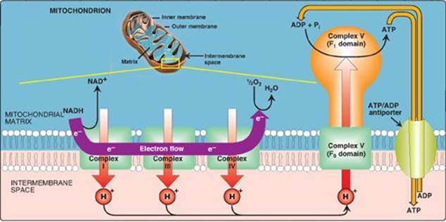
1. Proton pump: Electron transport is coupled to the phosphorylation of ADP by the pumping of protons across the inner mitochondrial membrane, from the matrix to the intermembrane space, at Complexes I, III, and IV. This process creates an electrical gradient (with more positive charges on the outside of the membrane than on the inside) and a pH gradient (the outside of the membrane is at a lower pH than the inside) as shown in Figure 6.13. The energy generated by this proton gradient is sufficient to drive ATP synthesis. Thus, the proton gradient serves as the common intermediate that couples oxidation to phosphorylation.
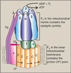
2. ATP synthase: The multisubunit enzyme ATP synthase (Complex V; see Figure 6.14) synthesizes ATP using the energy of the proton gradient. It contains a domain (Fo) that spans the inner mitochondrial membrane, and an extramembranous domain (F1) that appears as a sphere that protrudes into the mitochondrial matrix (see Figure 6.13). The chemiosmotic hypothesis proposes that after protons have been pumped to the cytosolic side of the inner mitochondrial membrane, they reenter the matrix by passing through a proton channel in the Fo domain, driving rotation of the c ring of Fo and, at the same time, dissipating the pH and electrical gradients. Fo rotation causes conformational changes in the β subunits of the F1 domain that allow them to bind ADP + Pi, phosphorylate ADP to ATP, and release ATP. [Note: ATP synthase is also called F1/Fo-ATPase because the isolated enzyme can catalyze the hydrolysis of ATP to ADP and Pi.]
Figure 6.12 Standard reduction potentials (Eo) of some reactions. NAD(H) = nicotinamide adenine dinucleotide; FMN(H2) = flavin mononucleotide.
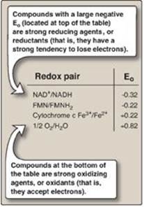
Figure 6.13 Electron transport chain shown in association with the transport of protons (H+). A total of ten H+ are pumped for each nicotinamide adenine dinucleotide (NADH) oxidized. [Note: H+ are not pumped at Complex II.]
a. Coupling in oxidative phosphorylation: In normal mitochondria, ATP synthesis is coupled to electron transport through the proton gradient. Increasing (or decreasing) one process has the same effect on the other. For example, hydrolysis of ATP to ADP and Pi in energy-requiring reactions increases the availability of substrates for ATP synthase and, thus, increases proton flow through the enzyme. Electron transport and proton pumping by the ETC increase to maintain the proton gradient. [Note: Increased oxidation of NADH at Complex I and, consequently, an increase in NADH-producing pathways of metabolism, such as the TCA cycle, results.]
b. Oligomycin: This drug binds to the Fo (hence the letter “o”) domain of ATP synthase, closing the proton channel and preventing reentry of protons into the matrix, thereby preventing phosphorylation of ADP to ATP. Because the pH and electrical gradients cannot be dissipated in the presence of this drug, electron transport stops because of the difficulty of pumping any more protons against the steep gradients. This dependency of cellular respiration on the ability to phosphorylate ADP to ATP is known as respiratory control and is the consequence of the tight coupling of these processes.
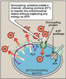
c. Uncoupling proteins: Uncoupling proteins (UCPs) occur in the inner mitochondrial membrane of mammals, including humans. These proteins form channels that allow protons to reenter the mitochondrial matrix without energy being captured as ATP (Figure 6.15). The energy is released as heat, and the process is called nonshivering thermogenesis. UCP1, also called thermogenin, is responsible for heat production in the brown adipocytes of mammals. In brown fat, unlike the more abundant white fat, almost 90% of its respiratory energy is used for thermogenesis in response to cold in the neonate and during arousal in hibernating animals. However, humans appear to have few concentrated deposits of brown fat (except in the newborn), and UCP1 does not appear to play a major role in energy balance. [Note: Uncoupling proteins UCP2–UCP5 have been found in other tissues, but their full significance remains unclear.]
Figure 6.14 ATP synthase (F1Fo-ATPase). [Note: The rotation of the ring of c subunits in the Fo domain results in conformational changes in the β subunits of the F1 domain that allow phosphorylation of adenosine diphosphate (ADP) to adenosine triphosphate (ATP). Pi = inorganic phosphate.
d. Synthetic uncouplers: Electron transport and phosphorylation can also be uncoupled by compounds that pick up protons in the intermembrane space and release them in the matrix, dissipating the gradient. The classic example is 2,4-dinitrophenol, a lipophilic proton carrier that readily diffuses through the mitochondrial membrane. This uncoupler causes electron transport to proceed at a rapid rate without establishing a proton gradient, much as do the UCPs (see Figure 6.15). Again, energy is released as heat rather than being used to synthesize ATP. [Note: In high doses, aspirin and other salicylates uncouple oxidative phosphorylation. This explains the fever that accompanies toxic overdoses of these drugs.]
Figure 6.15 Transport of protons across the mitochondrial membrane by an uncoupling protein. ADP = adenosine diphosphate; ATP = adenosine triphosphate.
B. Membrane transport systems
The inner mitochondrial membrane is impermeable to most charged or hydrophilic substances. However, it contains numerous transport proteins that permit passage of specific molecules from the cytosol (or more correctly, the intermembrane space) to the mitochondrial matrix.
1. ATP and ADP transport: The inner membrane requires specialized carriers to transport ADP and Pi from the cytosol (where ATP is hydrolyzed to ADP in many energy-requiring reactions) into mitochondria, where ATP can be resynthesized. An adenine nucleotide antiporter imports one ADP from the cytosol into the matrix, while exporting one ATP from the matrix into the cytosol (see Figure 6.13). A transporter moves Pi from the cytosol into mitochondria.
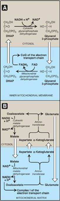
2. Transport of reducing equivalents: The inner mitochondrial membrane lacks an NADH transporter, and NADH produced in the cytosol (for example, in glycolysis; see p. 101) cannot directly enter the mitochondrial matrix. However, two electrons (reducing equivalents) of NADH are transported from the cytosol into the matrix using substrate shuttles. In the glycerophosphate shuttle (Figure 6.16A), two electrons are transferred from NADH to dihydroxyacetone phosphate by cytosolic glycerophosphate dehydrogenase. The glycerol 3-phosphate produced is oxidized by the mitochondrial isozyme as FAD is reduced to FADH2. CoQ of the ETC oxidizes the FADH2. The glycerophosphate shuttle, therefore, results in the synthesis of two ATPs for each cytosolic NADH oxidized. This contrasts with the malate-aspartate shuttle (Figure 6.16B), which produces NADH (rather than FADH2) in the mitochondrial matrix and, therefore, yields three ATPs for each cytosolic NADH oxidized by malate dehydrogenase as oxaloacetate is reduced to malate. A transport protein moves malate into the matrix.
C. Inherited defects in oxidative phosphorylation
Thirteen of the approximately 90 polypeptides required for oxidative phosphorylation are coded for by mtDNA and synthesized in mitochondria, whereas the remaining proteins are coded for by nuclear DNA, synthesized in the cytosol, and transported into mitochondria posttranslationally. Defects in oxidative phosphorylation are more likely a result of alterations in mtDNA, which has a mutation rate about 10 times greater than that of nuclear DNA. Tissues with the greatest ATP requirement (for example, central nervous system, skeletal and heart muscle, and liver) are most affected by defects in oxidative phosphorylation. Mutations in mtDNA are responsible for several diseases, including some cases of mitochondrial myopathies, and Leber hereditary optic neuropathy, a disease in which bilateral loss of central vision occurs as a result of neuroretinal degeneration, including damage to the optic nerve. [Note: mtDNA is maternally inherited because mitochondria from the sperm cell do not enter the fertilized egg.]
D. Mitochondria and apoptosis
The process of apoptosis, or programmed cell death, may be initiated through the intrinsic (mitochondrial-mediated) pathway by the formation of pores in the outer mitochondrial membrane. These pores allow cytochrome c to leave the intermembrane space and enter the cytosol. There, cytochrome c, in association with proapoptotic factors, activates a family of proteolytic enzymes (the caspases), causing cleavage of key proteins and resulting in the morphologic and biochemical changes characteristic of apoptosis.
Figure 6.16 Substrate shuttles for the transportof electrons across the inner mitochondrial membrane. A. Glycerophosphate shuttle. B. Malate-aspartate shuttle. DHAP = dihydroxyacetone phosphate; NAD(H) = nicotinamide adenine dinucleotide; FAD(H2) = flavin adenine dinucleotide; CoQ = coenzyme Q.
VII. CHAPTER SUMMARY
The change in free energy (∆G) occurring during a reaction predicts the direction in which that reaction will spontaneously proceed. If ∆G is negative (that is, the product has a lower free energy than the substrate), the reaction goes spontaneously. If ∆G is positive, the reaction does not go spontaneously. If ∆G = 0, the reactions are in equilibrium. The ∆G of the forward reaction is equal in magnitude but opposite in sign to that of the back reaction. The ∆Gs are additive in any sequence of consecutive reactions, as are the standard free energy changes (∆Gos). Therefore, reactions or processes that have a large, positive ∆G are made possible by couplingwith those that have a large, negative ∆G such as hydrolysis of adenosine triphosphate (ATP). The reduced coenzymes nicotinamide adenine dinucleotide (NADH) and flavin adenine dinucleotide (FADH2) each donate a pair of electrons to a specialized set of electron carriers, consisting of flavin mononucleotide (FMN), iron-sulfur centers, coenzyme Q, and a series of cytochromes, collectively called the electron transport chain. This pathway is present in the inner mitochondrial membrane (impermeable to most substances) and is the final common pathway by which electrons derived from different fuels of the body flow to O2, reducing it to water. The terminal cytochrome, cytochrome oxidase, is the only cytochrome able to bind O2. Electron transport results in the pumping of protons across the inner mitochondrial membrane from the matrix to the intermembrane space. This process creates electrical and pH gradients across the inner mitochondrial membrane. After protons have been transferred to the cytosolic side of the membrane, they reenter the matrix by passing through the Fo proton channel in ATP synthase (Complex V), dissipating the pH and electrical gradients and causing conformational changes in the β subunits of F1 that result in the synthesis of ATP from adenosine diphosphate + inorganic phosphate. Electron transport and phosphorylation are tightly coupled in oxidative phosphorylation (OXPHOS, Figure 6.17). Inhibition of one process inhibits the other. These processes can be uncoupled by uncoupling protein-1 of the inner mitochondrial membrane of cells in brown fat and by synthetic compounds such as 2,4-dinitrophenol and aspirin, all of which dissipate the proton gradient. In uncoupled mitochondria, the energy produced by the transport of electrons is released as heat rather than being used to synthesize ATP. Mutations in mitochondrial DNA, which is maternally inherited, are responsible for some cases of mitochondrial diseasessuch as Leber hereditary optic neuropathy. The release of cytochrome c into the cytoplasm and subsequent activation of proteolytic caspases results in apoptotic cell death.
Figure 6.17 Key concept map for oxidative phosphorylation (OXPHOS). [Note: Electron (e-) flow and ATP synthesis are envisioned as sets of interlocking gears to emphasize the idea of coupling.] TCA = tricarboxylic acid; NAD(H) = nicotinamide adenine dinucleotide; FAD(H2) = flavin adenine dinucleotide; FMN = flavin mononucleotide.
Study Questions
Choose the ONE best answer.
6.1 2,4-Dinitrophenol, an uncoupler of oxidative phosphorylation, was used as a weight-loss agent in the 1930s. Reports of fatal overdoses led to its discontinuation in 1939. Which of the following would most likely be true concerning individuals taking 2,4-dinitrophenol?
A. Adenosine triphosphate levels in the mitochondria are greater than normal.
B. Body temperature is elevated as a result of hypermetabolism.
C. Cyanide has no effect on electron flow.
D. The proton gradient across the inner mitochondrial membrane is greater than normal.
E. The rate of electron transport is abnormally low.
Correct answer = B. When phosphorylation is uncoupled from electron flow, a decrease in the proton gradient across the inner mitochondrial membrane and, therefore, impaired ATP synthesis is expected. In an attempt to compensate for this defect in energy capture, metabolism and electron flow to oxygen is increased. This hypermetabolism will be accompanied by elevated body temperature because the energy in fuels is largely wasted, appearing as heat. The electron transport chain will still be inhibited by cyanide.
6.2 Which of the following has the strongest tendency to gain electrons?
A. Coenzyme Q
B. Cytochrome c
C. Flavin adenine dinucleotide
D. Nicotinamide adenine dinucleotide
E. Oxygen
Correct answer = E. Oxygen is the terminal acceptor of electrons in the electron transport chain (ETC). Electrons flow down the ETC to oxygen because it has the highest (most positive) reduction potential (E0). The other choices precede oxygen in the ETC and have lower E0 values.
6.3 Explain why and how the malate-aspartate shuttle moves nicotinamide adenine dinucleotide reducing equivalents from the cytosol to the mitochondrial matrix.
There is no transporter for nicotinamide adenine dinucleotide (NADH) in the inner mitochondrial membrane. However, NADH can be oxidized to NAD+ by the cytoplasmic isozyme of malate dehydrogenase as oxaloacetate is reduced to malate. The malate is transported across the inner membrane, and the mitochondrial isozyme of malate dehydrogenase oxidizes it to oxaloacetate as mitochondrial NAD+ is reduced to NADH. This NADH can be oxidized by Complex I of the electron transport chain, generating three ATP through the coupled processes of oxidative phosphorylation.
6.4 Carbon monoxide binds to and inhibits Complex IV of the electron transport chain. What effect, if any, should this respiratory inhibitor have on phosphorylation of adenosine diphosphate to adenosine triphosphate?
Inhibition of the electron transport chain by respiratory inhibitors such as carbon monoxide results in an inability to maintain the proton gradient. Phosphorylation of ADP to ATP is, therefore, inhibited, as are ancillary reactions such as calcium uptake by mitochondria, because they also require the proton gradient.