Lippincott’s Illustrated Reviews: Biochemistr, Sixth Edition (2014)
UNIT II: Bioenergetics and Carbohydrate Metabolism
Chapter 9. Tricarboxylic Acid Cycle and Pyruvate Dehydrogenase Complex
I. OVERVIEW
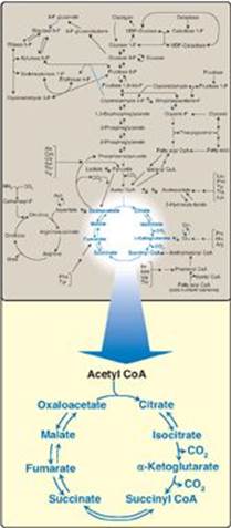
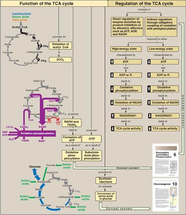
The tricarboxylic acid cycle ([TCA cycle] also called the citric acid cycle or the Krebs cycle) plays several roles in metabolism. It is the final pathway where the oxidative catabolism of carbohydrates, amino acids, and fatty acids converge, their carbon skeletons being converted to CO2 (Figure 9.1). This oxidation provides energy for the production of the majority of adenosine triphosphate (ATP) in most animals, including humans. The TCA cycle occurs totally in the mitochondria and is, therefore, in close proximity to the reactions of electron transport (see p. 73), which oxidize the reduced coenzymes (NADH and FADH2) produced by the cycle. The TCA cycle is an aerobic pathway, because O2 is required as the final electron acceptor. Reactions such as the catabolism of some amino acids generate intermediates of the cycle and are called anaplerotic (“filling up”) reactions. The TCA cycle also supplies intermediates for a number of important synthetic reactions. For example, the cycle functions in the formation of glucose from the carbon skeletons of some amino acids, and it provides building blocks for the synthesis of some amino acids (see p. 267) and heme (see p. 278). Therefore, this cycle should not be viewed as a closed circle but, instead, as a traffic circle with compounds entering and leaving as required.
Figure 9.1 The tricarboxylic acid cycle shown as a part of the essential pathways of energy metabolism. (See Figure 8.2, p. 92 for a more detailed view of the metabolic map.) CoA = coenzyme A.
II. REACTIONS OF THE CYCLE
In the TCA cycle, oxaloacetate is first condensed with an acetyl group from acetyl coenzyme A (CoA) and then is regenerated as the cycle is completed (Figure 9.1). Therefore, the entry of one acetyl CoA into one round of the TCA cycle does not lead to the net production or consumption of intermediates. [Note: Two carbons entering the cycle as acetyl CoA are balanced by two CO2 exiting.]
A. Oxidative decarboxylation of pyruvate
The major source of acetyl CoA, the two-carbon substrate for the TCA cycle, is the oxidative decarboxylation of pyruvate. Pyruvate, the end product of aerobic glycolysis, must be transported from the cytosol into the mitochondrion. This is accomplished by a specific transporter that facilitates movement of pyruvate across the inner mitochondrial membrane. Once in the mitochondrial matrix, pyruvate is converted to acetyl CoA by the pyruvate dehydrogenase complex (PDH complex), which is a multienzyme complex. [Note: Strictly speaking, the PDH complex is not part of the TCA cycle, but it supplies substrate for the cycle.]
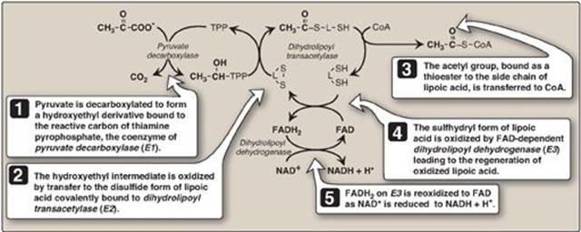
1. Component enzymes: The PDH complex is a protein aggregate of multiple copies of three enzymes, pyruvate carboxylase (E1, sometimes called pyruvate dehydrogenase), dihydrolipoyl transacetylase (E2), and dihydrolipoyl dehydrogenase (E3). Each catalyzes a part of the overall reaction (Figure 9.2). Their physical association links the reactions in proper sequence without the release of intermediates. In addition to the enzymes participating in the conversion of pyruvate to acetyl CoA, the complex also contains two tightly bound regulatory enzymes, pyruvate dehydrogenase kinase (PDH kinase) and pyruvate dehydrogenase phosphatase (PDH phosphatase).
2. Coenzymes: The PDH complex contains five coenzymes that act as carriers or oxidants for the intermediates of the reactions shown in Figure 9.2. E1 requires thiamine pyrophosphate (TPP), E2 requires lipoic acid and CoA, and E3 requires flavin adenine dinucleotide (FAD) and nicotinamide adenine dinucleotide (NAD+). [Note: TPP, lipoic acid, and FAD are tightly bound to the enzymes and function as coenzymes-prosthetic groups (see p. 54).]
Deficiencies of thiamine or niacin can cause serious central nervous system problems. This is because brain cells are unable to produce sufficient ATP (via the TCA cycle) if the PDH complex is inactive. Wernicke-Korsakoff, an encephalopathy-psychosis syndrome due to thiamine deficiency, may be seen with alcohol abuse.
Figure 9.2 Mechanism of action of the pyruvate dehydrogenase complex. [Note: All the coenzymes of the complex, except for lipoic acid, are derived from vitamins. TPP is from thiamine, FAD from riboflavin, NAD from niacin, and CoA from pantothenic acid.] TPP = thiamine pyrophosphate; L = lipoic acid; CoA = coenzyme A; FAD(H2) = flavin adenine dinucleotide; NAD(H) = nicotinamide adenine dinucleotide.
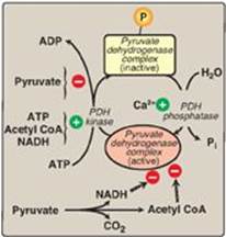
3. Regulation of the pyruvate dehydrogenase complex: Covalent modifications by the two regulatory enzymes that are part of the complex alternately activate and inactivate E1. The cyclic AMP–independent PDH kinasephosphorylates and, thereby, inactivates E1, whereas PDH phosphatase dephosphorylates and activates E1 (Figure 9.3). The kinase itself is allosterically activated by ATP, acetyl CoA, and NADH. Therefore, in the presence of these high-energy signals, the PDH complex is turned off. [Note: It is actually the rise in the ATP/ADP, NADH/NAD+, or acetyl CoA/CoA ratios that affects enzymic activity.] Pyruvate is a potent inhibitor of PDH kinase. Thus, if pyruvate concentrations are elevated, E1 will be maximally active. Calcium (Ca2+) is a strong activator of PDH phosphatase, stimulating E1 activity. This is particularly important in skeletal muscle, where release of Ca2+ during contraction stimulates the PDH complex and, thereby, energy production. [Note: Although covalent regulation by the kinase and phosphatase is primary, the complex is also subject to product (NADH and acetyl CoA) inhibition.]
Figure 9.3 Regulation of pyruvate dehydrogenase (PDH) complex. [![]() denotes product inhibition.]
denotes product inhibition.]
4. Pyruvate dehydrogenase complex deficiency: A deficiency in the activity of the α subunit of the dimeric E1 component of the PDH complex, although rare, is the most common biochemical cause of congenital lactic acidosis. This enzyme deficiency results in an inability to convert pyruvate to acetyl CoA, causing pyruvate to be shunted to lactate via lactate dehydrogenase (see p. 103). This creates particular problems for the brain, which relies on the TCA cycle for most of its energy and is particularly sensitive to acidosis. Symptoms are variable and include neurodegeneration; muscle spasticity; and, in the neonatal onset form, early death. The gene for the α subunit is X linked, and, because both males and females may be affected, the deficiency is classified as X-linked dominant. Although there is no proven treatment for PDH complex deficiency, dietary restriction of carbohydrate and supplementation with thiamine may reduce symptoms in select patients.
Leigh syndrome (subacute necrotizing encephalomyelopathy) is a rare, progressive, neurodegenerative disorder caused by defects in mitochondrial ATP production, primarily as a result of mutations in genes that code for proteins of the PDH complex, the electron transport chain, or ATP synthase. Both nuclear and mitochondrial DNA can be affected.
5. Mechanism of arsenic poisoning: As previously described (see p. 101), pentavalent arsenic (arsenate) can interfere with glycolysis at the glyceraldehyde 3-phosphate step, thereby decreasing ATP production. “Arsenic poisoning” is, however, due primarily to inhibition of enzymes that require lipoic acid as a coenzyme, including E2 of the PDH complex, α-ketoglutarate dehydrogenase (see below), and branched-chain α-keto acid dehydrogenase (see p. 266). Arsenite (the trivalent form of arsenic) forms a stable complex with the thiol (–SH) groups of lipoic acid, making that compound unavailable to serve as a coenzyme. When it binds to lipoic acid in the PDH complex, pyruvate (and, consequently, lactate) accumulates. As with PDH complex deficiency, this particularly affects the brain, causing neurologic disturbances and death.
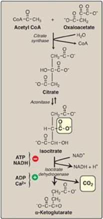
Figure 9.4 Formation of α-ketoglutarate from acetyl coenzyme A (CoA) and oxaloacetate. NAD(H) = nicotinamide adenine dinucleotide
B. Synthesis of citrate from acetyl coenzyme A and oxaloacetate
The condensation of acetyl CoA and oxaloacetate (OAA) to form citrate (a tricarboxylic acid) is catalyzed by citrate synthase (Figure 9.4). This aldol condensation has an equilibrium far in the direction of citrate synthesis. In humans, citrate synthase is not an allosteric enzyme. It is inhibited by its product, citrate. Substrate availability is another means of regulation for citrate synthase. The binding of OAA causes a conformational change in the enzyme that generates a binding site for acetyl CoA. [Note: Citrate, in addition to being an intermediate in the TCA cycle, provides a source of acetyl CoA for the cytosolic synthesis of fatty acids (see p. 183). Citrate also inhibits phosphofructokinase-1 (PFK-1), the rate-limiting enzyme of glycolysis (see p. 99), and activates acetyl CoA carboxylase (the rate-limiting enzyme of fatty acid synthesis; see p. 183).]
C. Isomerization of citrate
Citrate is isomerized to isocitrate by aconitase (aconitate hydratase), an Fe-S protein (see Figure 9.4). [Note: Aconitase is inhibited by fluoroacetate, a plant toxin that is used as a pesticide. Fluoroacetate is converted to fluoroacetyl CoA, which condenses with OAA to form fluorocitrate (a potent inhibitor of aconitase), resulting in citrate accumulation.]
D. Oxidative decarboxylation of isocitrate
Isocitrate dehydrogenase catalyzes the irreversible oxidative decarboxylation of isocitrate, yielding the first of three NADH molecules produced by the cycle and the first release of CO2 (see Figure 9.4). This is one of the rate-limiting steps of the TCA cycle. The enzyme is allosterically activated by ADP (a low-energy signal) and Ca2+ and is inhibited by ATP and NADH, levels of which are elevated when the cell has abundant energy stores.
E. Oxidative decarboxylation of α-ketoglutarate
The conversion of α-ketoglutarate to succinyl CoA is catalyzed by the α-ketoglutarate dehydrogenase complex, a protein aggregate of multiple copies of three enzymes (Figure 9.5). The mechanism of this oxidative decarboxylation is very similar to that used for the conversion of pyruvate to acetyl CoA by the PDH complex. The reaction releases the second CO2 and produces the second NADH of the cycle. The coenzymes required are TPP, lipoic acid, FAD, NAD+, and CoA. Each functions as part of the catalytic mechanism in a way analogous to that described for the PDH complex (see p. 110). The equilibrium of the reaction is far in the direction of succinyl CoA, a high-energy thioester similar to acetyl CoA. α-Ketoglutarate dehydrogenase complex is inhibited by its products, NADH and succinyl CoA, and activated by Ca2+. However, it is not regulated by phosphorylation/dephosphorylation reactions as described for PDH complex. [Note: α-Ketoglutarate is also produced by the oxidative deamination (see p. 252) and transamination of the amino acid glutamate (see p. 250).]
Figure 9.5 Formation of malate from α-ketoglutarate. NAD(H) = nicotinamide adenine dinucleotide; GDP = guanosine diphosphate; P = phosphate; CoA = coenzyme A; FAD(H2) = flavin adenine dinucleotide.
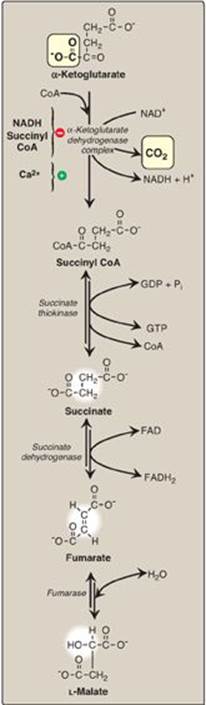
F. Cleavage of succinyl coenzyme A
Succinate thiokinase (also called succinyl CoA synthetase, named for the reverse reaction) cleaves the high-energy thioester bond of succinyl CoA (see Figure 9.5). This reaction is coupled to phosphorylation of guanosine diphosphate (GDP) to guanosine triphosphate (GTP). GTP and ATP are energetically interconvertible by the nucleoside diphosphate kinase reaction:
GTP + ADP ![]() GDP + ATP
GDP + ATP
The generation of GTP by succinate thiokinase is another example of substrate-level phosphorylation (see p. 102). [Note: Succinyl CoA is also produced from propionyl CoA derived from the metabolism of fatty acids with an odd number of carbon atoms (see p. 193), and from the metabolism of several amino acids (see pp. 265–266).]
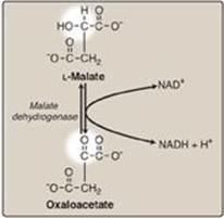
Figure 9.6 Formation (regeneration) of oxaloacetate from malate. NAD(H) = nicotinamide adenine dinucleotide.
G. Oxidation of succinate
Succinate is oxidized to fumarate by succinate dehydrogenase, as FAD (its coenzyme) is reduced to FADH2 (see Figure 9.5). Succinate dehydrogenase is the only enzyme of the TCA cycle that is embedded in the inner mitochondrial membrane. As such, it functions as Complex II of the electron transport chain (see p. 75). [Note: FAD, rather than NAD+, is the electron acceptor because the reducing power of succinate is not sufficient to reduce NAD+.]
H. Hydration of fumarate
Fumarate is hydrated to malate in a freely reversible reaction catalyzed by fumarase (fumarate hydratase; see Figure 9.5). [Note: Fumarate is also produced by the urea cycle (see p. 255), in purine synthesis (see p. 294), and during catabolism of the amino acids phenylalanine and tyrosine (see p. 263).]
I. Oxidation of malate
Malate is oxidized to oxaloacetate by malate dehydrogenase (Figure 9.6). This reaction produces the third and final NADH of the cycle. The standard free energy change (∆G0; see p. 70) of the reaction is positive, but the reaction is driven in the direction of OAA by the highly exergonic citrate synthase reaction. [Note: OAA is also produced by the transamination of the amino acid aspartic acid (see p. 250).]
III. ENERGY PRODUCED BY THE CYCLE
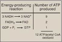
Two carbon atoms enter the cycle as acetyl CoA and leave as CO2. The cycle does not involve net consumption or production of OAA or of any other intermediate. Four pairs of electrons are transferred during one turn of the cycle: three pairs of electrons reducing three NAD+ to NADH and one pair reducing FAD to FADH2. Oxidation of one NADH by the electron transport chain leads to formation of approximately three ATP, whereas oxidation of FADH2yields approximately two ATP (see p. 77). The total yield of ATP from the oxidation of one acetyl CoA is shown in Figure 9.7. Figure 9.8 summarizes the reactions of the TCA cycle.
Figure 9.7 Number of ATP molecules produced from the oxidation of one molecule of acetyl coenzyme A (CoA) using both substrate-level and oxidative phosphorylation.
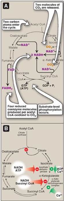
IV. REGULATION OF THE CYCLE
In contrast to glycolysis, which is regulated primarily by PFK-1, the TCA cycle is controlled by the regulation of several enzymes (see Figure 9.8). The most important of these regulated enzymes are those that catalyze reactions with highly negative ∆G0: citrate synthase, isocitrate dehydrogenase, and α-ketoglutarate dehydrogenase complex. Reducing equivalents needed for oxidative phosphorylation are generated by the PDH complex and the TCA cycle, and both processes are upregulated in response to a decrease in the ratio of ATP to ADP.
Figure 9.8 A. [Note: GTP and ATP are interconverted by nucleoside diphosphate kinase.] Production of reduced coenzymes, ATP, and CO2 in the citric acid cycle. B. Inhibitors and activators of the cycle.
V. CHAPTER SUMMARY
Pyruvate is oxidatively decarboxylated by pyruvate dehydrogenase (PDH) complex, producing acetyl coenzyme A (CoA), which is the major fuel for the tricarboxylic acid cycle ([TCA cycle] Figure 9.9). This multienzyme complex requires five coenzymes: thiamine pyrophosphate, lipoic acid, flavin adenine dinucleotide (FAD), nicotinamide adenine dinucleotide (NAD+), and CoA. PDH complex is regulated by covalent modification of E1(pyruvate decarboxylase) by PDH kinase and PDH phosphatase: phosphorylation inhibits E1. PDH kinase is allosterically activated by ATP, acetyl CoA, and NADH and inhibited by pyruvate. The phosphatase is activated by Ca2+. PDH complex deficiency is the most common biochemical cause of congenital lactic acidosis. The central nervous system is particularly affected in this X-linked dominant disorder. Arsenic poisoning causes inactivation of the PDH complex by binding to lipoic acid. Citrate is synthesized from oxaloacetate and acetyl CoA by citrate synthase. This enzyme is subject to product inhibition by citrate. Citrate is isomerized to isocitrate by aconitase (aconitate hydratase). Isocitrate is oxidatively decarboxylated by isocitrate dehydrogenase to α-ketoglutarate, producing CO2 and NADH. The enzyme is inhibited by ATP and NADH, and activated by ADP and Ca2+. α-Ketoglutarate is oxidatively decarboxylated to succinyl CoA by the α-ketoglutarate dehydrogenase complex, producing CO2 and NADH. The enzyme is very similar to the PDH complex and uses the same coenzymes. α-Ketoglutarate dehydrogenase complex is activated by Ca+2 and inhibited by NADH and succinyl CoA but is not covalently regulated. Succinyl CoA is cleaved by succinate thiokinase (also called succinyl CoA synthetase), producing succinate and GTP. This is an example of substrate-level phosphorylation. Succinate is oxidized to fumarate by succinate dehydrogenase, producing FADH2. Fumarate is hydrated to malateby fumarase (fumarate hydratase), and malate is oxidized to oxaloacetate by malate dehydrogenase, producing NADH. Three NADH, one FADH2, and one GTP (whose terminal phosphate can be transferred to ADP by nucleoside diphosphate kinase, producing ATP) are produced by one round of the TCA cycle. The generation of acetyl CoA by the oxidation of pyruvate via the PDH complex also produces an NADH. Oxidation of the NADH and FADH2 by the electron transport chain yields 14 ATP. An additional ATP (GTP) comes from substrate level phosphorylation in the TCA cycle. Therefore, a total of 15 ATP are produced from the complete mitochondrial oxidation of pyruvate to CO2.
Figure 9.9 Key concept map for the tricarboxylic acid (TCA) cycle. CoA = coenzyme A; NAD(H) = nicotinamide adenine dinucleotide; FAD(H2) = flavin adenine dinucleotide; GDP = guanosine diphosphate; GTP = guanosine triphosphate; ADP = adenosine diphosphate; Pi = inorganic phosphate.
Study Questions
Choose the ONE best answer.
9.1 The conversion of pyruvate to acetyl coenzyme A and CO2:
A. involves the participation of lipoic acid.
B. is activated when pyruvate decarboxylase of the pyruvate dehydrogenase (PDH) complex is phosphorylated by PDH kinase in the presence of ATP.
C. is reversible.
D. occurs in the cytosol.
E. requires the coenzyme biotin.
Correct answer = A. Lipoic acid is an intermediate acceptor of the acetyl group formed in the reaction. Pyruvate dehydrogenase complex catalyzes an irreversible reaction that is inhibited when the decarboxylase component is phosphorylated. The enzyme complex is located in the mitochondrial matrix. Biotin is utilized by carboxylases, not decarboxylases.
9.2 Which one of the following conditions decreases the oxidation of acetyl coenzyme A by the citric acid cycle?
A. A high availability of calcium
B. A high acetyl CoA/CoA ratio
C. A low ATP/ADP ratio
D. A low NAD+/NADH ratio
Correct answer = D. A low NAD+/NADH ratio limits the rates of the NAD+-requiring dehydrogenases. High availability of calcium and substrate (acetyl CoA), and a low ATP/ADP ratio stimulates the cycle.
9.3 The following is the sum of three steps in the citric acid cycle.
A + B + FAD + H2O → C + FADH2 + NADH
Choose the lettered answer that corresponds to the missing “A,” “B,” and “C” in the equation.
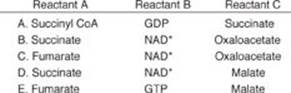
Correct answer = B. Succinate + NAD+ + FAD + H2O → oxaloacetate + NADH + FADH2
9.4 A 1-month-old male shows neurologic problems and lactic acidosis. Enzyme assay for pyruvate dehydrogenase (PDH) complex activity on extracts of cultured skin fibroblasts showed 5% of normal activity with a low concentration of thiamine pyrophosphate (TPP), but 80% of normal activity when the assay contained a thousand-fold higher concentration of TPP. Which one of the following statements concerning this patient is correct?
A. Administration of thiamine is expected to reduce his serum lactate level and improve his clinical symptoms.
B. A high carbohydrate diet would be expected to be beneficial for this patient.
C. Citrate production from aerobic glycolysis is expected to be increased.
D. PDH kinase, a regulatory enzyme of the PDH complex, is expected to be active.
Correct answer = A. The patient appears to have a thiamine-responsive pyruvate dehydrogenase (PDH) complex deficiency. The pyruvate decarboxylase (E1) component of the PDH complex fails to bind thiamine pyrophosphate at low concentration, but shows significant activity at a high concentration of the coenzyme. This mutation, which affects the Km of the enzyme for the coenzyme, is present in some, but not all, cases of PDH complex deficiency. Because the PDH complex is an integral part of carbohydrate metabolism, a diet low in carbohydrates would be expected to blunt the effects of the enzyme deficiency. Aerobic glycolysis generates pyruvate, the substrate of the PDH complex. Decreased activity of the complex decreases production of acetyl coenzyme A, a substrate for citrate synthase. PDH kinase is allosterically inhibited by pyruvate and, therefore, is inactive.
9.5 Which coenzyme-cosubstrate is used by the dehydrogenases of both glycolysis and the tricarboxylic acid cycle?
Oxidized nicotinamide adenine dinucleotide (NAD+) is used by glyceraldehyde 3-phosphate dehydrogenase of glycolysis and by isocitrate dehydrogenase, α-ketoglutarate dehydrogenase, and malate dehydrogenase of the tricarboxylic acid cycle.