Harper’s Illustrated Biochemistry, 29th Edition (2012)
SECTION I. Structures & Functions of Proteins & Enzymes
Chapter 6. Proteins: Myoglobin & Hemoglobin
Peter J. Kennelly, PhD & Victor W. Rodwell, PhD
OBJECTIVES
After studying this chapter, you should be able to:
![]() Describe the most important structural similarities and differences between myoglobin and hemoglobin.
Describe the most important structural similarities and differences between myoglobin and hemoglobin.
![]() Sketch binding curves for the oxygenation of myoglobin and hemoglobin.
Sketch binding curves for the oxygenation of myoglobin and hemoglobin.
![]() Identify the covalent linkages and other close associations between heme and globin in oxymyoglobin and oxyhemoglobin.
Identify the covalent linkages and other close associations between heme and globin in oxymyoglobin and oxyhemoglobin.
![]() Explain why the physiologic function of hemoglobin requires that its O2-binding curve be sigmoidal rather than hyperbolic.
Explain why the physiologic function of hemoglobin requires that its O2-binding curve be sigmoidal rather than hyperbolic.
![]() Explain the role of a hindered environment on the ability of hemoglobin to bind carbon monoxide.
Explain the role of a hindered environment on the ability of hemoglobin to bind carbon monoxide.
![]() Define P50 and indicate its significance in oxygen transport and delivery.
Define P50 and indicate its significance in oxygen transport and delivery.
![]() Describe the structural and conformational changes in hemoglobin that accompany its oxygenation and subsequent deoxygenation.
Describe the structural and conformational changes in hemoglobin that accompany its oxygenation and subsequent deoxygenation.
![]() Explain the role of 2,3-bisphosphoglycerate (BPG) in oxygen binding and delivery.
Explain the role of 2,3-bisphosphoglycerate (BPG) in oxygen binding and delivery.
![]() Outline the role of hemoglobin in CO2 and proton transport, and describe accompanying changes in the pKa of the relevant imidazolium group.
Outline the role of hemoglobin in CO2 and proton transport, and describe accompanying changes in the pKa of the relevant imidazolium group.
![]() Describe the structural consequences to HbS of lowering pO2.
Describe the structural consequences to HbS of lowering pO2.
![]() Identify the metabolic defect that occurs as a consequence of α- and -β thalassemias.
Identify the metabolic defect that occurs as a consequence of α- and -β thalassemias.
BIOMEDICAL IMPORTANCE
The heme proteins myoglobin and hemoglobin maintain a supply of oxygen essential for oxidative metabolism. Myoglobin, a monomeric protein of red muscle, stores oxygen as a reserve against oxygen deprivation. Hemoglobin, a tetrameric protein of erythrocytes, transports O2 to the tissues and returns CO2 and protons to the lungs. Cyanide and carbon monoxide kill because they disrupt the physiologic function of the heme proteins cytochrome oxidase and hemoglobin, respectively. The secondary-tertiary structure of the subunits of hemoglobin resembles myoglobin. However, the tetrameric structure of hemoglobin permits cooperative interactions that are central to its function. For example, 2,3-BPG promotes the efficient release of O2 by stabilizing the quaternary structure of deoxyhemoglobin. Hemoglobin and myoglobin illustrate both protein structure–function relationships and the molecular basis of genetic diseases such as sickle cell disease and the thalassemias.
HEME & FERROUS IRON CONFER THE ABILITY TO STORE & TO TRANSPORT OXYGEN
Myoglobin and hemoglobin contain heme, a cyclic tetrapyrrole consisting of four molecules of pyrrole linked by methyne bridges. This planar network of conjugated double bonds absorbs visible light and colors heme deep red. The substituents at the β-positions of heme are methyl (M), vinyl (V), and propionate (Pr) groups arranged in the order M, V, M, V, M, Pr, Pr, M (Figure 6–1). The atom of ferrous iron (Fe2+) resides at the center of the planar tetrapyrrole. Other proteins with metal-containing tetrapyrrole prosthetic groups include the cytochromes (Fe and Cu) and chlorophyll (Mg) (see Chapter 31). Oxidation and reduction of the Fe and Cu atoms of cytochromes are essential to their biologic function as carriers of electrons. By contrast, oxidation of the Fe2+ of myoglobin or hemoglobin to Fe3+ destroys their biologic activity.
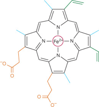
FIGURE 6–1 Heme. The pyrrole rings and methyne bridge carbons are coplanar, and the iron atom (Fe2+) resides in almost the same plane. The fifth and sixth coordination positions of Fe2+ are directly perpendicular to—and directly above and below—the plane of the heme ring. Observe the nature of the methyl (blue), vinyl (green), and propionate (orange) substituent groups on the β carbons of the pyrrole rings, the central iron atom (red), and the location of the polar side of the heme ring (at about 7 o’clock) that faces the surface of the myoglobin molecule.
Myoglobin Is Rich in α Helix
Oxygen stored in red muscle myoglobin is released during O2 deprivation (eg, severe exercise) for use in muscle mitochondria for aerobic synthesis of ATP (see Chapter 13). A 153-aminoacyl residue polypeptide (MW 17,000), myoglobin folds into a compact shape that measures 4.5 × 3.5 × 2.5 nm (Figure 6–2). An unusually high proportion, about 75%, of the residues are present in eight right-handed 7–20 residue α helices. Starting at the amino terminal, these are termed helices A–H. Typical of globular proteins, the surface of myoglobin is rich in amino acids bearing polar and potentially charged side chains, while—with only two exceptions—the interior contains only residues such as Leu, Val, Phe, and Met that possess nonpolar R groups. The exceptions are His E7 and His F8, the seventh and eighth residues in helices E and F, which lie close to the heme iron, where they function in O2 binding.
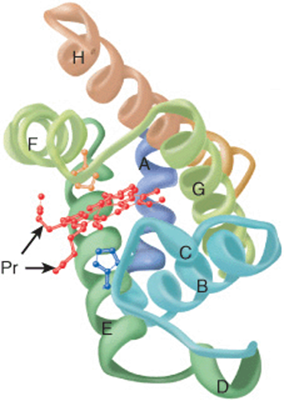
FIGURE 6–2 Three-dimensional structure of myoglobin. Shown is a ribbon diagram tracing the polypeptide backbone of myoglobin. The color of the polypeptide chain is graded along the visible spectrum from blue (N-terminal) to tan (C-terminal). The heme prosthetic group is red. The α-helical regions are designated A through H. The distal (E7) and proximal (F8) histidine residues are highlighted in blue and orange, respectively. Note how the polar propionate substituents (Pr) project out of the heme toward solvent. (Adapted from Protein Data Bank ID no. 1a6n.)
Histidines F8 & E7 Perform Unique Roles in Oxygen Binding
The heme of myoglobin lies in a crevice between helices E and F oriented with its polar propionate groups facing the surface of the globin (Figure 6–2). The remainder resides in the non-polar interior. The fifth coordination position of the iron is occupied by a nitrogen from the imidazole ring of the proximal histidine, His F8. The distal histidine, His E7, lies on the side of the heme ring opposite to His F8.
The Iron Moves Toward the Plane of the Heme when Oxygen Is Bound
The iron of unoxygenated myoglobin lies 0.03 nm (0.3 Å) outside the plane of the heme ring, toward His F8. The heme therefore “puckers” slightly. When O2 occupies the sixth coordination position, the iron moves to within 0.01 nm (0.1 Å) of the plane of the heme ring. Oxygenation of myoglobin thus is accompanied by motion of the iron, of His F8, and of residues linked to His F8.
Apomyoglobin Provides a Hindered Environment for the Heme Iron
When O2 binds to myoglobin, the bond between the first oxygen atom and the Fe2+ is perpendicular to the plane of the heme ring. The bond that links the first and second oxygen atoms lies at an angle of 121° to the plane of the heme, orienting the second oxygen away from the distal histidine (Figure 6–3, left). This permits maximum overlap between the iron and one of the lone pairs of electrons on the sp2 hybridized oxygen atoms, which lie at an angle of roughly 120° to the axis of the O=O double bond (Figure 6–4, left). Isolated heme binds carbon monoxide (CO) 25,000 times more strongly than oxygen. Since CO is present in small quantities in the atmosphere and arises in cells from the catabolism of heme, why is it that CO does not completely displace O2 from heme iron? The accepted explanation is that the apoproteins of myoglobin and hemoglobin create a hindered environment. When CO binds to isolated heme, all the three atoms (Fe, C, and O) lie perpendicular to the plane of the heme. This geometry maximizes the overlap between the lone pair of electrons on the sp hybridized oxygen of the CO molecule and the Fe2+ iron (Figure 6–4, right). However, in myoglobin and hemoglobin the distal histidine sterically precludes this preferred, high-affinity orientation of CO, but not that of O2. Binding at a less favored angle reduces the strength of the heme-CO bond to about 200 times that of the heme-O2 bond (Figure 6–3, right) at which level the great excess of O2 over CO normally present dominates. Nevertheless, about 1% of myoglobin typically is present combined with CO.
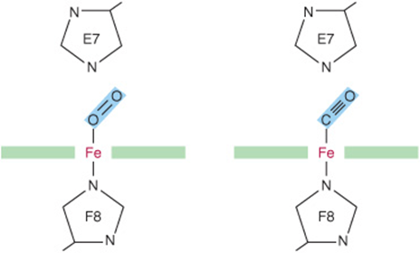
FIGURE 6–3 Angles for bonding of oxygen and carbon monoxide (CO) to the heme iron of myoglobin. The distal E7 histidine hinders bonding of CO at the preferred (90°) angle to the plane of the heme ring.
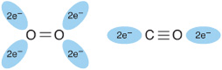
FIGURE 6–4 Orientation of the lone pairs of electrons relative to the ![]() and
and ![]() bonds of oxygen and carbon monoxide. In molecular oxygen, formation of the double bond between the two oxygen atoms is facilitated by the adoption of an sp2 hybridization state by the valence electron of each oxygen atom. As a consequence, the two atoms of the oxygen molecule and each lone pair of electrons are coplanar and separated by an angle of roughly 120° (left). By contrast, the two atoms of carbon monoxide are joined by a triple bond, which requires that the carbon and oxygen atoms adopt an sp hybridization state. In this state the lone pairs of electrons and triple bonds are arranged in a linear fashion, where they are separated by an angle of 180° (right).
bonds of oxygen and carbon monoxide. In molecular oxygen, formation of the double bond between the two oxygen atoms is facilitated by the adoption of an sp2 hybridization state by the valence electron of each oxygen atom. As a consequence, the two atoms of the oxygen molecule and each lone pair of electrons are coplanar and separated by an angle of roughly 120° (left). By contrast, the two atoms of carbon monoxide are joined by a triple bond, which requires that the carbon and oxygen atoms adopt an sp hybridization state. In this state the lone pairs of electrons and triple bonds are arranged in a linear fashion, where they are separated by an angle of 180° (right).
THE OXYGEN DISSOCIATION CURVES FOR MYOGLOBIN & HEMOGLOBIN SUIT THEIR PHYSIOLOGIC ROLES
Why is myoglobin unsuitable as an O2 transport protein but well suited for O2 storage? The relationship between the concentration, or partial pressure, of O2 (Po2) and the quantity of O2 bound is expressed as an O2 saturation isotherm (Figure 6–5). The oxygen-binding curve for myoglobin is hyperbolic. Myoglobin therefore loads O2 readily at the Po2 of the lung capillary bed (100 mm Hg). However, since myoglobin releases only a small fraction of its bound O2 at the Po2 values typically encountered in active muscle (20 mm Hg) or other tissues (40 mm Hg), it represents an ineffective vehicle for delivery of O2. When strenuous exercise lowers the Po2 of muscle tissue to about 5 mm Hg, myoglobin releases O2 for mitochondrial synthesis of ATP, permitting continued muscular activity.
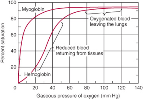
FIGURE 6–5 Oxygen-binding curves of both hemoglobin and myoglobin. Arterial oxygen tension is about 100 mm Hg; mixed venous oxygen tension is about 40 mm Hg; capillary (active muscle) oxygen tension is about 20 mm Hg; and the minimum oxygen tension required for cytochrome oxidase is about 5 mm Hg. Association of chains into a tetrameric structure (hemoglobin) results in much greater oxygen delivery than would be possible with single chains. (Modified, with permission, from Scriver CR et al (editors): The Molecular and Metabolic Bases of Inherited Disease, 7th ed. McGraw-Hill, 1995.)
THE ALLOSTERIC PROPERTIES OF HEMOGLOBINS RESULT FROM THEIR QUATERNARY STRUCTURES
The properties of individual hemoglobins are consequences of their quaternary as well as of their secondary and tertiary structures. The quaternary structure of hemoglobin confers striking additional properties, absent from monomeric myoglobin, which adapts it to its unique biologic roles. The allosteric (Gk allos “other,” steros “space”) properties of hemoglobin provide, in addition, a model for understanding other allosteric proteins (see Chapter 18).
Hemoglobin Is Tetrameric
Hemoglobins are tetramers composed of pairs of two different polypeptide subunits (Figure 6–6). Greek letters are used to designate each subunit type. The subunit composition of the principal hemoglobins are α2β2 (HbA; normal adult hemoglobin), α2γ2 (HbF; fetal hemoglobin), α2βs2, (HbS; sickle cell hemoglobin), and α2δ2 (HbA2; a minor adult hemoglobin). The primary structures of the β, γ, and δ chains of human hemoglobin are highly conserved.
FIGURE 6–6 Hemoglobin. Shown is the three-dimensional structure of deoxyhemoglobin with a molecule of 2,3-bisphosphoglycerate (dark blue) bound. The two α subunits are colored in the darker shades of green and blue, the two β subunits in the lighter shades of green and blue, and the heme prosthetic groups in red. (Adapted from Protein Data Bank ID no. 1b86.)
Myoglobin & the β Subunits of Hemoglobin Share Almost Identical Secondary and Tertiary Structures
Despite differences in the kind and number of amino acids present, myoglobin and the β polypeptide of hemoglobin A have almost identical secondary and tertiary structures. Similarities include the location of the heme and the helical regions, and the presence of amino acids with similar properties at comparable locations. Although it possesses seven rather than eight helical regions, the α polypeptide of hemoglobin also closely resembles myoglobin.
Oxygenation of Hemoglobin Triggers Conformational Changes in the Apoprotein
Hemoglobins bind four molecules of O2 per tetramer, one per heme. A molecule of O2 binds to a hemoglobin tetramer more readily if other O2 molecules are already bound (Figure 6–5). Termed cooperative binding, this phenomenon permits hemoglobin to maximize both the quantity of O2 loaded at the Po2 of the lungs and the quantity of O2 released at the Po2 of the peripheral tissues. Cooperative interactions, an exclusive property of multimeric proteins, are critically important to aerobic life.
P50 Expresses the Relative Affinities of Different Hemoglobins for Oxygen
The quantity P50, a measure of O2 concentration, is the partial pressure of O2 that half-saturates a given hemoglobin. Depending on the organism, P50 can vary widely, but in all instances, it will exceed the Po2 of the peripheral tissues. For example, the values of P50 for HbA and HbF are 26 and 20 mm Hg, respectively. In the placenta, this difference enables HbF to extract oxygen from the HbA in the mother’s blood. However, HbF is suboptimal postpartum since its high affinity for O2 limits the quantity of O2 delivered to the tissues.
The subunit composition of hemoglobin tetramers undergoes complex changes during development. The human fetus initially synthesizes a ξ2ε2 tetramer. By the end of the first trimester, ξ and ε subunits have been replaced by α and γ sub-units, forming HbF (α2γ2), the hemoglobin of late fetal life. While synthesis of β subunits begins in the third trimester, β subunits do not completely replace γ subunits to yield adult HbA (α2β2) until some weeks postpartum (Figure 6–7).
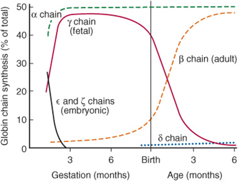
FIGURE 6–7 Developmental pattern of the quaternary structure of fetal and newborn hemoglobins. (Reproduced, with permission, from Ganong WF: Review of Medical Physiology, 20th ed. McGraw-Hill, 2001.)
Oxygenation of Hemoglobin Is Accompanied by Large Conformational Changes
The binding of the first O2 molecule to deoxyHb shifts the heme iron toward the plane of the heme ring from a position about 0.04 nm beyond it (Figure 6–8). This motion is transmitted to the proximal (F8) histidine and to the residues attached thereto, which in turn causes the rupture of salt bridges between the carboxyl terminal residues of all four subunits. As a result, one pair of α/β subunits rotates 15° with respect to the other, compacting the tetramer (Figure 6–9). Profound changes in secondary, tertiary, and quaternary structures accompany the O2-induced transition of hemoglobin from the low-affinity T (taut) state to the high-affinity R (relaxed) state. These changes significantly increase the affinity of the remaining unoxygenated hemes for O2, as subsequent binding events require the rupture of fewer salt bridges (Figure 6–10). The terms T and R also are used to refer to the low-affinity and high-affinity conformations of allosteric enzymes, respectively.
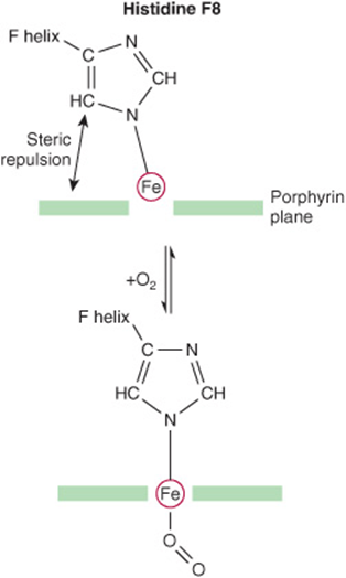
FIGURE 6–8 The iron atom moves into the plane of the heme on oxygenation. Histidine F8 and its associated residues are pulled along with the iron atom. (Slightly modified and reproduced, with permission, from Stryer L: Biochemistry, 4th ed. Freeman, 1995. Copyright © 1995 W. H. Freeman and Company.)
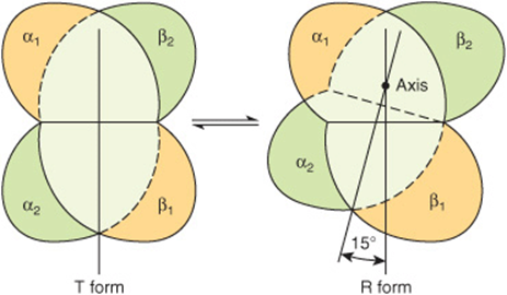
FIGURE 6–9 During transition of the T form to the R form of hemoglobin, the α2β2 pair of subunits (green) rotates through 15° relative to the pair of α1β1 subunits (yellow). The axis of rotation is eccentric, and the α2β2pair also shifts toward the axis somewhat. In the representation, the tan α1β1 pair is shown fixed while the green α2β2 pair of subunits both shifts and rotates.
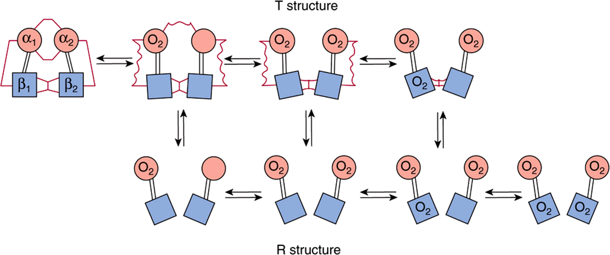
FIGURE 6–10 Transition from the T structure to the R structure. In this model, salt bridges (red lines) linking the subunits in the T structure break progressively as oxygen is added, and even those salt bridges that have not yet ruptured are progressively weakened (wavy red lines). The transition from T to R does not take place after a fixed number of oxygen molecules have been bound but becomes more probable as each successive oxygen binds. The transition between the two structures is influenced by protons, carbon dioxide, chloride, and BPG; the higher their concentration, the more oxygen must be bound to trigger the transition. Fully oxygenated molecules in the T structure and fully deoxygenated molecules in the R structure are not shown because they are unstable. (Modified and redrawn, with permission, from Perutz MF: Hemoglobin structure and respiratory transport. Sci Am [Dec] 1978;239:92.)
After Releasing O2 at the Tissues, Hemoglobin Transports CO2 & Protons to the Lungs
In addition to transporting O2 from the lungs to peripheral tissues, hemoglobin transports CO2, the byproduct of respiration, and protons from peripheral tissues to the lungs. Hemoglobin carries CO2 as carbamates formed with the amino terminal nitrogens of the polypeptide chains:
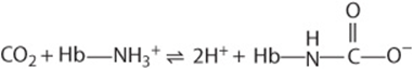
Carbamates change the charge on amino terminals from positive to negative, favoring salt bridge formation between α and β chains.
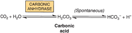
Hemoglobin carbamates account for about 15% of the CO2 in venous blood. Much of the remaining CO2 is carried as bicarbonate, which is formed in erythrocytes by the hydration of CO2 to carbonic acid (H2CO3), a process catalyzed by carbonic anhydrase. At the pH of venous blood, H2 CO3 dissociates into bicarbonate and a proton.
Deoxyhemoglobin binds one proton for every two O2 molecules released, contributing significantly to the buffering capacity of blood. The somewhat lower pH of peripheral tissues, aided by carbamation, stabilizes the T state and thus enhances the delivery of O2. In lungs, the process reverses. As O2 binds to deoxyhemoglobin, protons are released and combine with bicarbonate to form carbonic acid. Dehydration of H2CO3, catalyzed by carbonic anhydrase, forms CO2, which is exhaled. Binding of oxygen thus drives the exhalation of CO2 (Figure 6–11). This reciprocal coupling of proton and O2 binding is termed the Bohr effect. The Bohr effect is dependent upon cooperative interactions between the hemes of the hemoglobin tetramer. Myoglobin, a monomer, exhibits no Bohr effect.
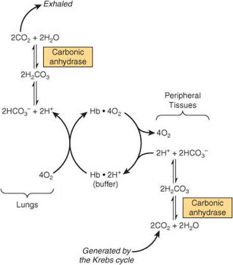
FIGURE 6–11 The Bohr effect. Carbon dioxide generated in peripheral tissues combines with water to form carbonic acid, which dissociates into protons and bicarbonate ions. Deoxyhemoglobin acts as a buffer by binding protons and delivering them to the lungs. In the lungs, the uptake of oxygen by hemoglobin releases protons that combine with bicarbonate ion, forming carbonic acid, which when dehydrated by carbonic anhydrase becomes carbon dioxide, which then is exhaled.
Protons Arise from Rupture of Salt Bridges When O2 Binds
Protons responsible for the Bohr effect arise from rupture of salt bridges during the binding of O2 to T-state hemoglobin. Conversion to the oxygenated R state breaks salt bridges involving β chain residue His 146. The subsequent dissociation of protons from His 146 drives the conversion of bicarbonate to carbonic acid (Figure 6–11). Upon the release of O2, the T structure and its salt bridges re-form. This conformational change increases the pKa of the β chain His 146 residues, which bind protons. By facilitating the re-formation of salt bridges, an increase in proton concentration enhances the release of O2 from oxygenated (R-state) hemoglobin. Conversely, an increase in Po2promotes proton release.
2,3- BPG Stabilizes the T Structure of Hemoglobin
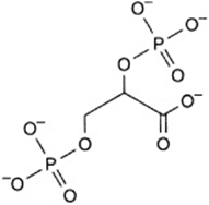
A low Po2 in peripheral tissues promotes the synthesis of 2,3-BPG in erythrocytes from the glycolytic intermediate 1,3-BPG.
The hemoglobin tetramer binds one molecule of BPG in the central cavity formed by its four subunits (Figure 6–6). However, the space between the H helices of the β chains lining the cavity is sufficiently wide to accommodate BPG only when hemoglobin is in the T state. BPG forms salt bridges with the terminal amino groups of both β chains via Val NA1 and with Lys EF6 and His H21 (Figure 6–12). BPG therefore stabilizes deoxygenated (T-state) hemoglobin by forming additional salt bridges that must be broken prior to conversion to the R state.
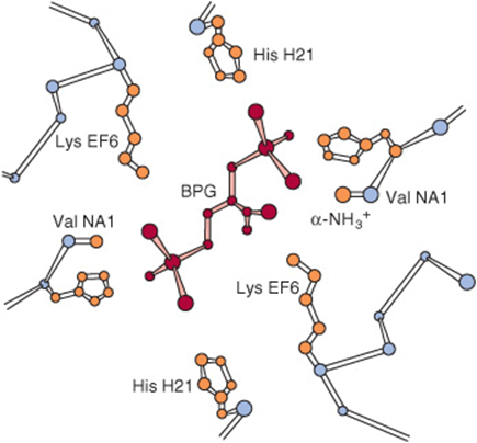
FIGURE 6–12 Mode of binding of 2,3-bisphosphoglycerate (BPG) to human deoxyhemoglobin. BPG interacts with three positively charged groups on each β chain. (Based on Arnone A: X-ray diffraction study of binding of 2,3-diphosphoglycerate to human deoxyhemoglobin. Nature 1972;237:146. Copyright © 1972. Adapted by permission from Macmillan Publishers Ltd.)
Residue H21 of the γ subunit of HbF is Ser rather than His. Since Ser cannot form a salt bridge, BPG binds more weakly to HbF than to HbA. The lower stabilization afforded to the T state by BPG accounts for HbF having a higher affinity for O2 than HbA.
Adaptation to High Altitude
Physiologic changes that accompany prolonged exposure to high altitude include an increase in the number of erythrocytes and in their concentrations of hemoglobin and of BPG. Elevated BPG lowers the affinity of HbA for O2(increases P50), which enhances the release of O2 at peripheral tissues.
NUMEROUS MUTATIONS AFFECTING HUMAN HEMOGLOBINS HAVE BEEN IDENTIFIED
Mutations in the genes that encode the α or β subunits of hemoglobin potentially can affect its biologic function. However, almost all of the over 1,100 known genetic mutations affecting human hemoglobins are both extremely rare and benign, presenting no clinical abnormalities. When a mutation does compromise biologic function, the condition is termed a hemoglobinopathy. It is estimated that more than 7% of the globe’s population are carriers for hemoglobin disorders. The URL http://globin.cse.psu.edu/ (Globin Gene Server) provides information about—and links for—normal and mutant hemoglobins. Selected examples are described below.
Methemoglobin & Hemoglobin M
In methemoglobinemia, the heme iron is ferric rather than ferrous. Methemoglobin thus can neither bind nor transport O2. Normally, the enzyme methemoglobin reductase reduces the Fe3+ of methemoglobin to Fe2+. Methemoglobin can arise by oxidation of Fe2+ to Fe3+ as a side effect of agents such as sulfonamides, from hereditary hemoglobin M, or consequent to reduced activity of the enzyme methemoglobin reductase.
In hemoglobin M, histidine F8 (His F8) has been replaced by tyrosine. The iron of HbM forms a tight ionic complex with the phenolate anion of tyrosine that stabilizes the Fe3+ form. In α-chain hemoglobin M variants, the R-T equilibrium favors the T state. Oxygen affinity is reduced, and the Bohr effect is absent. β-chain hemoglobin M variants exhibit R-T switching, and the Bohr effect is therefore present.
Mutations that favor the R state (eg, hemoglobin Chesapeake) increase O2 affinity. These hemoglobins therefore fail to deliver adequate O2 to peripheral tissues. The resulting tissue hypoxia leads to polycythemia, an increased concentration of erythrocytes.
Hemoglobin S
In HbS, the nonpolar amino acid valine has replaced the polar surface residue Glu6 of the β subunit, generating a hydrophobic “sticky patch” on the surface of the β subunit of both oxyHbS and deoxyHbS. Both HbA and HbS contain a complementary sticky patch on their surfaces that is exposed only in the deoxygenated T state. Thus, at low Po2, deoxyHbS can polymerize to form long, insoluble fibers. Binding of deoxy-HbA terminates fiber polymerization, since HbA lacks the second sticky patch necessary to bind another Hb molecule (Figure 6–13). These twisted helical fibers distort the erythro-cyte into a characteristic sickle shape, rendering it vulnerable to lysis in the interstices of the splenic sinusoids. They also cause multiple secondary clinical effects. A low Po2, such as that at high altitudes, exacerbates the tendency to polymerize. Emerging treatments for sickle cell disease include inducing HbF expression to inhibit the polymerization of HbS, stem cell transplantation, and, in the future, gene therapy.
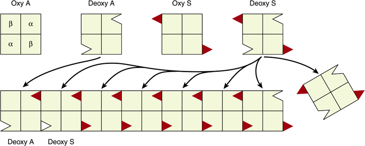
FIGURE 6–13 Representation of the sticky patch ![]() on hemoglobin S and its “receptor” (Δ) on deoxyhemoglobin A and deoxyhemoglobin S. The complementary surfaces allow deoxyhemoglobin S to polymerize into a fibrous structure, but the presence of deoxyhemoglobin A will terminate the polymerization by failing to provide sticky patches. (Modified and reproduced, with permission, from Stryer L: Biochemistry, 4th ed. Freeman, 1995. Copyright © 1995 W. H. Freeman and Company.)
on hemoglobin S and its “receptor” (Δ) on deoxyhemoglobin A and deoxyhemoglobin S. The complementary surfaces allow deoxyhemoglobin S to polymerize into a fibrous structure, but the presence of deoxyhemoglobin A will terminate the polymerization by failing to provide sticky patches. (Modified and reproduced, with permission, from Stryer L: Biochemistry, 4th ed. Freeman, 1995. Copyright © 1995 W. H. Freeman and Company.)
BIOMEDICAL IMPLICATIONS
Myoglobinuria
Following massive crush injury, myoglobin released from damaged muscle fibers colors the urine dark red. Myoglobin can be detected in plasma following a myocardial infarction, but assay of serum enzymes (see Chapter 7) provides a more sensitive index of myocardial injury.
Anemias
Anemias, reductions in the number of red blood cells or of hemoglobin in the blood, can reflect impaired synthesis of hemoglobin (eg, in iron deficiency; Chapter 50) or impaired production of erythrocytes (eg, in folic acid or vitamin B12 deficiency; Chapter 44). Diagnosis of anemias begins with spectroscopic measurement of blood hemoglobin levels.
Thalassemias
The genetic defects known as thalassemias result from the partial or total absence of one or more α or β chains of hemoglobin. Over 750 different mutations have been identified, but only three are common. Either the α chain (alpha thalassemias) or β chain (beta thalassemias) can be affected. A superscript indicates whether a subunit is completely absent (α0 or β0) or whether its synthesis is reduced (α– or β–). Apart from marrow transplantation, treatment is symptomatic.
Certain mutant hemoglobins are common in many populations, and a patient may inherit more than one type. Hemoglobin disorders thus present a complex pattern of clinical phenotypes. The use of DNA probes for their diagnosis is considered in Chapter 39.
Glycated Hemoglobin (HbA1c)
When blood glucose enters the erythrocytes, it glycates the ε-amino group of lysyl residues and the amino terminals of hemoglobin. The fraction of hemoglobin glycated, normally about 5%, is proportionate to blood glucose concentration. Since the half-life of an erythrocyte is typically 60 days, the level of glycated hemoglobin (HbA1c) reflects the mean blood glucose concentration over the preceding 6–8 weeks. Measurement of HbA1c therefore provides valuable information for management of diabetes mellitus.
SUMMARY
![]() Myoglobin is monomeric; hemoglobin is a tetramer of two subunit types (α2β2 in HbA). Despite having different primary structures, myoglobin and the subunits of hemoglobin have nearly identical secondary and tertiary structures.
Myoglobin is monomeric; hemoglobin is a tetramer of two subunit types (α2β2 in HbA). Despite having different primary structures, myoglobin and the subunits of hemoglobin have nearly identical secondary and tertiary structures.
![]() Heme, an essentially planar, slightly puckered, cyclic tetrapyrrole has a central Fe2+ linked to all four nitrogen atoms of the heme, to histidine F8, and, in oxyMb and oxyHb, also to O2.
Heme, an essentially planar, slightly puckered, cyclic tetrapyrrole has a central Fe2+ linked to all four nitrogen atoms of the heme, to histidine F8, and, in oxyMb and oxyHb, also to O2.
![]() The O2-binding curve for myoglobin is hyperbolic, but for hemoglobin it is sigmoidal, a consequence of cooperative interactions in the tetramer. Cooperativity maximizes the ability of hemoglobin both to load O2 at the Po2 of the lungs and to deliver O2 at the Po2 of the tissues.
The O2-binding curve for myoglobin is hyperbolic, but for hemoglobin it is sigmoidal, a consequence of cooperative interactions in the tetramer. Cooperativity maximizes the ability of hemoglobin both to load O2 at the Po2 of the lungs and to deliver O2 at the Po2 of the tissues.
![]() Relative affinities of different hemoglobins for oxygen are expressed as P50, the Po2 that half-saturates them with O2. Hemoglobins saturate at the partial pressures of their respective respiratory organ, eg, the lung or placenta.
Relative affinities of different hemoglobins for oxygen are expressed as P50, the Po2 that half-saturates them with O2. Hemoglobins saturate at the partial pressures of their respective respiratory organ, eg, the lung or placenta.
![]() On oxygenation of hemoglobin, the iron, histidine F8, and linked residues move toward the heme ring. Conformational changes that accompany oxygenation include rupture of salt bonds and loosening of the quaternary structure, facilitating binding of additional O2.
On oxygenation of hemoglobin, the iron, histidine F8, and linked residues move toward the heme ring. Conformational changes that accompany oxygenation include rupture of salt bonds and loosening of the quaternary structure, facilitating binding of additional O2.
![]() 2,3- BPG in the central cavity of deoxyHb forms salt bonds with the β subunits that stabilize deoxyHb. On oxygenation, the central cavity contracts, BPG is extruded, and the quaternary structure loosens.
2,3- BPG in the central cavity of deoxyHb forms salt bonds with the β subunits that stabilize deoxyHb. On oxygenation, the central cavity contracts, BPG is extruded, and the quaternary structure loosens.
![]() Hemoglobin also functions in CO2 and proton transport from tissues to lungs. Release of O2 from oxyHb at the tissues is accompanied by uptake of protons due to lowering of the pKa of histidine residues.
Hemoglobin also functions in CO2 and proton transport from tissues to lungs. Release of O2 from oxyHb at the tissues is accompanied by uptake of protons due to lowering of the pKa of histidine residues.
![]() In sickle cell hemoglobin (HbS), Val replaces the β6 Glu of HbA, creating a “sticky patch” that has a complement on deoxyHb (but not on oxyHb). DeoxyHbS polymerizes at low O2 concentrations, forming fibers that distort erythrocytes into sickle shapes.
In sickle cell hemoglobin (HbS), Val replaces the β6 Glu of HbA, creating a “sticky patch” that has a complement on deoxyHb (but not on oxyHb). DeoxyHbS polymerizes at low O2 concentrations, forming fibers that distort erythrocytes into sickle shapes.
![]() Alpha and beta thalassemias are anemias that result from reduced production of α and β subunits of HbA, respectively.
Alpha and beta thalassemias are anemias that result from reduced production of α and β subunits of HbA, respectively.
REFERENCES
Frauenfelder H, McMahon BH, Fenimore PW: Myoglobin: The hydrogen atom of biology and a paradigm of complexity. Proc Natl Acad Sci USA 2003;100:8615.
Hardison RC, Chui DH, Riemer C, et al: Databases of human hemoglobin variants and other resources at the globin gene server. Hemoglobin 2001;25:183.
Lukin JA, Ho C: The structure–function relationship of hemoglobin in solution at atomic resolution. Chem Rev 2004;104:1219.
Ordway GA, Garry DJ: Myoglobin: An essential hemoprotein in striated muscle. J Exp Biol 2004;207:3441.
Papanikolaou E, Anagnou NP: Major challenges for gene therapy of thalassemia and sickle cell dsease. Curr Gene Ther 2010;10:404.
Schrier SL, Angelucci E: New strategies in the treatment of the thalassemias. Annu Rev Med 2005;56:157.
Steinberg MH, Brugnara C: Pathophysiological-based approaches to treatment of sickle-cell disease. Annu Rev Med 2003;54:89.
Umbreit J: Methemoglobin—it’s not just blue: A concise review. Am J Hematol 207;82:134.
Weatherall DJ, Akinyanju O, Fucharoen S, et al: Inherited disorders of hemoglobin. In: Disease Control Priorities in Developing Countries, Jamison DT, Breman JG, Measham AR (editors). Oxford University Press and the World Bank, 2006;663–680.
Weatherall DJ, Clegg JB, Higgs DR, et al: The hemoglobinopathies. In: The Metabolic Basis of Inherited Disease, 8th ed. Scriver CR, Sly WS, Childs B, et al (editors). McGraw-Hill, 2000;4571.
Weatherall DJ, Clegg JD: The Thalassemia Syndromes. Blackwell Science, 2001.
Yonetani T, Laberge M: Protein dynamics explain the allosteric behaviors of hemoglobin. Biochim Biophys Acta 2008;1784:1146.