CHEMICAL BIOLOGY
Hypoxic Response and Associated Diseases
Emily Flashman, Christoph Loenarz, and Christopher J. Schofield, Chemistry Research Laboratory and Oxford Centre for Integrative Systems Biology, Mansfield Road, Oxford, U.K.
doi: 10.1002/9780470048672.wecb232
Animals must respond efficiently to changes in oxygen levels. This response is achieved via oxygen-dependent regulation of a heterodimeric hypoxia-inducible transcription factor (HIF), which enables upregulation of genes that assist in the hypoxic response. Posttranslational hydroxylation of the HIF-a subunit at either of two conserved prolyl residues enables binding to the von Hippel-Lindau protein elongin C/B complex that targets HIF-α for degradation via the ubiquitin proteasome pathway. Hydroxylation of an asparaginyl residue in the C-terminal transcriptional activation domain of HIF-α blocks its interaction with the transcriptional coactivator p300. The HIF prolyl and asparaginyl hydroxylases are oxygen dependent; therefore, their regulation of HIF directly links changes in oxygen concentration and the physiological response to hypoxia. The hypoxic response is implicated in a range of disease states, including cancer, ischemia, and heart disease. Manipulation of the HIF system for therapeutic advantage is therefore an area of medicinal interest.
In the late nineteenth century, Francois Viault observed that red blood cell production increased as humans moved from sea level to high altitude (1). The mechanism by which this response to an environment of limiting oxygen is induced has been a long-standing problem in physiology. A breakthrough in molecular understanding came when Semenza et al. (2) identified a transcription factor, hypoxia inducible factor (HIF), which is responsible for the regulation of erythropoietin (EPO). EPO is a primary modulator of red blood cell production and therefore oxygen capacity in mammals, and consequently it is upregulated in conditions of hypoxia (3). An enhancer region upstream (5’) of the EPO gene was identified and was found to bind HIF in an oxygen-dependent fashion (2, 4). HRE sequences have subsequently been found upstream of numerous genes that are upregulated in response to hypoxia, including those involved in erythropoiesis, angiogenesis, metabolism, and cell growth (5). This review summarizes research on the HIF system with a focus on our current understanding of the chemistry that determines how the HIF system senses changes in oxygen availability.
Overview of the HIF System
HIF is an α,β-heterodimeric transcription factor whose transcriptional activity is regulated via its oxygen-dependent, posttranslational hydroxylation. In the presence of sufficient oxygen, HIF-α in humans is hydroxylated in both its oxygen-dependent degradation domain (ODDD) and its C-terminal transactivation domain (C-TAD). Prolyl-4-hydroxylation in the ODDD allows HIF-α to bind to the von Hippel-Lindau protein elongin C/B complex (VCB), which targets HIF-α for degradation via the ubiquitin proteasome pathway. Asparaginyl hydroxylation at the C-TAD prevents HIF-α interacting with the p300 protein, which is part of the transcriptional coactivator complex for HIF target genes. All four identified human HIF hydroxylases are Fe(II)- and 2-oxoglutarate (2OG)-dependent oxygenases with an absolute requirement for oxygen as a cosubstrate. When oxygen availability is limited, HIF-α hydroxylation is incomplete or absent. HIF-α can then dimerize with HIF-fi, interact with the transcriptional coactivator complex via p300 binding, and bind to conserved pentanucleotide hypoxia response elements (HREs: TACGTC) to promote transcription of genes that enable a response to the challenge of hypoxic conditions. An overview of the HIF regulatory system is shown in Fig. 1.

Figure 1. Overview of the HIF dual regulatory system. In the presence of oxygen (O2), the HIF-α subunits are downregulated and inactivated by active HIF hydroxylases—the PHDs and FIH, respectively. The PHDs catalyze hydroxylation of HIFa prolyl residues, which enhances binding of HIF-α to the von Hippel-Lindau tumor suppressor complex (VCB) ~1000-fold and enables subsequent ubiquitin-mediated proteasomal degradation of HIF-α subunits. FIH catalyses hydroxylation of an HIF-α asparaginyl residue; this substance blocks p300 coactivator recruitment and results in transcriptional inactivation. Limiting oxygen levels (hypoxia) reduce HIF hydroxylase activity. This limitation permits heterodimerization of HIF-α with HIF-β subunits, translocation into the nucleus, and recruitment of transcriptional coactivators including p300. Subsequent binding to HREs on HIF target genes upregulates their transcription, enabling the body's hypoxic response.
HIF-α/β and Its Interaction with DNA
Both α and β subunits of HIF are basic helix-loop-helix PAS proteins (bHLH-PAS) (6, 7), as observed in other transcription- factor proteins (Fig. 2). The short, basic region of bHLH-PAS proteins is directly responsible for DNA binding, and the adjacent helix-loop-helix domain allows dimerization of the two HIF subunits after translocation of HIF-α to the nucleus. The PAS domain [PAS-A/PAS-B/PAC; PAS: PER (periodic circadian protein), aryl hydrocarbon receptor nuclear translocator (ARNT), single-minded protein (SIM) (7); PAC: motif C-terminal to PAS motifs) is involved in more specific interactions between the two subunits (for review see Reference 8). It is proposed that the specific interaction between the HIF-α and HIF-β subunits promotes folding and dimerization of the bHLH region that enables subsequent DNA binding (8). HIF-α is a Class I bHLH-PAS protein, which means that it can homodimerize and heterodimerize; HIF-β (also known as ARNT, which is a ubiquitously expressed protein) is a Class II bHLH-PAS protein, which heterodimerizes with Class I bHLH-PAS proteins (8, 9). The general PAS domain structure comprises a central β-sheet region flanked by several α-helices; NMR and mutation studies on HIF-2α have identified a hydrophobic region on the β-sheet as being responsible for the specific interaction with HIF-β, as well as homologous interactions between other PAS domains (10).
High-resolution structural data are not yet available on the interaction between the HIF heterodimer and the HRE. Interactions between bHLH proteins and DNA have shown that a conserved His-Glu-Arg triad in the bHLH binds in the DNA major groove (11). Modeling studies predict that the HIF-β domain binds to HREs in a similar fashion. HIF-α, however, only retains the Arg residue of this conserved triad, and the interaction of HIF with DNA is predicted to be mediated by other residues, specifically a serine and two alanines (12). These proposals are supported by mutagenesis studies (13).
HIF-α contains a large, central regulatory region. The oxygen-dependent degradation domain of HIF-α contains two sites of prolyl hydroxylation (Pro402 in the N-terminal ODDD and Pro564 in the C-terminal ODDD of HIF-1α) (14). If hydroxylation occurs at one or both of these sites, then HIF-α is targeted for ubiquitin-mediated proteasomal degradation (see below). In conditions of low oxygen however, HIF-α avoids hydroxylation and dimerizes with HIF-β. Once the two HIF subdomains have dimerized with each other and have bound to the HRE, cotranscriptional activators are necessary for the expression of the target genes. Optimal upregulation of HIF-target genes in eukaryotic cells involves the formation of a multiprotein coactivator complex to facilitate transcription. At least in eukaryotes, this process is likely to be tissue and cell-type dependent (15). p300/CBP (CREB binding protein) are two (related) key transcriptional coactivators that are histone acetyltransferases (16). p300/CBP binds directly to HIF-α at its C-terminal transactivation domain (C-TAD) in a hypoxia-dependent fashion (17, 18). Additonal interactions mediated by p300/CBP, as well as other coactivation mechanisms (15), are then responsible for optimal target gene expression.
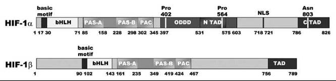
Figure 2. Domain architecture of human HIF-1α and HIF-1β/ARNT subunits. Both HIF-α and HIF-β subunits have similar bHLH domains and PAS domains responsible for DNA binding and protein dimerization. Only the C-terminus of HIF-α subunits contains regulatory domains involved in the hypoxic response: the oxygen-dependent degradation domain (ODDD), responsible for regulating HIF-α stability, and the N-TAD and C-TAD involved in regulating transactivation ability. These regulatory domains are controlled by enzymatic posttranslational hydroxylation if O2 is not limiting. Hydroxylation of HIF-1α at Pro402 and Pro564 within the ODDD enables proteasomal degradation. Hydroxylation of HIF-1α at Asn803 within the C-TAD blocks interaction with transcriptional coactivators after NLS-mediated translocation into the nucleus. PAC domain = motif C-terminal to PAS motifs. PAS domain = PER, AHR, ARNT, and SIM domains. Together, PAS and PAC motifs constitute the PAS structural domain fold. AHR, aryl-hydrocarbon receptor; NLS, nuclear localisation signal.
Types and Roles of Different HIF-α Isoforms
Three isoforms of HIF-α [HIF-1α, HIF-2α (endothelial PAS domain protein), and HIF-3α] have been defined in humans and are encoded by distinct loci (19, 20). HIF-1α and HIF-2α are more important than HIF-3α in the hypoxic response. Three isoforms of HIF-β [HIF-10 (ARNT), ARNT2 (21), and ARNT3 (22)] exist; HIF-1β (ARNT) is the most common form. Each HIF-α isoform can interact with any HIF-β subunit (23). mRNA levels of HIF-1α, HIF-2α, and HIF-1β are reportedly not affected by oxygen levels (7, 24, 25), but levels of HIF 3α mRNA increased after two hours of hypoxia (26, 27).
HIF-1α and HIF-2α have the same domain architecture and common mechanisms for DNA binding, dimerization, oxygen-dependent degradation (NODD and CODD domains), and transactivation domains. The sequences of these domains are related closely but contain differences that may have functional consequences (e.g., with respect to HIF hydroxylase selectivity). The interdomain sequences are related less closely, and the differences may enable differential binding of regulatory proteins to HIF-1α and HIF-2α. HIF-3α is more markedly different from the other two HIF-α isoforms as it apparently has no functional NODD domain, and only the CODD domain within the N-terminal TAD has been identified. Additionally, HIF-3α lacks the C-TAD that exists in both HIF-1α and HIF-2α. (28). Relatively little is known about the importance of HIF-3α as compared with the intensively investigated HIF-1/2α isoforms.
Five HIF-1α splice variants have been reported (29). These variants include HIF-1α736, which lacks the C-TAD but not the C-terminal nuclear localization signal (NLS) and is transcriptionally active under hypoxia (30), and two variants that lack the C-terminal ODD and act as dominant negative isoforms (31, 32). HIF-3α has at least six different splice variants (HIF-3α 1-6), of which only HIF-3α 1-3 contain the ODD domain (33). HIF-3α2 (IPAS) negatively regulates HIF-mediated gene expression in murine cornea by dimerizing with HIF-1α in the cytosol, maintaining an avascular phenotype under hypoxia (34), but it is upregulated by HIF-1α, which represents a negative feedback regulatory circuit (35).
Both HIF-1α and HIF-2α genes are expressed widely in human tissues but with different patterns of expression both in terms of organs and individual cell types within organs. For example, both HIF-1α and HIF-2α are expressed abundantly in the kidney; but in breast cancer cell lines, HIF-1α is the major isoform that is hypoxically induced (36). Importantly, HIF-1α and HIF-2α seem to have different profiles with respect to the genes that they regulate. Expression of carbonic anhydrase IX seems to be predominantly regulated by HIF-1α, whereas erythropoietin is predominantly regulated by HIF-2α (37, 38). HIF-1α and HIF-2α null mice also give different phenotypes that support proposals of different roles for the two isoforms (39-42). An important objective is to develop a molecular understanding of how the selectivity, which is apparent in terms of the different expression profiles for HIF-1α and HIF-2α, is achieved.
The Role of Hydroxylation
The direct interface between HIF-a regulation and oxygen is its posttranslational hydroxylation. In humans, in addition to constituting the crucial step in mammalian oxygen homeostasis (43), hydroxylation seems to be special (to date) amongst posttranslational modifications (PTMs) in that only a single atom is incorporated, as compared with other “additive” PTMs that entail sterically more demanding groups such as phosphorylation, acetylation, glycosylation, and ubiquitylation (44).
The dependence of HIF-1α stability on oxygen was demonstrated by the hydroxylation-dependent von Hippel-Lindau protein (pVHL) capture of HIF-α peptides, which gradually increased in vitro from ambient to hypoxic oxygen levels (45). In normoxia, HIF-1α protein is hydroxylated at Pro402 (in the N-terminal ODD) and/or Pro564 (in the C-terminal ODD). Both prolyl hydroxylation sites form part of a conserved motif, LXXLAP (14). Prolyl hydroxylation results in binding of pVHL and rapid degradation by the proteasome (46, 47). Under normoxia, HIF-α is thus essentially not detectable (7). The affinity of HIF-α ODD peptides for VCB increases about 1000-fold by trans-4-prolyl hydroxylation (48). Crystallographic analyses have revealed that the optimal arrangement of the new hydroxyproline alcohol group stabilizes a hydrogen bonding network in VCB between Ser111 and His115 of pVHL (Fig. 3) (48, 49). Notably, the precise positioning of the hydroxyl group may be induced by the stereoelectronic gauche effect within the vicinal N-C5-C4-OH arrangement, as the pyrrolidine ring of hydroxyprolyl 564 was observed in the C4-exo conformation. This conformation is the same as in collagen, in which hydroxyprolyl residues serve to stabilize the triple helix (50); a deficiency of collagen prolyl-hydroxylation leads to the disease scurvy (51). The α-domain (residues 155-213) of pVHL binds to elongin C, which nucleates a complex that contains elongin B, cullin-2 and Rbx1, forming the VCB-Cul2 RING-type E3 ubiquitin ligase (52). Provided oxygen is not limiting; the complex recruits specific proteins for the lysyl polyubiquitylation of prolyl-hydroxylated HIF-α subunits (53), which enables subsequent proteasomal degradation (54-56). Both the HIF-1α NODD and the CODD domains enable pVHL recognition after PHD-mediated hydroxylation in an independent and nonredundant manner (45).
In the presence of nonlimiting oxygen, posttranslational hydroxylation also occurs at the 4S position of an asparaginyl residue in the HIF-α C-TAD, which corresponds to Asn803 in HIF-1α, preventing the interaction of HIF-α with the cysteine/histidine rich (CH-1) domain of the transcriptional coactivator complex CBP/p300 (17, 28, 57). NMR studies indicate that Asn803 is part of an α-helix buried at the interface of the HIF-α-β300/CBP complex, which suggests its hydroxylation disrupts the hydrophobic binding interactions with CH-1 and/or the formation of the α-helix adopted by the HIF α C-TAD (Fig. 3) (58, 59).
Direct incorporation of 18O into the hydroxylation sites of asparaginyl and prolyl HIF-α substrates (60), and incorporation of the other dioxygen 18O2-derived oxygen into the succinate byproduct was confirmed by mass spectrometry (61). Thus, a very direct link exists between HIF modification and oxygen availability.

Figure 3. Dual effect of hydroxylation within the HIFa regulatory domains. (a) Hydroxylation of HIF-1α CODD domain at P564 enables binding to the von Hippel-Lindau tumor suppressor (pVHL), elongin C, elongin B, (VCB) complex. The newly formed hydroxyl group is optimally positioned for the hydrogen bonding network involving pVHL H115 and S111, as Hyp564 is observed in the C4-exo conformation. (b) Hydroxylation of HIF-1α C-TAD at N803 abrogates its binding to the p300 CH1/TAZ1 domain. N803 is deeply buried in the protein-protein interface, packed against I353 and the hydrophobic parts of the K349 and R350 side chains. Additionally, a network of N803 side-chain hydrogen-bonding interactions including with D799C_TAD and D346CHI is important for stabilization of C-TAD α-helices and the complex, but destabilized during β-hydroxylation of N803 (not shown). Hyp, (25,4R)-hydroxyproline;CH7, cysteine/histidine-rich domain-1;7AZ, transcriptional adapter zinc-binding motif.
The HIF Hydroxylases
The enzymatic, posttranslational prolyl and asparaginyl hydroxylation of HIF-α was found to be dependent on oxygen and Fe(II), enhanced by ascorbate, and inhibited by 2OG analogs (28, 62-64), all suggestive of the enzymes responsible being part of the large family of Fe(II)- and 2OG-dependent oxygenases. This finding led to the identification of three human HIF prolyl hydroxylases (PHDs or EGLNs 1-3) and FIH (factor inhibiting HIF) as the enzyme that catalyzed asparaginyl hydroxylation (45, 65). These substances all contained the iron-coordinating 2-histidine-1-carboxylate motif, which is characteristic of this enzyme family.
The Fe(II) and 2OG-dependent oxygenase family is a subset of the nonheme iron (II)-dependent oxygenases, and they catalyze a wide range of oxidative reactions, often involving cleavage of an otherwise unreactive C-H bond. Examples of reaction types catalyzed by this class of enzymes include oxidative cyclization, epimerization, desaturation, C-C bond cleavage, ring fragmentation, and hydroxylation (for reviews see References 66 and 67). These reactions are often not reproducible in nonenzyme catalyzed reactions. The enzymes and the substrates on which they act are found throughout nature. In plants and bacteria, they are known to act on small molecule substrates, for example, TauD in Escherichia coli can desulfonate taurine to provide a source of sulfur for growth (68) and anthocyanidin synthase in plants catalyses a desaturation step in flavonoid biosynthesis (69). The enzymes also play a role in the biosynthesis pathways of antibiotic production, including the medicinally important β-lactam antibiotics (70), and can catalyze chlorination of C-H bonds (71). In higher organisms, Fe(II)-/2OG-dependent oxygenases have been found to be involved in important biological processes such as DNA repair [e.g., ABH2/3 (72)] and collagen biosynthesis. Recently, a protein linked to increased fat mass in humans (FTO) has been found to be a Fe(II)-/2OG-dependent oxygenase, catalyzing DNA demethylation (73).
Catalytic activity of the HIF hydroxylases
A consensus mechanism for the Fe(II)-/2OG-dependent oxygenases has been proposed (Fig. 4). In most cases, iron is coordinated octahedrally at the active site by two histidines and the carboxylate group of a glutamate or an aspartate residue. The other three coordination positions are occupied by ligated water molecules. 2OG binds to the active site by ligating in a bidentate fashion to two coordination positions on the Fe(II) (replacing two water molecules) via its 1-carboxylate and 2-oxogroups. Binding of substrate (close to but not in contact with the Fe) causes a conformational change that results in alteration of the Fe(II) geometry to five-coordinate square pyramidal, and the last bound water molecule is released. This binding leaves a position free for an oxygen molecule to ligate to the Fe(II), possibly occupying the position whereby it is directed toward the target substrate. The reaction then seems likely to proceed via an Fe(III)-superoxo species, which attacks the 2-oxo group of the 2OG (susceptible to such an attack as it has been activated by the Lewis acidity of the Fe) to form a peroxide. This compound can then collapse to form carbon dioxide, iron coordinated succinate, and a highly reactive Fe(IV)-oxo species. This ferryl-oxo species is positioned adjacent to the target C-H bond such that either direct insertion of oxygen into the C-H bond occurs or the hydrogen atom is abstracted. Then, rapid rebound of the hydroxyl group is generated onto the carbon radical. Hydroxylated substrate and succinate are released from the enzyme active site, and the vacant coordination positions are once again occupied by water molecules.
Evidence to support this proposed mechanism comes from a variety of sources. The crystal structure of a cephalosporin synthase (74) showed the coordination of Fe(II) and 2OG at the active site before addition of oxygen or substrate. These complexes have subsequently been observed in multiple other structural studies (reviewed in Reference 66). EPR, circular dichroism, and UV-Vis spectroscopy studies on TfdA and CAS2 also confirmed the six-coordinate nature of the active site in solution, and they demonstrated the loss of water molecules and change in coordination environment promoted by substrate binding (75-77). Generation of data to support the later stages of the proposed reaction mechanism were more challenging, as the species involved are highly reactive and consequently unstable. Breakthrough experiments carried out on TauD however managed to demonstrate the existence of the postulated Fe(IV)-oxo species using rapid stopped-flow UV-Vis and freeze-quench Mossbauer and EPR techniques (78). The same species was later observed in a viral prolyl hydroxylase (79), which supports the hypothesis that this species is indeed the key reactive intermediate in a conserved mechanism of these enzymes.
The mechanism of the HIF hydroxylases is likely to be similar to the consensus mechanism of the Fe(II)-/2OG-dependent oxygenases described above. However, the role of these particular enzymes as oxygen sensors means that their mechanism of action is of interest, particularly in terms of their dependence on oxygen. Although the intermediates in the reaction cycles of FIH and the PHDs have not been characterized to date, studies on these enzymes provide clues as to how they conform and/or differ from other enzymes in this family. Steady-state kinetic work has monitored the Km of these enzymes with respect to their substrates and also to oxygen. These enzymes have found that in terms of oxygen sensitivity, the HIF hydroxylases (at least in an in vitro oxygen consumption assay) do not differ from other Fe(II)-/2OG-dependent oxygenases (specifically TauD and PAHX) (80). Different studies are not in agreement as to whether the PHDs are more sensitive to oxygen than FIH, but all studies have reported that these enzymes are suited to their roles as oxygen sensors (80-82). Differences in the range of reactions catalyzed by the PHDs are recognized (e.g., hydroxylating NODD and/or CODD in HIF-α 1-3); specific interactions are more or less likely to occur in different cell types and environments (14, 81, 83-91). For example, PHD3 has been shown to be more active on HIF-2α, which is predominantly expressed in the lung, endothelium, and carotid body (19), than on HIF-1α.
Other cellular factors are also likely to influence the catalytic activity of the HIF hydroxylases. Many Fe(II)-/2OG-dependent oxygenases are known to require ascorbate for optimal activity, and ascorbate has been shown to affect cellular HIF levels accordingly (92). The role of ascorbate in the reaction cycle is not clearly understood, although one possibility is that it reduces Fe(III) to Fe(II) when 2OG has been decarboxylated in the absence of prime substrate (uncoupled turnover), as is the case with collagen prolyl hydroxylase (93). Reactive oxygen species have also been proposed to reduce HIF hydroxylase activity, by oxidizing Fe(II) to Fe(III) via the Fenton reaction, decreasing its cellular availability (94). Ascorbate may “repair” this situation, thus playing a general role of ensuring maximum availability of active enzyme (94, 95). In the absence of 2OG, ascorbate and Fe(II)/Fe(III) can lead to oxidative damage to the active site of some 2OG oxygenases/related enzymes (96).
Evidence suggests that elevated levels of the tricarboxylic acid cycle intermediates fumarate and succinate (the latter a product of hydroxylase catalysis) in some tumors may lead to activation of the HIF system via hydroxylase inhibition (97, 98). Both succinate and fumarate are PHD2 inhibitors competing with 2OG for binding to Fe(II) (99, 100).
The interface between NO and the HIF system is complex and requires additional investigation (for review see Reference 101). Under hypoxic conditions, NO inhibits HIF-1α stabilization. However, under normoxic conditions, NO upregulation causes HIF-1α to accumulate. Because NO can act as an oxygen analog binding to 2OG oxygenases (102), it is tempting to speculate that the latter effect is caused by direct inhibition of the HIF hydroxylases. However, the far reaching effects of both NO and HIF on the cell biology mean that additional work is required to dissect the opposing effects of NO under normoxic and hypoxic conditions.
The situation is even more complex in the case of the interaction between reactive oxidizing species (ROS) and the HIF system (for review see References 103, 104). Quantifying the role of ROS, such as superoxide, is difficult because of their reactivity and ability to affect many cellular processes. Good evidence suggests that under stress conditions ROS can regulate HIF, possibly via interaction with the HIF hydroxylases (see e.g., References 94, 105, and 106), but the molecular mechanisms are unclear, as is the relevance of ROS regulation of HIF under normal physiological conditions.
The role of ascorbate, NO, and ROS, as well as other cellular factors, in oxygen sensing is not yet fully clear and requires more investigation. Additionally, other signaling pathways could likely be integrated with HIF regulation as indicated by studies showing that in HIF-1α, Thr-796 phosphorylation blocks Asn-803 hydroxylation by FIH (107).

Figure 4. Proposed outline reaction mechanism for the Fe(II)/2OG-dependent oxygenases.
Structural studies on the HIF hydroxylases
Since the first crystal structure of IPNS (108), which is an enzyme closely related to the Fe/2OG-dependent oxygenases, structures have been determined for many Fe(II)-/2OG-dependent oxygenases, all revealing a consensus core double-stranded β-helix (DSBH) fold or jelly-roll motif (66, 102). Typically this core consists of two four-stranded antiparallel β-sheets, with the major sheet supported by closely packed a-helices. The DSBH core is stabilized even more by internal hydrophobic interactions. The active site and HXD/E...H iron binding motif reside within this core structure. Additional or varying structural features from the DSBH domain define different structural subfamilies of this class of enzyme (66).
Crystal structures of FIH and PHD2 (catalytic domain) have been solved (Fig. 5) (109-112). These structures have shown that both enzymes contain the predicted core DSBH helix, but they belong to two different subfamilies of the Fe(II)-/2OG-dependent oxygenases. The C-terminus of FIH was shown to be involved in the formation of a functionally important dimer (107), whereas this finding was not the case for PHD2. Although in the absence of substrate, PHD2 crystallizes in a trimeric form, it does not reflect the situation in solution where PHD2 is monomeric (112). The entrance to the active site of PHD2 was also much narrower than that of FIH and other human Fe(II)-/2OG-dependent oxygenases. This finding may have functional implications for this particular enzyme (e.g., in terms of the kinetics of substrate binding) and also rationalizes the observed unusually strong interaction with 2OG and iron observed for PHD2 (113). The active sites of the two enzymes show that the iron binding triad is in a similar position; however, the 2OG 1-carboxylate group coordinates to the iron in a different position in the two enzymes. The residues that stabilize the 2OG 5-carboxylate group also differ, which is characteristic of their belonging to different subfamilies of the Fe(II)/2OG-dependent oxygenases.
The structure of FIH in complex with its HIF-α CAD peptide substrate shows that during substrate binding, the enzyme undergoes an induced-fit conformational change involving local ordering of specific amino acids that make contact with the substrate (110). Although structural data have not yet been published for PHD2 in complex with its substrate, it is likely that a flexible loop in PHD2 will be involved in NODD and CODD binding, perhaps changing conformation to act as a “lid” over the active site to enclose the substrate within it (112). In both PHD2 and FIH, it is necessary for substrate binding to place the target amino acid close to the active site iron, enabling efficient hydroxylation on oxygen binding.
Structures for the other human HIF hydroxylases, PHDs 1 and 3, are not yet reported, but sequence comparison and modeling studies suggest that they will be very similar to PHD2. The structure and functional implications of the N-terminal region of PHD2, which contains a MYND-like zinc finger (114), which reportedly can inhibit the hydroxylation activity of the catalytic domain, are unknown.

Figure 5. Comparison of the overall structures of FIH and PHD2 (upper panel) and differences at the active site of these enzymes (lower panel). Note the different orientation of the 2OG cosubstrate or cosubstrate analog, relative to the triad of Fe binding residues.
Alternative substrates for the HIF hydroxylases
Alternative substrates may exist for the PHDs; proposed examples include RNA polymerase II and IKB kinase-β (which is negatively regulated by PHD1) (115, 116). However, unequivocal evidence (e.g., demonstration of hydroxylation by mass spectrometry) has not yet been demonstrated for these proteins. In contrast, FIH has been shown to catalyze hydroxylation of ankyrin repeat domain (ARD) proteins from the NFKB (nuclear factor KB) and Notch family at highly conserved asparaginyl residues (117, 118). The ARD is a common protein motif, with over 200 human members of the ARD protein family being predicted. Evidence that ARD hydroxylation occurs frequently in human cells supports the assertion (117, 118) that posttranslational hydroxylation of cytoplasmic proteins in eukaryotes is actually more common than previously thought (119). The significance of ARD hydroxylation is unclear, and it may serve to stabilize the ARD fold in a similar way that prolyl-4-hydroxylation stabilizes the collagen triple helix (50). ARD proteins were found to compete with HIF-α for FIH binding, and it was thus proposed that the hydroxylation status of the pool of ARD that can interact with FIH regulates the amount of FIH that is “free” to hydroxylate HIF-α (120). Recently, FIH-mediated ARD hydroxylation has been observed in ASB4 (ankyrin repeat and SOCS box protein 4), allowing it to target substrate proteins for ubiquitin-mediated degradation (121) in an oxygen-dependent fashion. This observation indicates that hypoxic regulation by the HIF hydroxylases may extend beyond the HIF system.
Diseases Associated with the HIF System
The hypoxic response plays an important role in mammalian biology (122), and alterations in tissue oxygenation are hallmarks of many human diseases including stroke, heart disease, vascular disease, and cancer (123). Across a range of cancers, observed associations between aggressive phenotypes and micro-environmental hypoxia have focused attention on the molecular dissection of hypoxia pathways (124).
The most direct link (125) between genetic events predisposing to cancer and the HIF system occurs in von Hippel-Lindau (VHL) disease, which is an autosomal dominant inherited cancer syndrome affecting 1/35,000 humans (126). VHL disease is often regarded as a model syndrome among the oxygen-dependent diseases and is characterized by renal cell carcinomas (RCC), as well as tumors in the central nervous system, retina, adrenal glands, and pancreatic islet cells (127); malignant tumors also occur in the inner ear, pancreas, and epididymis (128). VHL syndrome is caused by mutations in the VHL gene that can result either in prematurely truncated protein transcription or in point mutations of specific amino acids. RCC are a common form of kidney cancer, and mutations in both alleles of the VHL gene cause ~80% of sporadic RCC (129). Mutations in the VHL gene affect the function of the pVHL protein, and the resultant disease phenotype has been partly rationalized by crystal structures of the pVHL E3 ligase complex with HIF-α (48, 49). VHL disease is associated with significant stabilization of HIF-α (130), as mutation of pVHL in either its a or p domain results in an inability to recruit other E3 ligase complex subunits or to recognize hydroxylated HIF-α, respectively. HIF overproduction and subsequent overexpression of HIF targets stimulates excessive blood vessel formation and nutrient uptake, which leads to formation of highly vascular tumors (131).
Another disease that develops from VHL mutations is the increase in red blood cell production, or polycythemia, endemic in the Chuvash population (132). The Chuvash disease does not manifest in tumors but is associated with venous abnormalities and a tendency toward arterial thrombosis (133). It is an autosomal recessive disorder in which mutations that affect pVHL (e.g., R200W) result in modulation of the HIF pathway (134-136).
RCC cells show a bias toward relative overexpression of HIF-2α rather than HIF-1α (137,138), and subsequent inhibition of HIF-2α was shown to be sufficient to suppress tumor growth (139). Consistently, it was proposed that their different transcriptional selectivity of HIF-1α and HIF-2α affects retardation or promotion of RCC tumor growth, respectively; the former promotes expression of proapoptotic factors, whereas the latter activates expression of protumorigenic genes such as cyclin D1, TGF-a, and VEGF (140). Recently, ablation of HIF-2α in mice after birth was reported to result in anemia, suggesting that HIF-2α may be the critical isoform that regulates EPO under physiological and stress conditions in adults (141).
Recent clinical findings underline the proposed role for PHD2 as the key oxygen sensor involved in HIF regulation in normoxic tissues (86). An inherited heterozygous PHD2 P317R was shown to be associated with familial erythro-cytosis and resulted in a marked decrease of enzyme activity (142). PHD2 Pro317 is located only two residues from the iron-binding Asp315 in a β-turn, and its proximity to the active site entrance suggests that P317 R may alter iron and/or substrate binding. This finding was substantiated by another erythrocytosis-associated PHD2 mutation, R371H, which caused decreased HIF binding and catalytic activity (143).
Changes in the levels of HIF are also associated with a range of other disease states. HIF-1α levels have been reported to increase as an early response to myocardial ischemia in patients undergoing coronary bypass surgery (144). Ischemic tissues induce the production of proangiogenic cytokines that stimulate blood vessel remodeling and growth (145). In elderly patients with atherosclerotic narrowing of blood vessels, these proangiogenic responses are impaired, which can lead to critical limb ischemia (146). Impaired recovery from this ischemia was attributed to age-related impairment of HIF-1α protein expression, as observed in mice; HIF-1α gene therapy was reported to counteract these pathogenic effects of aging (147).
Recently, complex interactions between HIF and proinflammatory signaling cascades have emerged. Oxygen delivery to sites of tissue damage, for example from trauma, inflammation, or infection, may be compromised or insufficient for metabolic demands. Recent work has provided direct evidence for the regulation of the innate immune response by the HIF/pVHL pathway (148), and the key pro-inflammatory transcription regulator NF-KB has been linked to HIF-1α-dependent and independent proinflammatory macrophage cytokine release (149). Intriguingly, both ankyrin repeats within the NF-KB repressor IKBa, and the p105 subunit of NF-KB itself have been shown to be posttranslationally hydroxylated by the asparaginyl hydroxylase FIH (117) that also hydroxylates HIF-α, blocking interaction with its transcriptional coactivators.
Therapeutic Possibilities
Therapeutic possibilities that involve the HIF system may be divided into those that upregulate or those that downregulate HIF target genes or proteins (for reviews see References 150-152 and others). Because strong evidence suggests that HIF-α upregulation occurs in many human tumors (reviewed in Reference 153), downregulation of HIF-α is an attractive anticancer strategy. The HIF target gene VEGF induces angiogenesis and is of particular interest with respect to tumor therapies; it has already been targeted successfully (154).
Several screens looking to identify small molecules that downregulate HIF-α have been conducted (155-157). For example, the mammalian target of rapamycin is involved in protein synthesis, and although it seems to be a generic target, specific inhibitors of this protein have been found to result in lower cellular levels of HIF and are in clinical development as anticancer agents (158). Curcumin, a constituent of the spice and colorant turmeric that has been linked to cancer treatment, has been found to minimize the availability of HIF by promoting degradation of HIF-β (159). Heat-shock protein 90 (HSP90) is known to stabilize HIF-1α by binding to the PAS B domain, preventing nonspecific degradation in hypoxia before nuclear translocation (160); HSP90 inhibitors (e.g., geldanamycin) have also been shown to reduce HIF-1α levels (161). Of course, promoting the activity of the HIF hydroxylases would also reduce the availability of HIF. To this end, treatment with 2OG has shown decreased VEGF production and angiogenesis in vitro (162), although it is unclear whether 2OG is likely to be limiting in vivo.
Anticancer drugs can also be designed to prevent HIF from performing its functions, for example targeting the dimerization of the α and β subunits, DNA binding, or preventing HIF from binding to its cotranscriptional activators, specifically p300. Some progress has been made taking advantage of the latter opportunity. For example, the interesting natural product chetomin, which is a disulphide that contains diketopiperazine, blocks binding of HIF-α to p300, resulting in abrogation of the transcription of hypoxically regulated genes both in vitro and in vivo (163). DNA-binding small molecules can also prevent HIF binding to the HRE, which results in decreased VEGF expression in cell cultures (164).
The hypoxic response can also be inhibited using gene therapy, by downregulating either HIF itself or, more specifically, the HIF-target genes such as erythropoietin and VEGF. However, the effects of HIF-silencing are not straightforward, and they do not necessarily correspond with a decrease in tumor size or vasculature (reviewed in Reference 165), as hypoxia-mediated cell death helps impede tumor growth.
EPO is very widely used for the treatment of anemia, which demonstrates the potential for upregulation of HIF target genes in medicine. HIF itself could be directly upregulated; a recent study has successfully shown that adenoviral transfer of constitutively active HIF-α was tolerated and shown to improve the condition of humans with peripheral limb ischemia (166). In aging mice, the same treatment resulted in improved blood flow in ischemic tissue (147). Alternatively, upregulation of the HIF-target genes can be therapeutically advantageous, and many studies have involved increasing VEGF levels with encouraging outcomes (reviewed in Reference 167).
Little evidence exists for most individuals that moderately limiting oxygen causes long-term health defects. Thus, the points at which oxygen availability is sensed by the HIF system (i.e., the HIF hydroxylases) are arguably preferred targets for therapeutic intervention. Very good precedent was set for targeting metalloenzymes by small-molecule inhibitors (e.g., cyclooxygenases), so significant efforts are being developed toward the generation of HIF hydroxylase inhibitors.
The requirement of the HIF hydroxylases for Fe(II) also suggests that competition for binding at the metal site by other metals may be a therapeutic possibility. Indeed, Co(II) ions have long been known to induce the hypoxic response (168). However, this strategy is likely to be nonspecific. Instead, following the demonstration that the 2OG analog, N-oxalylglycine, acts as a PHD inhibitor (45, 169), efforts have focused on identifying other organic inhibitors (45, 169-176). To date, all of those reported likely act as 2OG competitors and chelate to the active site Fe(II). In some cases, inhibitors have been shown to target the HIF hydroxylases in primates, which resulted in reportedly well-tolerated VEGF production (177).
It will be clearly desirable to develop HIF hydroxylase inhibitors that are selective for their targets (e.g., over other 2OG oxygenases). Another important question with targeting the HIF system is to what extent selectivity in terms of modulating the level of specific combinations of HIF target genes is desirable. For some diseases, it may be optimal to mimic the “natural” hypoxic response as closely as possible, whereas for others, possibly anemia, it may be best to target specific genes. The availability of HIF hydroxylase inhibitors as small-molecule probes, together with knockout mouse studies, should help to address questions as to the therapeutic use of targets within the HIF system.
Acknowledgments
We thank the E.P.S.R.C., the Rhodes Trust, the B.B.S.R.C., the Wellcome Trust and the National Institutes of Health for funding. We also thank all members of our laboratory and our close collaborators, especially the Ratcliffe/Pugh group, for their contributions..
References
1. Viault FG. Sur l’augmentation considerable du nombre globules rouges das le sang chez les habitants des hauts plateaux de l’Amerique du Sud. C.R. Acad. Sci. Paris. 1890; 111:917-918.
2. Semenza GL, Nejfelt MK, Chi SM, Antonarakis SE. Hypoxia-inducible nuclear factors bind to an enhancer element located 3’ to the human erythropoietin gene. Proc. Natl. Acad. Sci. U.S.A. 1991; 88:5680-5684.
3. Goldberg MA, Glass GA, Cunningham JM, Bunn HF. The regulated expression of erythropoietin by two human hepatoma cell lines. Proc. Natl. Acad. Sci. U.S.A. 1987; 84:7972-7976.
4. Semenza GL, Wang GL. A nuclear factor induced by hypoxia via de novo protein synthesis binds to the human erythropoietin gene enhancer at a site required for transcriptional activation. Mol. Cell Biol. 1992; 12:5447-5454.
5. Semenza GL. Targeting HIF-1 for cancer therapy. Nat. Rev. Cancer 2003; 3:721-732.
6. Wang GL, Semenza GL. Purification and characterization of hypoxia-inducible factor-1. J. Biol. Chem. 1995; 270:1230-1237.
7. Wang GL, Jiang B, Rue EA, Semenza GL. Hypoxia-Inducible factor 1 is a basic-helix-loop-helix-PAS Heterodimer regulated by cellular O2 tension. Proc. Natl. Acad. Sci. U.S.A. 1995; 92:5510-5514.
8. Kewley RJ, Whitelaw ML, Chapman-Smith A. The mammalian basic helix-loop-helix/PAS family of transcriptional regulators. Int. J. Biochem. Cell. Biol. 2004; 36:189-204.
9. Sablitzky F. Protein motifs: the helix-loop-helix motif. 2005. John Wiley & Sons, Ltd., New York.
10. Erbel PJ, Card PB, Karakuzu O, Bruick RK, Gardner KH. Structural basis for PAS domain heterodimerization in the basic helix-loop-helix-PAS transcription factor hypoxia-inducible factor. Proc. Natl. Acad. Sci. U.S.A. 2003; 100:15504-15509.
11. Shimizu T, Toumoto A, Ihara K, Shimizu M, Kyogoku Y, Ogawa N, Oshima Y, Hakoshima T. Crystal structure of PHO4 bHLH domain-DNA complex: flanking base recognition. EMBO J. 1997; 16:4689-4697.
12. Michel G, Minet E, Ernest I, Roland I, Durant F, Remacle J, Michiels C. A model for the complex between the hypoxia-inducible factor-1 (HIF-1) and its consensus DNA sequence. J. Biomol. Struct. Dyn. 2000; 18:169-179.
13. Michel G, Minet E, Mottet D, Remacle J, Michiels C. Site-directed mutagenesis studies of the hypoxia-inducible factor-1alpha DNA-binding domain. Biochim. Biophys. Acta 2002; 1578:73-83.
14. Masson N, Willam C, Maxwell PH, Pugh CW, Ratcliffe PJ. Independent function of two destruction domains in hypoxia-inducible factor-alpha chains activated by prolyl hydroxylation. EMBO J. 2001; 20:5197-5206.
15. Kasper LH, Brindle PK. Mammalian gene expression program resiliency: the roles of multiple coactivator mechanisms in hypoxia-responsive transcription. Cell Cycle 2006; 5:142-146.
16. Yang XJ, Ogryzko VV, Nishikawa J, Howard BH, Nakatani Y. A p300/CBP-associated factor that competes with the adenoviral oncoprotein E1A. Nature. 1996; 382:319-324.
17. Sang N, Fang J, Srinivas V, Leshchinsky I, Caro J. Carboxyl-terminal transactivation activity of hypoxia-inducible factor 1alpha is governed by a von Hippel-Lindau protein-independent, hydroxylation-regulated association with p300/CBP. Mol. Cell. Biol. 2002; 22:2984-2992.
18. Ebert BL, Bunn HF. Regulation of transcription by hypoxia requires a multiprotein complex that includes hypoxia-inducible factor 1, an adjacent transcription factor, and p300/CREB binding protein. Mol. Cell. Biol. 1998; 18:4089-4096.
19. Ema M, Taya S, Yokotani N, Sogawa K, Matsuda Y, Fujii- Kuriyama Y. A novel bHLH-PAS factor with close sequence similarity to hypoxia-inducible factor 1alpha regulates the VEGF expression and is potentially involved in lung and vascular development. Proc. Natl. Acad. Sci. U.S.A. 1997; 94:4273-4278.
20. Gu YZ, Moran SM, Hogenesch JB, Wartman L, Bradfield CA. Molecular characterization and chromosomal localization of a third alpha-class hypoxia inducible factor subunit, HIF3alpha. Gene Expr. 1998; 7:205-213.
21. Hirose K, Morita M, Ema M, Mimura J, Hamada H, Fujii H, Saijo Y, Gotoh O, Sogawa K, Fujii-Kuriyama Y. cDNA cloning and tissue-specific expression of a novel basic helix-loop-helix/PAS factor (Arnt2) with close sequence similarity to the aryl hydrocarbon receptor nuclear translocator (Arnt). Mol. Cell. Biol. 1996; 16:1706-1713.
22. Takahata S, Sogawa K, Kobayashi A, Ema M, Mimura J, Ozaki N, Fujii-Kuriyama Y. Transcriptionally active heterodimer formation of an Arnt-like PAS protein, Arnt3, with HIF-1α, HLF, and clock. Biochem. Biophys. Res. Commun. 1998; 248:789-794.
23. Semenza GL. Regulation of mammalian O2 homeostasis by hypoxiainducible factor 1. Annu. Rev. Cell Dev. Biol. 1999; 15:551-578.
24. Kallio PJ, Pongratz I, Gradin K, McGuire J, Poellinger L. Activation of hypoxia-inducible factor 1alpha: posttranscriptional regulation and conformational change by recruitment of the Arnt transcription factor. Proc. Natl. Acad. Sci. U.S.A. 1997; 94:5667-5672.
25. Wiesener MS, Turley H, Allen WE, Willam C, Eckardt KU, Talks KL, Wood SM, Gatter KC, Harris AL, Pugh CW, Ratcliffe PJ, Maxwell PH. Induction of endothelial PAS domain protein-1 by hypoxia: characterization and comparison with hypoxia-inducible factor-1 alpha. Blood. 1998; 92:2260-2268.
26. Li QF, Wang XR, Yang YW, Lin H. Hypoxia upregulates hypoxia inducible factor (HIF)-3α expression in lung epithelial cells: characterization and comparison with HIF-1α. Cell Res. 2006; 16:548-558.
27. Heidbreder M, Frohlich F, Johren O, Dendorfer A, Qadri F, Dominiak, P. Hypoxia rapidly activates HIF-3α mRNA expression. FASEB J. 2003; 17:1541-1543.
28. Lando D, Peet DJ, Whelan DA, Gorman JJ, Whitelaw ML. Asparagine hydroxylation of the HIF transactivation domain: a hypoxic switch. Science 2002; 295:858-861.
29. Lee JW, Bae SH, Jeong JW, Kim SH, Kim KW. Hypoxia-inducible factor (HIF-1)alpha: its protein stability and biological functions. Exp Mol Med. 2004; 36:1-12.
30. Gothie E, Richard DE, Berra E, Pages G, Pouyssegur J. Identification of alternative spliced variants of human hypoxia-inducible factor-1alpha. J. Biol. Chem. 2000; 275:6922-6927.
31. Chun YS, Choi EJ, Yeo EJ, Lee JH, Kim MS, Park JW. A new HIF-1 alpha variant induced by zinc ion suppresses HIF-1-mediated hypoxic responses. J. Cell. Sci. 2001; 114:4051-4061.46.
32. Chun Y-S, Choi E, Kim T-Y, Kim M-S, Park J-W. A dominantnegative isoform lacking exons 11 and 12 of the human hypoxia- inducible factor-1alpha gene. Biochem. J. 2002; 362:71-79.
33. Maynard MA, Qi H, Chung J, Lee EHL, Kondo Y, Hara 47. S, Conaway RC, Conaway JW, Ohh M. Multiple splice variants of the human HIF-3alpha locus are targets of the von Hippel-Lindau E3 ubiquitin ligase complex. J. Biol. Chem. 48. 2003; 278:11032-11040.
34. Makino Y, Cao RH, Svensson K, Bertilsson GR, Asman M, Tanaka H, Cao YH, Berkenstam A, Poellinger L. Inhibitory PAS domain protein is a negative regulator of hypoxia-inducible gene 49. expression. Nature 2001; 414:550-554.
35. Makino Y, Uenishi R, Okamoto K, Isoe T, Hosono O, Tanaka H, Kanopka A, Poellinger L, Haneda M, Morimoto C. Transcriptional up-regulation of inhibitory PAS domain protein gene expression by hypoxia-inducible factor 1 (HIF-1): a negative feedback regulatory circuit in HIF-1-mediated signaling in hypoxic cells. J. Biol. Chem. 2007; 282:14073-14082.
36. Blancher C, Moore JW, Talks KL, Houlbrook S, Harris AL. Relationship of hypoxia-inducible factor (HIF)-1α and HIF-2α expression to vascular endothelial growth factor induction and hypoxia survival in human breast cancer cell lines. Cancer Res. 2000; 60:7106-7113.
37. Sowter HM, Raval RR, Moore JW, Ratcliffe PJ, Harris AL. Predominant role of hypoxia-inducible transcription factor (HIF)-1alpha versus HIF-2alpha in regulation of the transcriptional 55. response to hypoxia. Cancer Res. 2003; 63:6130-6134.
38. Warnecke C, Zaborowska Z, Kurreck J, Erdmann VA, Frei U, Wiesener M, Eckardt KU. Differentiating the functional 56. role of hypoxia-inducible factor (HIF)-1alpha and HIF-2alpha (EPAS-1) by the use of RNA interference: erythropoietin is a HIF-2alpha target gene in Hep3B and Kelly cells. FASEB J. 2004; 18:1462-1464.
39. Helton R, Cui J, Scheel JR, Ellison JA, Ames C, Gibson C, Blouw B, Ouyang L, Dragatsis I, Zeitlin S, Johnson RS, Lipton SA, Barlow C. Brain-specific knock-out of hypoxia-inducible factor-1alpha reduces rather than increases hypoxic-ischemic damage. J. Neurosci. 2005; 25:4099-4107.
40. Kotch LE, Iyer NV, Laughner E, Semenza GL. Defective vascularization of HIF-1alpha-null embryos is not associated with VEGF deficiency but with mesenchymal cell death. Dev. Biol. 1999; 209:254-267.
41. Scortegagna M, Ding K, Oktay Y, Gaur A, Thurmond F, Yan LJ, Marck BT, Matsumoto AM, Shelton JM, Richardson JA, Bennett MJ, Garcia JA. Multiple organ pathology, metabolic abnormalities and impaired homeostasis of reactive oxygen species in Epas1-/-mice. Nat. Genet. 2003; 35:331-340.
42. Scortegagna M, Morris MA, Oktay Y, Bennett M, Garcia JA. The HIF family member EPAS1/HIF-2alpha is required for normal hematopoiesis in mice. Blood 2003; 102:1634-1640.
43. Maxwell PH. Oxygen homeostasis and cancer: insights from a rare disease. Clin. Med. 2002; 2:356-362.
44. Walsh CT, Garneau-Tsodikova S, Gatto GJ Jr. Protein posttranslational modifications: the chemistry of proteome diversifications. Angewandte Chemie Internat. Ed. 2005; 44:7342-7372.
45. Epstein ACR, Gleadle JM, McNeill LA, Hewitson KS, O’Rourke J, Mole DR, Mukherji M, Metzen E, Wilson MI, Dhanda A, Tian YM, Masson N, Hamilton DL, Jaakkola P, Barstead R, Hodgkin J, Maxwell PH, Pugh CW, Schofield CJ, Ratcliffe PJ. C-elegans EGL-9 and mammalian homologs define a family of dioxygenases that regulate HIF by prolyl hydroxylation. Cell 2001; 107:43-54.
46. Srinivas V, Zhang L-P, Zhu X-H, Caro J. Characterization of an oxygen/redox-dependent degradation domain of hypoxiainducible factor a (HIF-a) proteins. Biochem. Biophys. Res. Commun. 1999; 260:557-561.
47. Masson N, Ratcliffe PJ. HIF prolyl and asparaginyl hydroxylases in the biological response to intracellular O2 levels. J. Cell. Sci. 2003; 116:3041-3049.
48. Hon WC, Wilson MI, Harlos K, Claridge TDW, Schofield CJ, Pugh CW, Maxwell PH, Ratcliffe PJ, Stuart DI, Jones EY. Structural basis for the recognition of hydroxyproline in HIF-1 alpha by pVHL.Nature 2002; 417:975-978.
49. Min JH, Yang HF, Ivan M, Gertler F, Kaelin WG, Pavletich NP. Structure of an HIF-1 alpha-pVHL complex: hydroxyproline recognition in signaling. Science 2002; 296:1886-1889.
50. Jenkins CL, Raines RT. Insights on the conformational stability of collagen. Nat. Prod. Rep. 2002; 19:49-59.
51. Myllyharju J, and Kivirikko KI. Collagens and collagen-related diseases. Ann. Med. 2001; 33(1):7-21.
52. Ivan M, Kaelin WG. The von Hippel-Lindau tumor suppressor protein. Curr. Opin. Genet. Dev. 2001; 11:27-34.
53. Kamura T, Sato S, Iwai K, Czyzyk-Krzeska M, Conaway RC, Conaway JW. Activation of HIF1alpha ubiquitination by a reconstituted von Hippel-Lindau (VHL) tumor suppressor complex. Proc. Natl. Acad. Sci. U.S.A. 2000; 97:10430-10435.
54. Ohh M. Ubiquitin pathway in VHL cancer syndrome. Neoplasia 2006; 8:623-629.
55. Kim W, Kaelin WG Jr. The von Hippel-Lindau tumor suppressor protein: new insights into oxygen sensing and cancer. Curr. Opin. Genet. Dev. 2003; 13:55-60.
56. Maxwell PH, Pugh CW, Ratcliffe PJ. The pVHL-HIF-1 system. A key mediator of oxygen homeostasis. Adv. Exp. Med. Biol. 2001; 502:365-376.
57. Freedman SJ, Sun Z-YJ, Poy F, Kung AL, Livingston DM, Wagner G, Eck MJ. Structural basis for recruitment of CBP/p300 by hypoxia inducible factor-1a. Proc. Natl. Acad. Sci. U.S.A. 2002; 99:5367-5372.
58. McNeill LA, Hewitson KS, Claridge TD, Seibel JF, Horsfall LE, Schofield CJ. Hypoxia-inducible factor asparaginyl hydroxylase (FIH-1) catalyses hydroxylation at the beta-carbon of asparagine-803. Biochem. J. 2002; 367:571-575.
59. Dames SA, Martinez-Yamout M, De Guzman RN, Dyson HJ, Wright PE. Structural basis for HIF-la/CBP recognition in the cellular hypoxic response. Proc. Natl. Acad. Sci. U.S.A. 2002; 99:5271-5276.
60. McNeill LA, Hewitson KS, Gleadle J, Horsfall LE, Oldham NJ, Maxwell P, Pugh CW, Ratcliffe PJ, Schofield CJ. The use of dioxygen by HIF prolyl hydroxylase (PHD1). Bioorg. Med. Chem. Lett. 2002; 12:1547-1550.
61. Welford RW, Kirkpatrick JM, McNeill LA, Puri M, Oldham NJ, Schofield CJ. Incorporation of oxygen into the succinate co-product of iron(II) and 2-oxoglutarate dependent oxygenases from bacteria, plants and humans. FEBS Lett. 2005; 579:5170-5174.
62. Ivan M, Kondo K, Yang HF, Kim W, Valiando J, Ohh M, Salic A, Asara JM, Lane WS, Kaelin WG. HIF alpha targeted for VHL-mediated destruction by proline hydroxylation: implications for O2sensing. Science 2001; 292:464-468.
63. Jaakkola P, Mole DR, Tian YM, Wilson MI, Gielbert J, Gaskell SJ, von Kriegsheim A, Hebestreit HF, Mukherji M, Schofield CJ, Maxwell PH, Pugh CW, Ratcliffe PJ. Targeting of HIF-alpha to the von Hippel-Lindau ubiquitylation complex by O2-regulated prolyl hydroxylation. Science 2001; 292:468-472.
64. Hewitson KS, McNeill LA, Riordan MV, Tian YM, Bullock AN, Welford RWD, Elkins JM, Oldham NJ, Battacharya S, Gleadle J, Ratcliffe PJ, Pugh CW, Schofield CJ. Hypoxia inducible factor (HIF) asparagine hydroxylase is identical to factor inhibiting HIF (FIH) and is related to the cupin structural family. J. Biol. Chem. 2002; 277:26351-26355.
65. Bruick RK, McKnight SL. A conserved family of prolyl-4-hydroxylases that modify HIF. Science 2001; 294:1337-1340.
66. Clifton IJ, McDonough MA, Ehrismann D, Kershaw NJ, Gra- natino N, Schofield CJ. Structural studies on 2-oxoglutarate oxygenases and related double-stranded beta-helix fold proteins. J. Inorg. Biochem. 2006; 100:644-669.
67. Hausinger RP. Fe(II)/a-ketoglutarate-dependent hydroxylases and related enzymes. Crit. Rev. Biochem. Mol. 2004; 39:21-68.
68. Eichhorn E, van der Ploeg JR, Kertesz MA, Leisinger T. Characterization of a-Ketoglutarate-dependent taurine dioxygenase from Escherichia coli. J. Biol. Chem. 1997; 272:23031-23036.
69. Saito K, Kobayashi M, Gong Z, Tanaka Y, Yamazaki M. Direct evidence for anthocyanidin synthase as a 2-oxoglutarate-dependent oxygenase: molecular cloning and functional expression of cDNA from a red forma of Perilla frutescens. Plant J. 1999; 17:181-189.
70. Kershaw NJ, Caines ME, Sleeman MC, Schofield CJ. The enzymology of clavam and carbapenem biosynthesis. Chem. Com- mun. 2005; (34):4251-4263.
71. Vaillancourt FH, Yeh E, Vosburg DA, O’Connor SE, Walsh CT. Cryptic chlorination by a non-haem iron enzyme during cyclopropyl amino acid biosynthesis. Nature 2005; 436:1191-1194.
72. Duncan T, Trewick SC, Koivisto P, Bates PA, Lindahl T, Sedgwick B. Reversal of DNA alkylation damage by two human dioxygenases. Proc. Natl. Acad. Sci. U.S.A. 2002; 99:16660-16665.
73. Gerken T, Girard CA, Tung YC, Webby CJ, Saudek V, Hewitson KS, Yeo GS, McDonough MA, Cunliffe S, McNeill LA, Galvanovskis J, Rorsman P, Robins P, Prieur X, Coll AP, Ma M, Jovanovic Z, Farooqi IS, Sedgwick B, Barroso I, Lindahl T, Ponting CP, Ashcroft FM, O’Rahilly S, Schofield CJ. The obesity-associated FTO gene encodes a 2-oxoglutarate dependent nucleic acid demethylase. Science 2007; 318:1469-1472.
74. Valegard K, van Scheltinga ACT, Lloyd MD, Hara T, Ra- maswamy S, Perrakis A, Thompson A, Lee HJ, Baldwin JE, Schofield CJ, Hajdu J, Andersson I. Structure of a cephalosporin synthase. Nature 1998; 394:805-809.
75. Zhou J, Gunsior M, Bachmann BO, Townsend CA, Solomon EI. Substrate binding to the α-ketoglutarate-dependent non-heme iron enzyme clavaminate synthase 2: coupling mechanism of oxidative decarboxylation and hydroxylation. J. Am. Chem. Soc. 1998; 120:13539-13540.
76. Pavel EG, Zhou J, Busby RW, Gunsior M, Townsend CA, Solomon EI. Circular dichroism and magnetic circular dichroism spectroscopic studies of the non-heme ferrous active site in clavaminate synthase and its interaction with α-ketoglutarate cosubstrate. J. Am. Chem. Soc. 1998; 120:743-753.
77. Whiting AK, Que L, Saari RE, Hausinger RP, Fredrick MA, McCracken J. Metal coordination environment of a Cu(II)- substituted α-keto acid-dependent dioxygenase that degrades the herbicide 2,4-D. J. Am. Chem. Soc. 1997; 119:3413-3414.
78. Price JC, Barr EW, Tirupati B, Bollinger JM Jr, Krebs C. The first direct characterization of a high-valent iron intermediate in the reaction of an alpha-ketoglutarate-dependent dioxygenase: a high-spin FeIV complex in taurine/alpha-ketoglutarate dioxygenase (TauD) from Escherichia coli. Biochemistry 2003; 42:7497-7508.
79. Hoffart LM, Barr EW, Guyer RB, Bollinger JM Jr, Krebs C. Direct spectroscopic detection of a C-H-cleaving high-spin Fe(IV) complex in a prolyl-4-hydroxylase. Proc. Natl. Acad. Sci. U. S.A. 2006; 103:14738-14743.
80. Ehrismann D, Flashman E, Genn DN, Mathioudakis N, Hewitson KS, Ratcliffe PJ, Schofield CJ. Studies on the activity of the hypoxia-inducible-factor hydroxylases using an oxygen consumption assay. Biochem. J. 2007; 401:227-234.
81. Hirsila M, Koivunen P, Gunzler V, Kivirikko KI, Myllyharju J. Characterization of the human prolyl 4-hydroxylases that modify the hypoxia-inducible factor. J. Biol. Chem. 2003; 278:30772-30780.
82. Koivunen P, Hirsila M, Gunzler V, Kivirikko KI, Myllyharju J. Catalytic properties of the asparaginyl hydroxylase (FIH) in the oxygen sensing pathway are distinct from those of its prolyl 4-hydroxylases. J. Biol. Chem. 2004; 279:9899-9904.
83. Flashman E, Bagg EAL, Chowdhury R, Mecinovic J, Loenarz C, McDonough MA, Hewitson KS, Schofield CJ. Kinetic rationale for selectivity towards the N- and C-terminal oxygen dependent degradation domain substrates mediated by a loop region of HIF prolyl hydroxylases. J. Biol. Chem. 2008; 582:434-438.
84. Villar D, Vara-Vega A, Landazuri MO, Del Peso L. Identification of a region on HIF prolyl 4-hydroxylases that determines their specificity for the oxygen degradation domains. Biochem. J. 2007; 408:231-240.
85. Appelhoff RJ, Tian YM, Raval RR, Turley H, Harris AL, Pugh CW, Ratcliffe PJ, Gleadle JM. Differential function of the prolyl hydroxylases, PHD1, 2 and 3 in the regulation of hypoxia inducible factor (HIF). J. Biol. Chem. 2004; 279:38458-38465.
86. Berra E, Benizri E, Ginouves A, Volmat V, Roux D, Pouyssegur J. HIF prolyl-hydroxylase 2 is the key oxygen sensor setting low steady-state levels of HIF-1α in normoxia. EMBO J. 2003; 22:4082-4090.
87. Chan DA, Sutphin PD, Yen SE, Giaccia AJ. Coordinate regulation of the oxygen-dependent degradation domains of hypoxia- inducible factor 1 alpha. Mol. Cell. Biol. 2005; 25:6415-6426.
88. Huang J, Zhao Q, Mooney SM, Lee FS. Sequence determinants in hypoxia-inducible factor-la for hydroxylation by the prolyl hydroxylases PHD1, PHD2 and PHD3. J. Biol. Chem. 2002; 277:39792-39800.
89. Landazuri MO, Vara-Vega A, Viton M, Cuevas Y, del Peso L. Analysis of HIF-prolyl hydroxylases binding to substrates. Biochem. Biophys. Res. Commun. 2006; 351:313-320.
90. Metzen E, Berchner-Pfannschmidt U, Stengel P, Marxsen JH, Stolze I, Klinger M, Huang WQ, Wotzlaw C, Hellwig-Burgel T, Jelkmann W, Acker H, Fandrey J. Intracellular localisation of human HIF-1 alpha hydroxylases: implications for oxygen sensing. J. Cell. Sci. 2003; 116:1319-1326.
91. Tuckerman JR, Zhao Y, Hewitson KS, Tian YM, Pugh CW, Ratcliffe PJ, Mole DR. Determination and comparison of specific activity of the HIF-prolyl hydroxylases. FEBS Lett. 2004; 576:145-150.
92. Knowles HJ, Raval RR, Harris AL, Ratcliffe PJ. Effect of ascorbate on the activity of hypoxia-inducible factor in cancer cells. Cancer Res. 2003; 63:1764-1768.
93. Myllyla R, Majamaa K, Gunzler V, Hanauskeabel HM, Kivirikko KI. Ascorbate is consumed stoichiometrically in the uncoupled reactions catalyzed by prolyl 4-hydroxylase and lysyl hydroxylase. J. Biol. Chem. 1984; 259:5403-5405.
94. Gerald D, Berra E, Frapart YM, Chan DA, Giaccia AJ, Mansuy D, Pouyssegur J, Yaniv M, Mechta-Grigoriou F. JunD reduces tumor angiogenesis by protecting cells from oxidative stress. Cell 2004; 118:781-794.
95. Page EL, Chan DA, Giaccia AJ, Levine M, Richard DE. Hypoxia-inducible factor-1α stabilization in nonhypoxic conditions: role of oxidation and intracellular ascorbate depletion. Mol. Biol. Cell 2008; 19:86-94.
96. Zhang ZH, Barlow JN, Baldwin JE, Schofield CJ. Metal-catalyzed oxidation and mutagenesis studies on the iron(II) binding site of 1-aminocyclopropane-1-carboxylate oxidase. Biochemistry 1997; 36:15999-16007.
97. Isaacs JS, Jung YJ, Mole DR, Lee S, Torres-Cabala C, Chung YL, Merino M, Trepel J, Zbar B, Toro J, Ratcliffe PJ, Linehan WM, Neckers L. HIF overexpression correlates with biallelic loss of fumarate hydratase in renal cancer: novel role of fumarate in regulation of HIF stability. Cancer Cell 2005; 8:143-153.
98. Selak MA, Armour SM, MacKenzie ED, Boulahbel H, Watson DG, Mansfield KD, Pan Y, Simon MC, Thompson CB, Gottlieb E. Succinate links TCA cycle dysfunction to oncogenesis by inhibiting HIF-alpha prolyl hydroxylase. Cancer Cell 2005; 7:77-85.
99. Hewitson KS, Lienard BM, McDonough MA, Clifton IJ, Butler D, Soares AS, Oldham NJ, McNeill LA, Schofield CJ. Structural and mechanistic studies on the inhibition of the hypoxia-inducible transcription factor hydroxylases by tricarboxylic acid cycle intermediates. J. Biol. Chem. 2007; 282:3293-3301.
100. Koivunen P, Hirsila M, Remes AM, Hassinen IE, Kivirikko KI, Myllyharju J. Inhibition of hypoxia-inducible factor (HIF) hydroxylases by citric acid cycle intermediates: possible links between cell metabolism and stabilization of HIF. J. Biol. Chem. 2007; 282:4524-4532.
101. Brune B, Zhou J. Hypoxia-inducible factor-1alpha under the control of nitric oxide. Methods Enzymol. 2007; 435:463-478.
102. Schofield CJ, and Zhang ZH. Structural and mechanistic studies on 2-oxoglutarate-dependent oxygenases and related enzymes. 117. Curr. Opin. Struct. Biol. 1999; 9:722-731.
103. Cash TP, Pan Y, Simon MC. Reactive oxygen species and cellular oxygen sensing. Free Radic. Biol. Med. 2007; 43:1219-1225.
104. Gorlach A, Kietzmann T. Superoxide and derived reactive oxygen species in the regulation of hypoxia-inducible factors. Methods Enzymol. 2007; 435:421-446.
105. Chandel NS, McClintock DS, Feliciano CE, Wood TM, Melendez JA, Rodriguez AM, Schumacker PT. Reactive oxygen species generated at mitochondrial complex III stabilize hypoxia-inducible factor-1alpha during hypoxia: a mechanism of O2 sensing. J. Biol. Chem. 2000; 275:25130-25138.
106. Richard DE, Berra E, Pouyssegur J. Nonhypoxic pathway mediates the induction of hypoxia-inducible factor 1alpha in vascular smooth muscle cells. J. Biol. Chem. 2000; 275:26765-26771.
107. Lancaster DE, McNeill LA, McDonough MA, Aplin RT, Hewitson KS, Pugh CW, Ratcliffe PJ, Schofield CJ. Disruption of dimerisation and substrate phosphorylation inhibit factor inhibiting hypoxia-inducible factor (FIH) activity. Biochem. J. 2004; (383):429-427.
108. Roach PL, Clifton IJ, Fulop V, Harlos K, Barton GJ, Hajdu J, Andersson I, Schofield CJ, Baldwin JE. Crystal-structure of isopenicillin N-synthase is the first from a new structural family of enzymes. Nature 1995; 375:700-704.
109. Dann III CE, Bruick RK, Deisenhofer J. Structure of factor-inhibiting hypoxia-inducible factor 1: an asparaginyl hydroxylase involved in the hypoxic response pathway. Proc. Natl. Acad. Sci. U.S.A. 2002; 99:15351-15356.
110. Elkins JM, Hewitson KS, McNeill LA, Seibel JF, Schlemminger I, Pugh CW, Ratcliffe PJ, Schofield CJ. Structure of factor-inhibiting hypoxia-inducible factor (HIF) reveals mechanism of oxidative modification of HIF-1α. J. Biol. Chem. 2003; 278:1802-1806.
111. Lee C, Kim SJ, Jeong DG, Lee SM, Ryu SE. Structure of human FIH-1 reveals a unique active site pocket and interaction sites for HIF-1 and von Hippel-Lindau. J. Biol. Chem. 2003; 278:7558-7563.
112. McDonough MA, Li V, Flashman E, Chowdhury R, Mohr C, Lienard BM, Zondlo J, Oldham NJ, Clifton IJ, Lewis J, McNeill LA, Kurzeja RJ, Hewitson KS, Yang E, Jordan S, Syed RS, Schofield CJ. Cellular oxygen sensing: crystal structure of hypoxia-inducible factor prolyl hydroxylase (PHD2). Proc. Natl. Acad. Sci. U.S.A. 2006; 103:9814-9819.
113. McNeill LA, Flashman E, Buck MR, Hewitson KS, Clifton IJ, Jeschke G, Claridge TD, Ehrismann D, Oldham NJ, Schofield CJ. Hypoxia-inducible factor prolyl hydroxylase 2 has a high affinity for ferrous iron and 2-oxoglutarate. Mol. Biosyst. 2005; 1:321-324.
114. Choi KO, Lee T, Lee N, Kim JH, Yang EG, Yoon JM, Kim JH, Lee TG, Park H. Inhibition of the catalytic activity of hypoxia-inducible factor-1alpha-prolyl-hydroxylase 2 by a MYND-type zinc finger. Mol. Pharmacol. 2005; 68:1803-1809.
115. Cummins EP, Berra E, Comerford KM, Ginouves A, Fitzgerald KT, Seeballuck F, Godson C, Nielsen JE, Moynagh P, Pouyssegur J, Taylor CT. Prolyl hydroxylase-1 negatively regulates IkappaB kinase-beta, giving insight into hypoxia-induced NFkappaB activity. Proc. Natl. Acad. Sci. U.S.A. 2006; 103:18154-18159.
116. Kuznetsova AV, Meller J, Schnell PO, Nash JA, Ignacak ML, Sanchez Y, Conaway JW, Conaway RC, Czyzyk-Krzeska MF. von Hippel-Lindau protein binds hyperphosphorylated large subunit of RNA polymerase II through a proline hydroxylation motif and targets it for ubiquitination. Proc. Natl. Acad. Sci. U.S.A. 2003; 100:2706-2711.
117. Cockman ME, Lancaster DE, Stolze IP, Hewitson KS, McDonough MA, Coleman ML, Coles CH, Yu X, Hay RT, Ley SC, Pugh CW, Oldham NJ, Masson N, Schofield CJ, Ratcliffe PJ. Posttranslational hydroxylation of ankyrin repeats in IKB proteins by the hypoxia-inducible factor (HIF) asparaginyl hydroxylase, factor inhibiting HIF (FIH). Proc. Natl. Acad. Sci. U.S.A. 2006; 103:14767-14772.
118. Coleman ML, Ratcliffe PJ. Oxygen sensing and hypoxia-induced responses. Essays Biochem. 2007; 43:1-15.
119. Walsh CT. Posttranslational Modification of Proteins. Expanding Nature’s Inventory. 2005. Roberts & Co., Greenwood Village, CO.
120. Coleman ML, McDonough MA, Hewitson KS, Coles C, Mecinovic J, Edelmann M, Cook KM, Cockman ME, Lancaster DE, Kessler BM, Oldham NJ, Ratcliffe PJ, Schofield CJ. As- paraginyl hydroxylation of the Notch ankyrin repeat domain by factor inhibiting hypoxia-inducible factor. J. Biol. Chem. 2007; 282:24027-24038.
121. Ferguson JE 3rd, Wu Y, Smith K, Charles P, Powers K, Wang H, Patterson C. ASB4 is a hydroxylation substrate of FIH and promotes vascular differentiation via an oxygen-dependent mechanism. Mol. Cell. Biol. 2007; 27:6407-6419.
122. Wiesener MS, Maxwell PH. HIF and oxygen sensing; as important to life as the air we breathe? Ann.Med. 2003; 35:183-190.
123. Semenza GL, Agani F, Feldser D, Iyer N, Kotch L, Laughner E, Yu A. Hypoxia, HIF-1, and the pathophysiology of common human diseases. Adv. Exp. Med. Biol. 2000; 475:123-130.
124. Harris AL. Hypoxia - a key regulatory factor in tumour growth. Nat. Rev. Cancer. 2002; 2:38-47.
125. Maxwell PH, Wiesener MS, Chang GW, Clifford SC, Vaux EC, Cockman ME, Wykoff CC, Pugh CW, Maher ER, Ratcliffe PJ. The tumour suppressor protein VHL targets hypoxia-inducible factors for oxygen-dependent proteolysis. Nature 1999; 399:271-275.
126. Kaelin WG Jr. The von Hippel-Lindau protein, HIF hydroxylation, and oxygen sensing. Biochem. Biophys. Res. Commun. 2005; 338:627-638.
127. Iida K, Okimura Y, Takahashi K, Inomata S, Iguchi G, Kaji H, Chihara K. A variety of phenotype with R161Q germline mutation of the von Hippel-Lindau tumor suppressor gene in Japanese kindred. Int. J. Mol. Med. 2004; 13:401-404.
128. Lonser RR, Glenn GM, Walther M, Chew EY, Libutti SK, Linehan WM, Oldfield EH. von Hippel-Lindau disease. The Lancet 2003; 361:2059-2067.
129. Sufan RI, Jewett MAS, Ohh M. The role of von Hippel-Lindau tumor suppressor protein and hypoxia in renal clear cell carcinoma. Am. J. Physiol. Renal Physiol. 2004; 287:F1-6.
130. Clifford SC, Cockman ME, Smallwood AC, Mole DR, Woodward ER, Maxwell PH, Ratcliffe PJ, Maher ER. Contrasting effects on HIF-1α regulation by disease-causing pVHL mutations correlate with patterns of tumourigenesis in von Hippel-Lindau disease. Hum. Mol. Genet. 2001; 10:1029-1038.
131. Czyzyk-Krzeska MF, Meller J. von Hippel-Lindau tumor suppressor: not only HIF’s executioner. Trends Mol. Med. 2004; 10:146-149.
132. Percy MJ, McMullin MF, Jowitt SN, Potter M, Treacy M, Watson WH, Lappin TRJ. Chuvash-type congenital polycythemia in 4 families of Asian and Western European ancestry. Blood 2003; 102:1097-1099.
133. Gordeuk VR, Sergueeva AI, Miasnikova GY, Okhotin D, Voloshin Y, Choyke PL, Butman JA, Jedlickova K, Prchal JT, Polyakova LA. Congenital disorder of oxygen sensing: association of the homozygous Chuvash polycythemia VHL mutation with thrombosis and vascular abnormalities but not tumors. Blood 2004; 103:3924-3932.
134. Ang SO, Chen H, Hirota K, Gordeuk VR, Jelinek J, Guan Y, Liu E, Sergueeva AI, Miasnikova GY, Mole D, Maxwell PH, Stockton DW, Semenza GL, Prchal JT. Disruption of oxygen homeostasis underlies congenital Chuvash polycythemia. Nat. Genet. 2002; 32:614-621.
135. Cario H, Schwarz K, Jorch N, Kyank U, Petrides PE, Schneider DT, Uhle R, Debatin KM, Kohne E. Mutations in the von Hippel-Lindau (VHL) tumor suppressor gene and VHL-haplotype analysis in patients with presumable congenital erythrocytosis. Haematologica 2005; 90:19-24.
136. Gordeuk VR, Stockton DW, Prchal JT. Congenital polycythemias/ erythrocytoses. Haematologica 2005; 90:109-116.
137. Rosenberger C, Mandriota S, Jurgensen JS, Wiesener MS, Horstrup JH, Frei U, Ratcliffe PJ, Maxwell PH, Bachmann S, Eckardt K-U. Expression of hypoxia-inducible factor-1α and -2α in hypoxic and ischemic rat kidneys. J. Am. Soc. Nephrol. 2002; 13:1721-1732.
138. Krieg M, Haas R, Brauch H, Acker T, Flamme I, Plate KH. Up-regulation of hypoxia-inducible factors HIF-1alpha and HIF-2alpha under normoxic conditions in renal carcinoma cells by von Hippel-Lindau tumor suppressor gene loss of function. Oncogene 2000; 19:5435-5443.
139. Kondo K, Kim WY, Lechpammer M, Kaelin WG. Inhibition of HIF2α is sufficient to suppress pVHL-defective tumor growth. PLoS Biol. 2003; 1:e83.
140. Raval RR, Lau KW, Tran MGB, Sowter HM, Mandriota SJ, Li J-L, Pugh CW, Maxwell PH, Harris AL, Ratcliffe PJ. Contrasting properties of hypoxia-inducible factor 1 (HIF-1) and HIF-2 in von Hippel-Lindau-associated renal cell carcinoma. Mol. Cell. Biol. 2005; 25:5675-5686.
141. Gruber M, Hu C-J, Johnson RS, Brown EJ, Keith B, Simon MC. Acute postnatal ablation of Hif-2alpha results in anemia. Proc. Natl. Acad. Sci. U.S.A. 2007; 104:2301-2306.
142. Percy MJ, Zhao Q, Flores A, Harrison C, Lappin TR, Maxwell PH, McMullin MF, Lee FS. A family with erythrocytosis establishes a role for prolyl hydroxylase domain protein 2 in oxygen homeostasis. Proc. Natl. Acad. Sci. U.S.A. 2006; 103:654-659.
143. Percy MJ, Furlow PW, Beer PA, Lappin TR, McMullin MF, Lee FS. A novel erythrocytosis-associated PHD2 mutation suggests the location of a HIF binding groove. Blood 2007; 110:2193-2196.
144. Lee SH, Wolf PL, Escudero R, Deutsch R, Jamieson SW, Thistlethwaite PA. Early expression of angiogenesis factors in acute myocardial ischemia and infarction. N. Engl. J. Med. 2000; 342:626-633.
145. Losordo DW, Dimmeler S. Therapeutic angiogenesis and vasculogenesis for ischemic disease: part I: angiogenic cytokines. Circulation 2004; 109:2487-2491.
146. Hirsch AT. Critical limb ischemia and stem cell research: anchoring hope with informed adverse event reporting. Circulation 2006; 114:2581-2583.
147. Bosch-Marce M, Okuyama H, Wesley JB, Sarkar K, Kimura H, Liu YV, Zhang H, Strazza M, Rey S, Savino L, Zhou YF, McDonald KR, Na Y, Vandiver S, Rabi A, Shaked Y, Kerbel R, Lavallee T, Semenza GL. Effects of aging and hypoxia-inducible factor-1 activity on angiogenic cell mobilization and recovery of perfusion following limb ischemia. Circ. Res. 2007; 101:1310-1318.
148. Walmsley SR, Cowburn AS, Clatworthy MR, Morrell NW, Roper EC, Singleton V, Maxwell P, Whyte MKB, Chilvers ER. Neutrophils from patients with heterozygous germline mutations in the von Hippel Lindau protein (pVHL) display delayed apoptosis and enhanced bacterial phagocytosis. Blood 2006; 108:3176-3178.
149. Peyssonnaux C, Cejudo-Martin P, Doedens A, Zinkernagel AS, Johnson RS, Nizet V. Cutting edge: essential role of hypoxia inducible factor-1α in development of lipopolysaccharide-induced sepsis. J. Immunol. 2007; 178:7516-7519.
150. Hewitson KS, Schofield CJ. The HIF pathway as a therapeutic target. Drug Discov. Today 2004; 9:704-711.
151. Semenza GL. Development of novel therapeutic strategies that target HIF-1. Expert Opin. Ther. Targets. 2006; 10:267-280.
152. Melillo G. Targeting hypoxia cell signaling for cancer therapy. Cancer Metastasis Rev. 2007; 26:341-352.
153. Melillo G. Inhibiting hypoxia-inducible factor 1 for cancer therapy. Mol. Cancer Res. 2006; 4:601-605.
154. Herbst RS. Therapeutic options to target angiogenesis in human malignancies. Expert Opin. Emerg. Drugs 2006; 11:635-650.
155. Jones DT, Harris AL. Identification of novel small-molecule inhibitors of hypoxia-inducible factor-1 transactivation and DNA binding. Mol. Cancer Ther. 2006; 5:2193-2202.
156. Chau NM, Rogers P, Aherne W, Carroll V, Collins I, McDonald E, Workman P, Ashcroft M. Identification of novel small molecule inhibitors of hypoxia-inducible factor-1 that differentially block hypoxia-inducible factor-1 activity and hypoxia-inducible factor-1alpha induction in response to hypoxic stress and growth factors. Cancer Res. 2005; 65:4918-4928.
157. Rapisarda A, Uranchimeg B, Scudiero DA, Selby M, Sausville EA, Shoemaker RH, Melillo G. Identification of small molecule inhibitors of hypoxia-inducible factor 1 transcriptional activation pathway. Cancer Res. 2002; 62:4316-4324.
158. Thomas GV, Tran C, Mellinghoff IK, Welsbie DS, Chan E, Fueger B, Czernin J, Sawyers CL. Hypoxia-inducible factor determines sensitivity to inhibitors of mTOR in kidney cancer. Nat. Med. 2006; 12:122-127.
159. Choi H, Chun YS, Kim SW, Kim MS, Park JW. Curcumin inhibits hypoxia-inducible factor-1 by degrading aryl hydrocarbon receptor nuclear translocator: a mechanism of tumor growth inhibition.Mol. Pharmacol. 2006; 70:1664-1671.
160. Katschinski DM, Le L, Schindler SG, Thomas T, Voss AK, Wenger RH. Interaction of the PAS B domain with HSP90 accelerates hypoxia-inducible factor-1alpha stabilization. Cell. Physiol. Biochem. 2004; 14:351-360.
161. Hur E, Kim HH, Choi SM, Kim JH, Yim S, Kwon HJ, Choi Y, Kim DK, Lee MO, Park H. Reduction of hypoxia-induced transcription through the repression of hypoxia-inducible factor- 1alpha/aryl hydrocarbon receptor nuclear translocator DNA binding by the 90-kDa heat-shock protein inhibitor radicicol. Mol. Pharmacol. 2002; 62:975-982.
162. Matsumoto K, Imagawa S, Obara N, Suzuki N, Takahashi S, Nagasawa T, Yamamoto M. 2-Oxoglutarate downregulates expression of vascular endothelial growth factor and erythropoietin through decreasing hypoxia-inducible factor-1alpha and inhibits angiogenesis. J. Cell. Physiol. 2006; 209:333-340.
163. Kung AL, Zabludoff SD, France DS, Freedman SJ, Tanner EA, Vieira A, Cornell-Kennon S, Lee J, Wang B, Wang J, Memmert K, Naegeli HU, Petersen F, Eck MJ, Bair KW, Wood AW, Livingston DM. Small molecule blockade of transcriptional coactivation of the hypoxia-inducible factor pathway. Cancer Cell 2004; 6:33-43.
164. Olenyuk BZ, Zhang GJ, Klco JM, Nickols NG, Kaelin WG Jr, Dervan PB. Inhibition of vascular endothelial growth factor with a sequence-specific hypoxia response element antagonist. Proc. Natl. Acad. Sci. U.S.A. 2004; 101:16768-16773.
165. Carroll VA, Ashcroft M. Targeting the molecular basis for tumour hypoxia. Expert Rev. Mol. Med. 2005; 7:1-16.
166. Rajagopalan S, Olin J, Deitcher S, Pieczek A, Laird J, Grossman PM, Goldman CK, McEllin K, Kelly R, Chronos N. Use of a constitutively active hypoxia-inducible factor-1alpha transgene as a therapeutic strategy in no-option critical limb ischemia patients: phase I dose-escalation experience. Circulation 2007; 115:1234-1243.
167. Yoon YS, Johnson IA, Park JS, Diaz L, Losordo DW. Therapeutic myocardial angiogenesis with vascular endothelial growth factors. Mol. Cell. Biochem. 2004; 264:63-74.
168. Maxwell P, Salnikow K. HIF-1: an oxygen and metal responsive transcription factor. Cancer Biol. Ther. 2004; 3:29-35.
169. Schlemminger I, Mole DR, McNeill LA, Dhanda A, Hewitson KS, Tian YM, Ratcliffe PJ, Pugh CW, Schofield CJ. Analogues of dealanylalahopcin are inhibitors of human HIF prolyl hydroxylases. Bioorg. Med. Chem. Lett. 2003; 13:1451.
170. Banerji B, Conejo-Garcia A, McNeill LA, McDonough MA, Buck MR, Hewitson KS, Oldham NJ, Schofield CJ. The inhibition of factor inhibiting hypoxia-inducible factor (FIH) by beta-oxocarboxylic acids. Chem. Commun (Camb). 2005; (43):5438-5440.
171. Ivan M, Haberberger T, Gervasi DC, Michelson KS, Gunzler V, Kondo K, Yang H, Sorokina I, Conaway RC, Conaway JW, Kaelin WG. Biochemical purification and pharmacological inhibition of a mammalian prolyl hydroxylase acting on hypoxia-inducible factor. Proc. Natl. Acad. Sci. U.S.A. 2002; 99:13459-13464.
172. McDonough MA, McNeill LA, Tilliet M, Papamicael CA, Chen QY, Banerji B, Hewitson KS, Schofield CJ. Selective inhibition of factor inhibiting hypoxia-inducible factor. J. Am. Chem. Soc. 2005; 127:7680-7681.
173. Warshakoon NC, Wu S, Boyer A, Kawamoto R, Renock S, Xu K, Pokross M, Evdokimov AG, Zhou S, Winter C, Walter R, Mekel M. Design and synthesis of a series of novel pyrazolopyridines as HIF-1alpha prolyl hydroxylase inhibitors. Bioorg. Med. Chem. Lett. 2006; 16:5687-5690.
174. Warshakoon NC, Wu S, Boyer A, Kawamoto R, Sheville J, Bhatt RT, Renock S, Xu K, Pokross M, Zhou S, Walter R, Mekel M, Evdokimov AG, East S. Design and synthesis of substituted pyridine derivatives as HIF-1alpha prolyl hydroxylase inhibitors. Bioorg. Med. Chem. Lett. 2006; 16:5616-5620.
175. Warshakoon NC, Wu S, Boyer A, Kawamoto R, Sheville J, Renock S, Xu K, Pokross M, Evdokimov AG, Walter R, Mekel M. A novel series of imidazo[1,2-α]pyridine derivatives as HIF-1alpha prolyl hydroxylase inhibitors. Bioorg. Med. Chem. Lett. 2006; 16:5598-5601.
176. Warshakoon NC, Wu S, Boyer A, Kawamoto R, Sheville J, Renock S, Xu K, Pokross M, Zhou S, Winter C, Walter R, Mekel M, Evdokimov AG. Structure-based design, synthesis, and SAR evaluation of a new series of 8-hydroxyquinolines as HIF-1alpha prolyl hydroxylase inhibitors. Bioorg. Med. Chem. Lett. 2006; 16:5517-5522.
177. Asikainen TM, Waleh NS, Schneider BK, Clyman RI, White CW. Enhancement of angiogenic effectors through hypoxia-inducible factor in preterm primate lung in vivo. Am. J. Physiol. Lung Cell. Mol. Physiol. 2006; 291:L588-595.
Further Reading
Oxygen Biology and Hypoxia. Methods Enzymol. 2007; 435 (entire issue).
Oxygen Sensing. Methods Enzymol. 2004; 381 (entire issue).
See Also
Approaches to Enzyme Inhibition
Post-Translational Modifications, Chemical Biology of Diseases Related to
Metallo-Enzymes and Metallo-Proteins, Chemistry of*
Oxygen-Activating Enzymes, Chemistry of
Ubiquitin Proteasome Pathway