CHEMICAL BIOLOGY
Lipids, Phase Transitions of
Rumiana Koynova and Boris Tenchov, Northwestern University, Evanston, Illinois
doi: 10.1002/9780470048672.wecb287
Lipids constitute a diverse group of biomolecules of varied biologic roles. Their major function is to serve as building blocks of the biologic membranes. According to the fluid-mosaic model, the biomembranes are liquid-crystalline lipid bilayers with embedded proteins. This model includes two references to the lipid phase state—liquid crystalline and bilayer—both of which are of critical importance for the proper membrane functioning. The majority of lipids are amphiphilic compounds, which, similarly to other surfactants, also can form a large variety of other, non-liquid-crystalline and nonbilayer phases by transforming into each other via different kinds of phase transitions. In particular, the impressive variety of lipids in the biomembranes includes a large fraction of species that in isolation prefer to adopt curved, hexagonal, or cubic phases rather than the lamellar phase. The physiologic importance of the lipid diversity and mesomorphic behavior stems from the possibility of finely tuning and optimizing the properties of the biomembranes by regulating their lipid composition.
Lipids constitute a diverse and important group of biomolecules. Most lipids can behave as lyotropic liquid crystals. In the presence of water, they self-assemble in a variety of phases with different structure and geometry. The lipid polymorphic and mesomorphic behavior, i.e., their ability to form various ordered, crystalline, gel, or liquid-crystalline phases as a function of water content, temperature, and composition, is one of the most intriguing features of lipid-water systems. The mutual transformations between these phases and their physiologic implications are the subject of this article.
Biologic Background
Lipids have varied biologic roles: in energy storage and fat digestion, as enzyme cofactors, electron carriers, light-absorbing pigments, intracellular messengers, hormones, constituents of the pulmonary surfactant and the skin stratum corneum, and so forth. However, the issue of their phase behavior in water is particularly relevant and most often has been discussed in relation to their major structural function—as building blocks of the biologic membranes.
The major discovery in the field of biologic membranes is undoubtedly the finding that the biomembrane is a liquid-crystalline lipid bilayer with embedded proteins (1-3). This so-called fluid-mosaic model (1) has been the central paradigm in membrane biology for more than three decades and has been very successful in rationalizing a large body of experimental observations. The model includes two references to the lipid phase state—liquid crystalline and bilayer—both of which are of vital importance for the proper biomembrane functioning. The liquid-crystalline bilayers can arrange in stacks with interbilayer aqueous spaces typically ~1-2 nm and thus build up the best-known lipid phase—the lamellar liquid-crystalline phase La. However, in addition to the La phase, the membrane lipids also can form a large variety of other phases (subgel, gel, cubic, hexagonal, and micellar). All these phases are interrelated and transform into each other via different kinds of phase transitions. On the temperature scale, the existence range of the La phase typically is limited from above by lamellar-nonlamellar transitions into cubic, hexagonal, and micellar phases and limited from below by fluid-solid transitions into gel and subgel phases. A remarkable property of the lipid dispersions from a biologic viewpoint is that the transition temperatures that limit the stability ranges of the different phases can be altered by tens of degrees by varying the composition of the lipid-water system. The possibility of modulating the lipid phase behavior in very broad limits by varying the lipid composition seems to represent the basis of important regulatory mechanisms involved in the biomembrane responses to external stimuli, such as changes in the environmental conditions, and the basis for the regulation of various membrane-associated processes (see the sections that follow).
Nonlamellar lipid phases and biologic membranes
Several classes of lipids common for the biomembranes can form inverted nonlamellar phases under physiologic conditions (4). The principle ones are phosphatidylethanolamines and monogalactosyldiglycerides. Also, cardiolipins and phosphatidic acids can form inverted phases in the presence of divalent cations, and phosphatidylserines and phosphatidic acids both form inverted phases at low pH. Moreover, biomembrane lipid extracts and membrane-mimicking lipid compositions form nonlamellar phases if heated above physiologic temperatures, dehydrated, or treated with divalent cations (5-7).
Recent developments show that the ability of lipids to form nonlamellar structures seems to be a prerequisite for important membrane-associated cell processes (5). It has been demonstrated, for example, that prokaryotic organisms maintain a delicately adjusted balance between lamellar-forming and nonlamellar-forming lipids (6). Growing evidence suggests that nonlamellar-forming membrane lipids play essential roles in many aspects of membrane functioning. Short-lived nonbilayer structures are supposed to mediate the processes of fusion and fission, and long-lived bilayer structures with a small radius of curvature occur in some types of biologic membranes (e.g., endoplasmic reticulum, inner mitochondrial membrane, and prolamellar bodies). Membrane phase transitions take place in the course of some cellular processes (8-11). For example, the action of anesthetic agents is believed to correlate with a lamellar-cubic transition in membranes (12). The prolamellar bodies in the etioplasts of dark-grown seedlings are organized into a cubic lipid phase; they undergo a light-induced phase transition to a lamellar phase—the thylakoid membranes of chloroplasts. Cubic patterns have been inferred from the electron micrographs of many cytomembranes (13, 14). Thus, the study of the roles played by membrane lipids, the functional lipidomics (see Lipidomics), is becoming increasingly important in membrane biology.
Modulation of membrane protein activity
A biologic reason for the abundance of nonlamellar lipids in membranes is that they possess the ability to modulate the activities of membrane proteins (15, 16). It has been recognized that membranes exist in a state of curvature frustration, which may be sufficiently large to have significant effect on certain protein conformations (17). Many examples show that the lipid bilayer elastic curvature stress indeed couples to conformational changes of membrane proteins (15, 18, 19). Protein kinase C is one such example of an enzyme activated by lipids that exhibit a propensity for nonlamellar phase formation (20). The activity of Ca2+ -ATPase from sarcoplasmic reticulum membranes also strongly correlates with the occurrence of nonbilayer lipids in the membrane and increases with the increase of their amount. It is noteworthy that the protein activity does not depend on the chemical structure of the lipids but only on their phase propensity; thus specific binding interactions are ruled out. The list of proteins with activities that depend on the phase properties of the surrounding lipid includes rhodopsin, dolichylphospho-mannose synthase, mycoplasma Mg2+-ATPase, mitochondrial ubiquinol-cytochrome C reductase, and H+-ATPase (21, 22).
The transition from liquid-crystalline to gel phase, which results in a marked change in the physical properties of the lipid bilayer, also strongly affects the activities of membrane proteins. Some membrane proteins, such as the Ca2+-ATPase, show low activity in gel-phase bilayers because of the effects of the gel phase on the protein conformation (22). An increase in the bilayer thickness during a liquid-crystalline-gel transition has been shown also to affect the activity of membrane proteins, for example, that of the diacylglycerol kinase (18).
Membrane fusion
Membrane fusion is an essential process in the life of cells found in a variety of key extracellular and intracellular events, for example, exocytosis, vesicular transport, fertilization, neurosecretion, and viral infection (see Membrane fusion, mechanisms of). This process also is of intense biomedical interest, especially for drug delivery systems (see Drug delivery).
The propensity of membranes to fuse correlates with the fraction of inverted phase-forming lipids; conversely, membrane fusability is reduced with an increase of the lipid fraction that inhibits inverted phase formation. Substantial evidence suggests that the mechanism of lipid membrane fusion is related to the mechanism of lamellar/inverted phase transitions (23). The intermediates that form in membrane fusion seem to be identical to those that form during the transformations between lamellar, bicontinuous inverted cubic and inverted hexagonal lipid liquid-crystalline phases, and these transitions can be used successfully as a model for studying the lipid membrane fusion mechanism and kinetics.
Proposed more than 20 years ago, the stalk intermediate—a highly curved lipid structure—provides the most plausible description of the initial fusion stage currently available. The related stalk-pore mechanism (23-25) of fusion is viewed favorably by most researchers. It shows the close relation between fusion and the transition from lamellar into bilayer cubic and hexagonal phases (see Fig. 4 in the section entitled “Formation of nonlamellar phases in membrane lipids”). Studies on the rhombohedral phase formed in partially dehydrated lipids provide another insight into the possible structure of fusion stalks (26).
Nonuniform lipid distribution in membranes: lipid rafts
Growing awareness regarding the nonuniform lateral and transmembrane organization of lipids in membranes (9, 27, 28) has given rise to new developments of the fluid-mosaic biomembrane model (5). A remarkable recent advance is the widely discussed formation of membrane rafts, which represents a specific case of a lipid demixing transition (29) (see Lipid rafts). Rafts are microdomains, enriched in certain kinds of lipids such as cholesterol, sphingomyelin, saturated glycerophospholipids, and glycosphingolipids, that reside in the so-called liquid-ordered phase, which is immiscible with the regular liquid-crystalline (disordered) phase. Whereas the in vivo existence of lipid rafts still remains a controversial topic (30), the liquid-ordered microdomains and the cases of liquid-liquid immiscibility (31) have attracted much attention both as novel features in the membrane models and for their potential importance in phenomena such as membrane signaling and differential trafficking of various membrane components.
Membrane adaptation and protection in extreme external conditions
External temperature variations, for example, seasonal variations, pose a severe challenge for the maintenance of normal physiologic activity in poikilothermic organisms, which cannot regulate body temperature, and in plants and bacteria. The dominant hypothesis that accounts for acquired protection to low temperature damage relates to membrane lipid composition. Cold conditioning generally leads to an increased proportion of unsaturated fatty acids in the major phospholipid classes (10, 32). Because increased chain unsaturation strongly reduces the gel-liquid-crystalline phase transition temperature of lipids (see the section entitled “Gel-liquid-crystalline phase transition”), the increase in the content of unsaturated fatty acids serves as an adaptation mechanism that allows the membrane to maintain its physiologic liquid-crystalline state also at much lower temperatures.
In many cases, the preservation of the membrane homeostasis during strong changes in the environmental conditions, such as low temperatures, high salinity, and dehydration, is attained by means of synthesis of water-soluble compounds. Low-molecular compounds with various chemical structures—saccharides, polyols, and amino acids—are known to be natural protectors and osmoregulators. It is important to note that all protectors modulate the membrane phase behavior in a virtually identical way. Despite their different chemical structures, all natural cryo- and lyoprotectors are kosmotropic agents (water-structure makers) that have been shown to stabilize the inverted hexagonal nonlamellar phase at the expense of the lamellar phase by shifting down the lamellar-nonlamellar phase transition temperature (34). This paradoxical result becomes more meaningful in the context of reports that show that the biomembranes of various (prokaryotic) organisms display optimum activity when they reside in close proximity to their lamellar-nonlamellar phase boundary (6). This way, during a large drop of environmental temperatures, by decreasing the lamellar-nonlamellar transition temperature, the accumulating kosmotropic cryoprotective compounds maintain the membrane state in the proximity of the lamellar-nonlamellar phase boundary.
Skin stratum corneum lipid phase behavior
The barrier function of skin is attributed predominantly to its outermost layer, the stratum corneum, which protects the body from a percutaneous penetration of compounds and from desiccation, thus permitting terrestrial mammals to control their internal aqueous balance and to survive in a nonaqueous environment. Stratum corneum lipids have highly specific composition and organization and play an important role in the skin barrier function. Phospholipids are almost absent, and major lipid constituents are ceramides, cholesterol, and free long-chain fatty acids. They exhibit phase behavior that is quite different from that of phospholipids and is characterized by the presence of a unique, unusually thick, lamellar solid phase of high crystallinity in stratum corneum. The repeating units contain two bilayers, with cholesterol asymmetrically distributed across the bilayers, with rather tight chain packing and with extremely narrow fluid spaces between adjacent bilayers that limit water diffusion and guarantee the barrier function of skin (35-37).
Fat digestion
Dietary fats are absorbed in the small intestine. To be absorbed through the intestinal wall, the ingested triacylglycerols are converted from insoluble fat particles into finely dispersed microscopic micelles. For this purpose, bile salts are synthesized from cholesterol in the liver and released into the small intestine. They act as biologic detergents and convert dietary fats into mixed micelles of bile salts and triacylglycerols. The transformation of the solid lipid particles into micelles enormously increases the fraction of lipid molecules accessible to the action of water-soluble lipases. The enzymatic conversion of triglycerides into monoglycerides and fatty acids is accompanied by a sequence of transformations between lipid cubic phases of different morphology along the fat digestion pathway (38).
Lung surfactant
Lung surfactant forms a layer at the surface of lung alveoli that strongly reduces the surface tension at the air-water interface, thereby preventing alveolar collapse. The alveolar surfactant consists of about 90% lipids, mainly phospholipids and cholesterol. After secretion from the epithelial cells, the surfactant lipids form lamellar bodies with spherically concentric bilayers. During their release into the alveolar space, the lamellar bodies swell, reorganize, and transform into a characteristic cross-hatched structure termed tubular myelin. It exhibits complex topology, fitting an infinite periodic surface with a tetragonal structure (39). This structure supplies lipids to the surface, thus regulating the surface tension of lung alveoli.
Phase Transitions in Lipid-Water Systems
Lipids are amphiphilic molecules, which self-assemble in a variety of different phases depending on their molecular structure and shape (chain length, branching and unsaturation, and backbone and head group structure) and on external variables such as water content, temperature, pressure, aqueous phase, and lipid mixture compositions. The lipid phases are made of aggregates of different architecture (Fig. 1), with the aggregation process being driven by the hydrophobic effect. The physical principles that govern the lipid self-organization in aqueous media are identical for all surfactants.
An overwhelming majority of the studies on the membrane lipid phase transitions have been performed with simplified model systems. The rationale for the model approach stems from the circumstance that biomembranes are heterogeneous multicomponent structures with complex and labile molecular organization. In contrast, model lipid systems (liposomes, monolayers, and oriented films) are much more simple and stable. The latter systems are amenable to studies by physical and physico-chemical methods and have been used for several decades to investigate membrane-related processes and characteristics. It would not be an exaggeration to state that virtually all of our understanding of the lipid phase behavior results from the model approach.

Figure 1. Examples of various lipid phases: I. Lamellar phases: (A) subgel, Lc; (B) gel, untilted chains, Lβ; (C) gel, tilted chains, Lβ’; (D) rippled gel, Pβ’; (E) fully interdigitated gel, Lβint; (F) partially interdigitated gel; (G) mixed interdigitated gel; (H) liquid crystalline, La. II. Mesomorphic (liquid crystalline) aggregates of various topology: (A) spherical micelles; (B) cylindrical micelles (tubules); (C) disks; (D) inverted micelles; (E) a fragment of a rhombohedral phase; (F) lamellae (G) inverted hexagonal phase; (H) inverted micellar cubic phase; (J) bilayer cubic Im3m phase; (K) bilayer cubic Pn3m phase; (L) bilayer cubic Ia3d phase.
Lipid phase nomenclature
Lipid polymorphic and mesomorphic phases generally are characterized by their 1) symmetry in one, two, or three dimensions, 2) hydrocarbon chain ordering and specific chain arrangements in the ordered gel and crystalline phases, and 3) type (normal or inverted) for the curved mesomorphic phases. For four decades, the nomenclature introduced by Luzzati (40) has been used to designate lipid phases. Because of the rapid growth of the number of new lipid phases identified in the 1980s and 1990s, the introduction of new simplified notation has been attempted as part of the Lipid Data Bank project (41), but still the Luzzati scheme is used mostly in the literature. In the latter scheme, lattice periodicity is characterized by uppercase Latin letters: L for a one-dimensional lamellar lattice; H for a two-dimensional hexagonal lattice; P for a two-dimensional oblique or rectangular lattice; T, R, and Q for the three-dimensional rectangular, rhombohedral, and cubic lattices, with space groups specified according to the International Tables (42). A lowercase Greek or Latin subscript is used as a descriptor for the chain conformation: a for disordered (liquid crystalline), P for ordered (gel), P’ for ordered tilted, and C for crystalline (subgel). Roman numerals are used to designate the aggregate type: I for the oil-in-water (normal) type, and II for the water-in-oil (inverted) type.
Phase transition types
Temperature and water content are primary variables in the lipid-water systems, responsible for their thermotropic and lyotropic phase behavior, respectively. An illustration of a lipid-water phase diagram, in which the transitions are driven by water content, is shown in Fig. 2 (43). A similar phase sequence can be produced by changes in temperature as well, and phospholipid phase diagrams generally exhibit pronounced temperature dependence. A generalized phase sequence of thermotropic phase transitions for the typical membrane lipids can be defined (44):
![]()
A lamellar crystalline (subgel) Lc phase transforms into a lamellar gel Lβ phase at a higher temperature; the latter phase undergoes a melting transition into the lamellar liquid-crystalline La phase. With an additional increase of temperature, a series of mesomorphic phase transitions follow the sequence: bilayer cubic QBII-inverted hexagonal Hll-micellar cubic QMII-micellar MII. Some lipids can form two or more modifications of a given phase, for example, gel phases of different structures (interdigitated, noninterdigitated, tilted, rippled, etc., see Fig. 1-I) and mesomorphic cubic phases of different topology (Im3m, Pn3m, etc., see Fig. 1-II j, k, and 1). Intermediate lipid phases have been reported as well, for example, the liquid ordered phase has attracted much attention in recent years because of its relevance to the functional lipid rafts in membranes (29) (see Lipid rafts).
From a biologic viewpoint, of greatest interest are the transitions that involve the physiologically important lamellar liquid-crystalline phase, namely, the gel- liquid-crystalline (melting) transition, and the lamellar-nonlamellar mesomorphic transitions.

Figure 2. Illustrative lyotropic lipid-water phase diagram for phospholipid amphiphiles with transitions between the phase ranges driven by the water content. Hatched areas indicate two-phase regions (reproduced from (52) with permission from Elsevier).
Gel-liquid-crystalline phase transition
The lamellar gel-lamellar liquid-crystalline (Lβ-La) phase transition, frequently also referred to as (chain-)melting, order-disorder, solid-fluid, or main transition, is the major energetic event in the lipid bilayers and takes place with a large enthalpy change. It is associated with rotameric disordering of the hydrocarbon chains, increased headgroup hydration, and increased intermolecular entropy (45). The energy required to expand the hydrocarbon chain region against attractive van der Waals interactions (volume expansion) and to increase the bilayer area (increased hydrophobic exposure at the polar-apolar interface) contributes to the large transition enthalpy change.
The melting gel-liquid-crystalline transitions in fully hydrated lipids are accompanied by large increases in lipid surface area (~25%) and specific volume (~4%). In calorimetric measurements they manifest as sharp, narrow heat capacity peaks with an enthalpy of ~20-40 kJ/mol (46, 47). Also, large volume fluctuations give rise to a strong increase of the isothermal bilayer compressibility at the melting transition temperature (48). Because of a dramatic increase of the bending elasticity, large bilayer undulations (anomalous swelling) have been observed at the melting transition (49-51).
The temperature of the chain-melting transition is determined largely by the hydrocarbon chains: The longer and more saturated they are, the higher the transition temperature (Fig. 3a) (33, 34 52-54). For lipids with unsaturated chains, the position and type of the chain double bonds substantially modulate the melting temperature (Fig. 3c). Additionally, the melting temperature is affected also by chain branching and by the chemical link between the chains and the polar headgroup. Anhydrous lipids with identical hydrocarbon chains exhibit melting phase transitions at nearly identical temperatures. In aqueous dispersions, however, the headgroup interactions and the lipid-water interactions largely modify the lipid phase behavior (see, e.g., the effect of progressive N-methylation of the phosphoethanolamine headgroup in Fig. 3b). A summary of the phase transition temperatures of the major membrane lipid classes with different chain lengths is given in Table 1 (33, 46, 52, 55, 56). Most membrane lipids have two different hydrocarbon chains, usually one saturated and one unsaturated; most common are the glycerophospholipids with a saturated sn-1 chain typically 16-18 carbon atoms long and an unsaturated sn-2 chain typically 18-20 carbon atoms long. The gel-liquid-crystalline (Lβ→La) transition temperatures of mixed-chain phosphatidylcholines are summarized in Table 2. It is evident from these data that altering the lipid chain length and unsaturation modulates the lipid phase state in very broad limits and therefore provides the basis of a mechanism for membrane adaptation to large fluctuations in the environmental temperatures (see the section entitled “Membrane adaptation and protection in extreme external conditions”).
Table 1. Gel — liquid-crystalline and lamellar — nonlamellar phase transition temperatures [° C] of fully hydrated lipids as a function of the lipid polar head group and hydrocarbon chain length (33, 46, 52, 55, 56)

aLβ → HII;
bLa → QII;
CQII → HII;
dLc → HII; transition; PC, diacylphosphatidylcholines; PE, diacylphosphatidylethanolamines; PG, diacylphosphatidylglycerols; PS, diacylphosphatidylserines; PA, diacylphosphatidis acids; PI, diacylphosphatidylinositols; CL, cardiolipins; Glc, diacylglucosylglycerols; Gal, diacylgalactosylglycerols; Mal, dialkylmaltosylglycerols; N-Sph PC, sphingomyelins; N-Sph Gal, galactocerebrosides (for the sphingolipids, chain length refers to the single fatty acid chain).
Table 2. Decrease of the gel-liquid-crystalline (Lβ→La) transition temperatures of fully hydrated acyl-chain phosphatidylcholines with increasing sn-2 chain unsaturation (33)
|
Chains, sn-1/sn-2 |
Temperature [°C] |
|
16:0/16:0 |
41.7 |
|
16:0/16:1c9 |
30.0 |
|
16:0/18:0 |
49.0 |
|
16:0/18:1c9 |
- 2.5 |
|
16:0/18:2c9,12 |
- 19.6 |
|
16:0/20:0 |
51.3 |
|
16:0/20:4c5,8,11,14 |
- 22.5 |
|
16:0/22:0 |
52.8 |
|
16:0/22:1c13 |
11.5 |
|
16:0/22:6c4,7,10,13,16,19 |
- 3.0 |
|
18:0/18:0 |
54.5 |
|
18:0/18:1c9 |
6.9 |
|
18:0/18:2c9,12 |
- 14.4 |
|
18:0/18:3c9,12,15 |
- 12.3 |
|
18:0/20:0 |
60.4 |
|
18:0/20:1c11 |
13.2 |
|
18:0/20:2c11,14 |
- 5.4 |
|
18:0/20:3c8,11,14 |
- 9.3 |
|
18:0/20:4c5,8,11,14 |
- 12.9 |
|
18:0/20:5c5,8,11,14,17 |
- 10.4 |
|
18:0/22:0 |
61.9 |
|
18:0/22:1c13 |
19.6 |
|
18:0/22:4c7,10,13,16 |
- 8.5 |
|
18:0/22:5c4,7,10,13,16 |
- 6.4 |
|
18:0/22:6c4,7,10,13,16,19 |
- 3.8 |
|
18:0/24:0 |
62.7 |
|
18:0/24:1c15 |
31.8 |
|
20:0/18:0 |
57.5 |
|
20:0/18:1c9 |
11.5 |
|
20:0/20:0 |
65.3 |
|
20:0/20:1c11 |
20.5 |
|
20:0/20:2c11,14 |
5.4 |
|
20:0/20:3c11,14,17 |
1.8 |
|
20:0/20:4c5,8,11,14 |
- 7.5 |
|
20:0/22:0 |
69.6 |
|
20:0/22:1c13 |
29.2 |
|
20:0/24:0 |
70.6 |
|
20:0/24:1c15 |
36.6 |
|
22:0/18:0 |
58.6 |
|
22:0/18:1c9 |
15.1 |
|
22:0/20:0 |
67.7 |
|
22:0/20:1c11 |
22.9 |
|
22:0/22:0 |
73.6 |
|
22:0/22:1c13 |
32.8 |
|
22:0/24:0 |
77.1 |
|
22:0/24:1c15 |
41.7 |
|
24:0/18:0 |
58.9 |
|
24:0/18:1c9 |
20.7 |
|
24:0/20:0 |
68.4 |
|
24:0/20:1c11 |
24.5 |
The first cis-double bond causes the biggest transition temperature drop to occur, whereas additional increases of chain unsaturation have much smaller effects.

Figure 3. Dependence of the phase transitions temperature on lipid chemical structure and aqueous phase composition: (a) hydrocarbon chain length dependence of the Lβ-La (black squares) and La—HII (open circles) phase transition temperatures in saturated diacyl phosphatidylethanolamines (53, 73); (b) dependence of the Lβ-La (black squares) and La—HII (open circles) phase transition temperatures on the degree of N-methylation of the polar headgroup of ditetradecylphosphatidylethanolamine (reproduced with permission from (73);copyright (1983) American Chemical Society);(c) dependence of the Lβ—La phase transition temperature on the double-bond position for dioctadecenoyl phosphatidylcholine bilayers (58);(d) dependence of the phase transition temperatures of dihexadecyl phosphatidylethanolamine on the concentration of sucrose (open squares) and NaSCN (full circles) (reproduced from (33) with kind permission of Springer Science and Business Media);(e) pH dependence of the chain-melting transition temperature of dimyristoylglycerophospholipid bilayers (superscripts give the lipid charge;abbreviations as in Table 1; MPA, methylphosphatidic acid) (reproduced with permission from (67));(f) pH dependence of the gel-fluid (circles) and lamellar-hexagonal (squares) phase transition temperatures of didodecylphosphatidylethanolamine; the dashed line indicates the appearance of additional lines in the region of the lamellar-hexagonal transition (reproduced with permission from (68);copyright (1983) American Chemical Society).
Formation of nonlamellar phases in membrane lipids
Dispersions of double-chain nonlamellar membrane lipids most frequently display a lamellar-inverted hexagonal, La-HII, phase transition. In some instances, they also can form inverted phases of cubic symmetry. The membrane elastic energy plays an important role in lamellar-nonlamellar transformations (15, 23).
The La-HII transition may be considered a result of competition between the spontaneous tendency of the lipid layers to bend and the resulting hydrocarbon chain packing strain; thus, membranes exist in a state of frustrated curvature stress (17). Respectively, the La-HII transition is believed to be driven by the relaxation of the curvature of the lipid monolayers toward their spontaneous curvature. Conversely to the Lβ-La transition, the La-HII transition temperature decreases with the hydrocarbon chain length increase (Fig. 3a). At sufficiently long chains, the La phase is eliminated completely, and direct Lβ-HII transitions take place on heating. Such direct transitions have been observed for diacyl PEs of 22-carbon chains and monogly- cosyldiacylglycerols of 19-20 carbon chains (Table 1). With long-chain glycolipids, a direct Lc-HII transition even is observed in which both the Lβ and La phases are eliminated from the phase sequence. Interestingly, intermediate phases missing on heating may intervene in the cooling phase sequence (see the section entitled “Reversibility of the phase transitions”; Table 3).
Table 3. Examples of heating, cooling, and isothermal phase sequences in lipid dispersions (44)
|
Lipid |
Scan direction |
Phase sequence |
|
DPPC |
heating cooling isothermal equilibration < 8° C |
|
|
DLPE |
Ist heating cooling isothermal equilibration 2° C, 9 days isothermal equilibration 26° C, 15h isothermal equilibration 32° C, 15h |
|
|
DMPE |
Ist heating cooling isothermal equilibration 2° C, 9 days |
|
|
DOPE |
heating cycling (n > 100) deep cooling (< — 20° C) |
|
|
DOPE-Me |
heating ( > 1° C/h) heating ( < 1° C/h) isothermal equilibration 55° C, 20h deep cooling ( < -20° C) |
|
|
14-Gal |
Ist heating cooling 2nd heating |
|
|
18-Gal |
Ist heating cooling 2nd heating isothermal equilibration 60 — 70° C, 1 h |
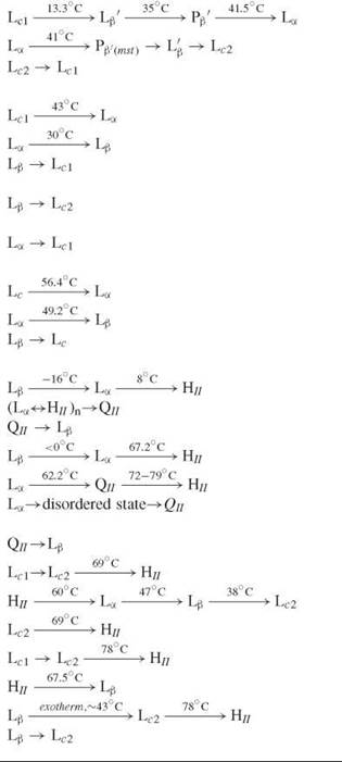
DOPE-Me, dioleoyl-N-methylethanolamine; 14-Gal, ditetradecylgalactosylglycerol; 18-Gal, dioctadecylgalactosylglycerol.
Among the seven cubic phases so far identified in lipids, of greatest interest are the inverted bicontinuous or bilayer cubic phases with space groups Q224(Pn3m),Q229(Im3m), and Q230(Ia3d) (43, 57, 58) (Fig. 1-II j, k, and l). Whenever present, the bilayer cubic phases are located in a temperature range between the La and the HII phases. However, direct La-QIIB transitions are rare in membrane lipid dispersions and mainly observed for short-chain PEs and monoglycosyldiacylglycerols (52, 55). In many cases, a QIIB phase can be induced by means of temperature cycling through the La-HIItransition or by a cooling of the HII phase (59, 60). A transformation from a lamellar into a bilayer cubic phase may be considered as a cooperative act of multiple fusion events, whereby a set of initially separate, parallel bilayers fuse into a single bilayer of specific topology (Fig. 4). The lamellar-cubic transitions have very small, if any, latent heats. Although energetically inexpensive, these transitions typically are rather slow. The slow formation, hysteretic behavior, and extended metastability ranges of the cubic phases create significant difficulties in their study and applications.
Inverted micellar cubic phases have been observed mainly in mixtures of double-chain polar lipids with fatty acids or diacylglycerols but also in some single-component dispersions of glycolipids (61). The most frequently observed inverted micellar cubic phase in lipids is of space group Q227(Fd3m). For medium-chain lipids (≥16C atoms), it typically forms via an HII-QIIM transition; however, the Lβ gel phase of diC19-xylopyranosyl has been found to melt directly into the Fd3m cubic phase (61).

Figure 4. The modified stalk mechanism of membrane fusion and inverted phase formation. (a) planar lamellar (La) phase bilayers; (b) the stalk intermediate; the stalk is cylindrically-symmetrical about the dashed vertical axis; (c) the TMC (trans monolayer contact) or hemifusion structure; the TMC can rupture to form a fusion pore, referred to as interlamellar attachment, I LA (d); (e) If ILAs accumulate in large numbers, they can rearrange to form QII phases. (f) For systems close to the La/HII phase boundary, TMCs can also aggregate to form HII precursors and assemble into HII domains. The balance between QII and HII phase formation is dictated by the value of the Gaussian curvature elastic modulus of the bilayer (reproduced from (25) with permission of the Biophysical Society); The stalk in (b) is structural unit of the rhombohedral phase; (b') electron density distribution for the stalk fragment of the rhombohedral phase, along with a cartoon of a stalk with two lipid monolayers merged to form a hourglass structure (reproduced from (26) with permission of the Biophysical Society).
Polymorphic transitions between solid lipid phases
At temperatures below the main transition, a basic equilibrium structure is the subgel (crystalline) Lc phase. Its formation usually requires prolonged low-temperature incubation. In addition to the Lc phase, many intermediate stable, metastable, and transient lamellar gel structures are adopted by different lipid classes—with perpendicular or tilted chains with respect to the bilayer plane, with fully interdigitated, partially interdigitated, or noninterdigitated chains, rippled bilayers with various ripple periods, and so forth. (Fig. 1). Several polymorphic phase transitions between these structures have been reported. Well-known examples of polymorphic transitions are the subtransition (Lc-Lβ) and the pretransition (Lβ’-Pβ’) in phosphatidylcholines (33). Recently, a polymorphic transition that included rapid, reversible transformation of the usual gel phase into a metastable, more ordered gel phase with orthorhombic hydrocarbon chain-packing (so-called Y-transition) was reported to represent a common pathway of the bilayer transformation into a subgel (crystalline) Lc phase (62).
Reversibility of the phase transitions: formation of metastable phases
Because of long relaxation times, especially in the transition vicinity, lipid phase transitions often are not reversible and end up with the formation of metastable phases, which replace the equilibrium phases in cooling scans. The metastable phases can be very long-lived and display no spontaneous conversion to the ground state in sensible time scales.
Many rate-limiting factors have been suggested as physical reasons that lead to the formation of metastable phases: long hydration/dehydration times, slow reformation of hydrogen-bond networks, restricted molecular motion in the low-temperature solid-solid transformations, relative stability of the interfaces between solid and fluid domains, large spatial rearrangements in lamellar-nonlamellar transitions, low rate of appearance of critical-size nuclei of the nascent phase, and arrestment in local free-energy minima. A comparison between heating and cooling phase sequences observed in aqueous dispersions of lipids shows the frequent occurrence of additional, metastable phases, which only form in a cooling direction (Table 3).
Phase transitions in lipid mixtures: phase diagrams
Composition is another important variable that strongly modulates the lipid phase behavior. The phase properties of a lipid mixture are best presented by means of a temperature-composition phase diagram. Such diagrams may be constructed by using various experimental techniques (63); a most appropriate one is the differential scanning calorimetry. In addition to being nonperturbing, it also has the advantage of recording not only the temperature but also the enthalpy of the phase transitions.
Various types of lipid phase diagrams reported in the literature are shown in Fig. 5 (64-73). The lens-like diagram in Fig. 5a is characteristic for lipid mixtures that are completely miscible in both gel and liquid-crystalline phases. To display such complete miscibility, the two components must be very similar structurally. This kind of diagram is typical for lipid species with the same headgroup, differing by not more than two methylene groups in their hydrocarbon chains, such as the DMPC/DPPC binary (Fig. 5a). A usual complication of the lens-type diagram is the frequently occurring solid-state miscibility gap, where the mixture separates into two solid phases of different composition. In mixtures of lipids with sufficiently different structures, the miscibility gap may overlap with the region of the solid-liquid-crystalline phase coexistence and give rise to eutectic (Fig. 5b and c) or peritectic phase diagrams (Fig. 5d), in which single three-phase points exist. Horizontal solidus lines, reporting for such kind of behavior, have been observed for numerous lipid mixtures. Miscibility gaps also may occur in the liquid-crystalline phase of certain lipid mixtures. A phase diagram with a liquid-liquid immiscibility region is the monotectic phase diagram shown in Fig. 5e, with a monotectic triple point of coexistence of one solid and two liquid phases.
Deviations from ideal mixing may occur not only with a tendency for clustering of the like molecules and eventually leading to phase separation but also when contacts between unlike molecules are preferred - when the nearest-neighbor pairs tend to be made up of unlike molecules (a “chessboard” arrangement). Such mixtures often display phase diagrams with an upper isoconcentration (azeotropic) point, such as the DPPC/palmitic acid diagram shown in Fig. 5g. Except for the phosphatidylcholine/fatty acid mixtures, phase diagrams with an upper isoconcentration point are typical for mixtures that contain a charged lipid. An example of a phase diagram with a lower isoconcentration point is shown in Fig. 5f.
The DPPC/cholesterol phase diagram in Fig. 5h contains a critical point. It is related to the existence of a peculiar, liquid ordered (lo) phase in the mixtures, which is believed to be the prototype of the lipid rafts (see Lipid rafts).
Figs. 5i—l illustrate the phase behavior in lipid/water mixtures. Aqueous phase diagrams of monoacylglycerols of various chain length and saturation show the effect of molecular geometry on the lipid phase behavior. Increasing the chain length from C8:0 to C20:0 introduces cubic and hexagonal phases between the lamellar liquid-crystalline and the liquid L2 phase (72). In the unsaturated C18:1glyceryl monooleate, two cubic phases, Ia3d (G) and Pn3m (D), and the inverted hexagonal HII phase form (73) (Fig. 5l).
Ternary phase diagrams are another important tool for the characterization of the phase properties of complex lipid mixtures. This kind of diagram has proven especially useful recently in the analysis of domains in model systems. Ternary mixtures of one phospholipid that has a relatively high melting temperature and another phospholipid that has a relatively low melting temperature together with cholesterol are viewed as useful models for the outer leaflet of animal-cell plasma membranes. An example of a ternary phase diagram is shown in Fig. 6 (74). It illustrates the rich phase behavior displayed by ternary lipid mixtures represented in this particular case by four regions of two-phase coexistence and one region of three-phase coexistence.

Figure 5. Types of lipid phase diagrams: (a) close to ideal phase diagram of the DMPC/DPPC mixture. (Reproduced from Reference 64 with author's permission.) (b) Eutectic phase diagram of the 1-stearoyl-2-caprylphosphatidylcholine/DMPC mixture. (Reproduced from Reference 65 with permission from Elsevier.) (c) Phase diagram of the DPPC/C12 C24PC mixture exhibiting eutectic and peritectic points. (Reproduced from Reference 66 with permission from Elsevier.) (d) Peritectic phase diagram of the DMPE/DSPE mixture. (Reproduced from Reference 67 with kind permission of Springer Science and Business Media.) (e) Monotectic phase diagram of the DEPC/DPPE mixture. (Reproduced with permission from Reference 68; copyright 1975 American Chemical Society.) (f) Phase diagram with a lower isoconcentration (azeotropic) point) of the DEPC/DMPC mixture. (Reproduced from Reference 69 with permission from Elsevier.) (g) Phase diagram of the DPPC/palmitic acid mixture, combining upper isoconcentration (azeotropic) point with eutectic and peritectic points. (Reproduced from Reference 70 with permission from Elsevier.) (h) Phase diagram with a critical point of the DPPC/Cholesterol mixture. (Reproduced from Reference 71 with permission of the Biophysical Society.). The main features of the phase diagrams of saturated monoacylglycerol/water systems: (i) C8:0-C12:0; (j) C14:0-C18:0; (k) C20:0. (Reproduced from Reference 72 with permission.) (l) Phase diagram of glyceryl monooleate/water system. (Reproduced from Reference 73 with permission.)

Figure 6. Phase diagram of the ternary mixture distearoylphosphatidylcholine (DSPC)/dioleoylphosphatidylcholine (DOPC)/cholesterol at 23° C, showing four regions of two-phase coexistence: liquid crystalline and gel (La + Lβ), liquid ordered and gel (L0 + Lβ), liquid crystalline and liquid ordered (La + L0), and liquid ordered and crystals of cholesterol monohydrate; also one region of three-phase coexistence exists, liquid crystalline, gel and liquid ordered (La + Lβ + L0). (Reproduced from Reference 74 with permission.)
Role of the aqueous phase composition
The Hofmeister Effect
The interactions of the lipid polar groups with water have an important contribution to the energy balance of a given phase. The relatively high hydration, typical for the membrane lipids, is responsible in particular for their ability to form liquid-crystalline bilayers separated by aqueous spaces. Many lipid phase transitions take place with large changes in the lipid surface area and consequently in the amount of bound water. The lipid hydration is determined by the chemical structure of the polar groups, but it is essential to note that in fully hydrated systems with water in excess, the extent of the polar group hydration depends also on the state of the bulk water. On the other side, various low-molecular solutes are known for their ability to modulate strongly the bulk water structure: “water-structure makers” (kosmotropes) and “water-structure breakers” (chaotropes). It thus turns possible that, even without direct interaction with the lipid polar heads, solutes largely can modulate the properties of the lipid-water interface and hence the lipid phase behavior. Changes in the aqueous phase composition can shift substantially the temperature regions of stability of the different lipid phases and induce or suppress the formation of certain phases. Indirect solute effects of such kind on the interfacial properties, generally termed the Hofmeister effect, have been found in many lipid-water phases (34). Many studies on the nature of the Hofmeister effect indicate that it results from an interplay of electrostatics, dispersion forces, thermal motion, fluctuations, hydration, polarizability, ion size effects, and the impact of interfacial water (75, 76).
According to their effect on the lipid phase transitions, the Hofmeister solutes fall into two categories: i) chaotropic solutes that favor the formation of the lamellar liquid-crystalline phase La at the expense of the neighboring HII and Lβ phases and 2) kosmotropic solutes that favor the formation of the HII and Lβ phase at the expense of the La phase. Their effects are described correctly by an equation of the Clapeyron-Clausius type between phase transition temperature and solute concentration (34). The sign and magnitude of the transition shifts induced by the different solutes depend on the solute ability to distribute unevenly between interlamellar and free water. Kosmotropic solutes tend to minimize the area of the lipid-water contact. They suppress the La phase, as it has the largest surface area in contact with water. At a high enough concentration of kosmotropic solutes, the latter phase may disappear completely from the phase diagram. This disappearance is precisely what is seen with sucrose, trehalose, proline, and some salts and is consistent with the opposite effect caused by chaotropic solutes (Fig. 3 d). The addition of chaotropic solutes also can induce the appearance of missing liquid-crystalline phases from the general lipid phase sequence (1).
Effect of pH
Changes in pH modulate the lipid phase behavior as a consequence of protonation/deprotonation of the lipid headgroups, which results in a change of the surface charge of the membrane (77). They also modify the surface polarity and hydration. Typically, protonation decreases lipid hydration and increases the main transition temperature (53). The effects of pH titration on the chain-melting transition temperature Tm of dimyristoyl phospholipids is illustrated in Fig. 3e, which shows that single protonation increases the melting transition temperature by about 5-15° C.
The shifts of the lamellar-hexagonal transition during titration are greater and in the opposite direction relative to changes of Tm. Thus, for didodecyl PE, the lamellar-hexagonal transition decreases by 41° C during phosphate protonation (pK~1.9) and by 50° C during amine protonation (pK~9.3), whereas for Tm these shifts are 6° C and 15° C, respectively, in the opposite direction (Fig. 3f) (54).
Tools and Techniques
From the analysis of the data in the LIPIDAT database (41), more than 150 different methods and method modifications have been used to collect data related to the lipid phase transitions. Almost 90% of the data is accounted for by less than 10 methods. Differential scanning calorimetry strongly dominates the field with two thirds of all phase transition records. From the other experimental techniques, various fluorescent methods account for~ 10% of the information records. X-ray diffraction, nuclear magnetic resonance (NMR), Raman spectroscopy, electron spin resonance (ESR), infrared (IR) spectroscopy, and polarizing microscopy each contribute to about or less than 2-3% of the phase transition data records in the database. Especially useful in gaining insight into the mechanism and kinetics of lipid phase transitions has been time-resolved synchrotron X-ray diffraction (62, 78-81).
References
1. Singer SJ, Nicolson GL. Fluid mosaic model of structure of cell-membranes. Science 1972; 175:720-731.
2. Shimshick EJ, McConnel HM. Lateral phase separation in phospholipid membranes. Biochemistry 1973; 12:2351-2360.
3. Trauble H, Sackmann E. Studies of crystalline-liquid crystalline phase-transition of lipid model membranes.3. Structure of a steroid-lecithin system below and above lipid-phase transition. J. Am. Chem. Soc. 1972; 94:4499-4510.
4. Lewis RNAH, Mannock DA, McElhaney RN. Membrane lipid molecular structure and polymorphism. In: Current Topics in Membranes, Volume 44. Lipid Polymorphism and Membrane Properties. Epand RM, ed. 1997. Academic Press, San Diego. pp. 25-102
5. Mouritsen OG, Andersen OS, eds. In Search of a New Biomembrane Model. 1998. Munksgaard, Copenhagen.
6. Rilfors L, Lindblom G. Regulation of lipid composition in biological membranes-Biophysical studies of lipids and lipid synthesizing enzymes. colloids surfaces B-biointerfaces 2002; 26:112-124.
7. Koynova R, MacDonald RC. Natural lipid extracts and biomembrane-mimicking lipid compositions are disposed to form nonlamellar phases, and they release DNA from lipoplexes most efficiently. Biochim. Biophys. Acta-Biomemb. 2007; 1768:23732382.
8. Biltonen RL. A statistical-thermodynamic view of cooperative structural-changes in phospholipid-bilayer membranes-Their potential role in biological function. J. Chem. Thermodyn. 1990; 22:1-19.
9. Bloom M, Evans E, Mouritsen OG. Physical-properties of the fluid lipid-bilayer component of cell-membranes-A perspective. Q. Rev. Biophys. 1991; 24:293-397.
10. Hazel Jr. Thermal adaptation in biological-membranes-Is homeo-viscous adaptation the explanation. Annu. Rev. Physiol. 1995; 57:19-42.
11. Heimburg T, Jackson AD. The thermodynamics of general anesthesia. Biophys. J. 2007; 92: 3159-3165.
12. Larsson K. Anesthetic effect and a lipid bilayer transition involving periodic curvature. Langmuir 1988; 4:215-217.
13. Landh T. From entangled membranes to eclectic morphologies-Cubic membranes as subcellular space organizers. FEBS Lett. 1995; 369:13-17.
14. Almsherqi ZA, Kohlwein SD, Deng Y. Cubic membranes: a legend beyond the flatland of cell membrane organization. J. Cell Biol. 2006; 173:839-844.
15. Gruner SM. Coupling between bilayer curvature elasticity and membrane-protein activity. In: Biomembrane Electrochemistry, Volume 235. Blank M, Vodyanoy I, eds. 1994. American Chemical Society, Washington, DC. pp. 129-149.
16. McIntosh TJ, Simon SA. Roles of bilayer material properties in function and distribution of membrane proteins. Annu. Rev. Biophys. Biomol. Struct. 2006; 35:177-198.
17. Gruner SM. Intrinsic curvature hypothesis for biomembrane lipid-composition-A role for nonbilayer lipids. Proc. Natl. Acad. Sci. U. S. A. 1985; 82:3665-3669.
18. Lee AG. How lipids affect the activities of integral membrane proteins. Biochim. Biophys. Acta-Biomemb. 2004; 1666:62-87.
19. DeKruijff B. Biomembranes - Lipids beyond the bilayer. Nature 1997; 386: 129-130.
20. Kinnunen PKJ. On the molecular-level mechanisms of peripheral protein-membrane interactions induced by lipids forming inverted non-lamellar phases. Chem. Phys. Lipids 1996; 81:151-166.
21. Botelho AV, Gibson NJ, Thurmond RL, Wang Y, Brown MF. Conformational energetics of rhodopsin modulated by nonlamellar-forming lipids. Biochemistry 2002; 41:6354-6368.
22. Lee AG. Lipid-protein interactions in biological membranes: a structural perspective. Biochim. Biophys. Acta-Biomemb. 2003; 1612:1-40.
23. Siegel DP. The relationship between bicontinuous inverted cubic phases and membrane fusion. In: Bicontinuous Liquid Crystals. Lynch ML, Spicer PT, eds. 2005. Taylor & Francis Group, CRC Press, Boca Raton, FL. pp. 59-98.
24. Kozlov MM, Markin VS. Possible mechanism of membrane-fusion. Biofizika 1983; 28:242-247.
25. Siegel DP. The modified stalk mechanism of lamellar/inverted phase transitions and its implications for membrane fusion. Biophys. J. 1999; 76:291-313.
26. Yang L, Huang HW. A rhombohedral phase of lipid containing a membrane fusion intermediate structure. Biophys. J. 2003; 84:1808-1817.
27. Tenchov BG. Nonuniform lipid distribution in membranes. Prog. Surf. Sci. 1985; 20:273-340.
28. Mouritsen OG, Jorgensen K. Microscale, nanoscale and mesoscale heterogeneity of lipid bilayers and its influence on macroscopic membrane-properties. Mol. Membr. Biol. 1995; 12:15-20.
29. Simons K, Ikonen E. Functional rafts in cell membranes. Nature 1997; 387: 569-572.
30. Lagerholm BC, Weinreb GE, Jacobson K, Thompson NL. Detecting microdomains in intact cell membranes. Annu. Rev. Phys. Chem. 2005; 56:309-336.
31. McConnell HM, Vrljic M. Liquid-liquid immiscibility in membranes. Annu. Rev. Biophys. Biomol. Struct. 2003; 32:469-492.
32. Cossins AR. Homeoviscous adaptation of biological membranes and its functional significance. In: Temperature Adaptation of Biological Membranes. Cossins AR, ed. 1994. Portland Press, London. pp. 63-76.
33. Koynova R, Caffrey M. Phases and phase transitions of the phosphatidylcholines. Biochim. Biophys. Acta-Rev. Biomembranes 1998; 1376:91-145.
34. Koynova R, Brankov J, Tenchov B. Modulation of lipid phase behavior by kosmotropic and chaotropic solutes - Experiment and thermodynamic theory. Eur. Biophys. J. Biophys. Lett. 1997; 25:261-274.
35. White SH, Mirejovsky D, King GI. Structure of lamellar lipid domains and corneocyte envelopes of murine stratum-corneum - An X-ray-diffraction study. Biochemistry 1988; 27:3725-3732.
36. McIntosh TJ. Organization of skin stratum corneum extracellular lamellae: diffraction evidence for asymmetric distribution of cholesterol. Biophys. J. 2003; 85:1675-1681.
37. de Jager MW, Gooris GS, Ponec M, Bouwstra JA. Lipid mixtures prepared with well-defined synthetic ceramides closely mimic the unique stratum corneum lipid phase behavior. J. Lipid Res. 2005; 46:2649-2656.
38. Luzzati V. Biological significance of lipid polymorphism: the cubic phases-commentary. Curr. Opin. Struct. Biol. 1997; 7:661-668.
39. Larsson K. Aqueous dispersions of cubic lipid-water phases. Curr. Opin. Colloid Interface Sci. 2000; 5:64-69.
40. Luzzati V. X-Ray Diffraction studies of lipid-water systems. In: Biological Membranes, Volume 1. Chapman D, ed. 1968. Academic Press, London. pp. 71-123.
41. Caffrey M, Koynova R, Hogan J, Moynihan D. LIPIDAT: a database of lipid phase transition temperatures and enthalpy changes. In: Handbook of Nonmedical Applications of Liposomes, Volume 2. Barenholz Y, Lasic DD, eds. 1996. CRC Press, Boca Raton, FL. pp. 85-104.
42. Kasper JS, Lonsdale K. International tables for x-ray crystallography. 1985. Riedel Publishing Company, Dordrecht, The Netherlands.
43. Seddon JM. Structure of the inverted hexagonal (HII) phase, and non-lamellar phase-transitions of lipids. Biochim. Biophys. Acta 1990; 1031:1-69.
44. Tenchov B. On the reversibility of the phase-transitions in lipid-water systems. Chem. Phys. Lipids 1991; 57:165-177.
45. Nagle JF. Theory of the main lipid bilayer phase-transition. Annu. Rev. Phys. Chem. 1980; 31:157-195.
46. Marsh D. Handbook of Lipid Bilayers. 1990. CRC Press, Boca Raton, FL.
47. Albon N, Sturtevant JM. Nature of Gel to Liquid-Crystal Transition of Synthetic Phosphatidylcholines. Proc. Natl. Acad. Sci. U. S. A. 1978; 75(5):2258-2260.
48. Schrader W, Ebel H, Grabitz P, Hanke E, Heimburg T, Hoeckel M, Kahle M, Wente F, Kaatze U. Compressibility of lipid mixtures studied by calorimetry and ultrasonic velocity measurements. J. Phys. Chem. B 2002; 106:6581-6586.
49. Honger T, Mortensen K, Ipsen JH, Lemmich J, Bauer R, Mouritsen OG. Anomalous swelling of multilamellar lipid bilayers in the transition region by renormalization of curvature elasticity. Phys. Rev. Lett. 1994; 72:3911-3914.
50. Chu N, Kucerka N, Liu YF, Tristram-Nagle S, Nagle JF. Anomalous swelling of lipid bilayer stacks is caused by softening of the bending modulus. Phys. Rev. E 2005; 71:041904.
51. Pabst G, Amenitsch H, Kharakoz DP, Laggner P, Rappolt M. Structure and fluctuations of phosphatidylcholines in the vicinity of the main phase transition. Phys. Rev. E 2004; 70:021908.
52. Koynova R, Caffrey M. Phases and phase-transitions of the hydrated phosphatidylethanolamines. Chem. Phys. Lipids 1994; 69:1-34.
53. Cevc G, Marsh D. Phospholipid Bilayers. 1987. J. Wiley & Sons, Inc., New York.
54. Seddon JM, Cevc G, Marsh D. Calorimetric studies of the gel-fluid (L-Beta-L-Alpha) and lamellar-inverted hexagonal (L-Alpha-HII) phase-transitions in dialkyl and diacylphosphatidylethanolamines. Biochemistry 1983; 22:1280-1289.
55. Koynova R, Caffrey M. Phases and phase-transitions of the glyco-glycerolipids. Chem. Phys. Lipids 1994; 69:181-207.
56. Koynova R, Caffrey M. Phases and phase-transitions of the sphin- golipids. Biochim. Biophys. Acta-Lipids Lipid Metab. 1995; 1255:213-236.
57. Lindblom G, Rilfors L. Nonlamellar phases formed by membrane-lipids. Adv. Colloid Interface Sci. 1992; 41:101-125.
58. Luzzati V, Delacroix H, Gulik A, GulikKrzywicki T, Mariani P, Vargas R. The cubic phases of lipids. In: Lipid Polymorphism and Membrane Properties.Current Topics in Membranes, Volume 44. Epand RM, ed. 1997. Academic Press. pp. 3-24.
59. Shyamsunder E, Gruner SM, Tate MW, Turner DC, So PTC, Tilcock CPS. Observation of inverted cubic phase in hydrated di- oleoylphosphatidylethanolamine membranes. Biochemistry 1988; 27:2332-2336.
60. Tenchov B, Koynova R, Rapp G. Accelerated formation of cubic phases in phosphatidylethanolamine dispersions. Biophys. J. 1998; 75:853-866.
61. Seddon JM, Robins J, Gulik-Krzywicki T, Delacroix H.nInverse micellar phases of phospholipids and glycolipids. Phys. Chem. Chem. Phys. 2000; 2:4485-4493.
62. Tenchov B, Koynova R, Rapp G. New ordered metastable phases between the gel and subgel phases in hydrated phospholipids. Biophys. J. 2001; 80:1873-1890.
63. Koynova R, Caffrey M. An index of lipid phase diagrams. Chem. Phys. Lipids 2002; 115: 107-219.
64. Mabrey S, Sturtevant JM. Investigation of phase-transitions of lipids and lipid mixtures by high sensitivity differential scanning calorimetry. Proc. Natl. Acad. Sci. U.S.A. 1976; 73:3862-3866.
65. Lin HN, Huang CH. Eutectic Phase-behavior of 1-stearoyl-2-caprylphosphatidylcholine and dimyristoylphosphatidylcholine mixtures. Biochim. Biophys. Acta 1988; 946:178-184.
66. Gardam M, Silvius Jr. Intermixing of dipalmitoylphosphatidyl-choline with phospholipids and sphingolipids bearing highly asymmetric hydrocarbon chains. Biochim. Biophys. Acta 1989; 980:319-325.
67. Dorfler HD. Relationships between miscibility behavior and chemical structure of phospholipids in pseudobinary systems. Colloid Polym. Sci. 2000; 278:130-136.
68. Wu SHW, McConnell HM. Phase separations in phospholipid membranes. Biochemistry 1975; 14:847-854.
69. VanDijck PWM, Kaper AJ, Oonk HAJ, DeGier J. Miscibility properties of binary phosphatidylcholine mixtures-calorimetric study. Biochim. Biophys. Acta 1977; 470:58-69.
70. Inoue T, Yanagihara S, Misono Y, Suzuki M. Effect of fatty acids on phase behavior of hydrated dipalmitoylphosphatidylcholine bilayer: saturated versus unsaturated fatty acids. Chem. Phys. Lipids 2001; 109:117-133.
71. Ipsen JH, Mouritsen OG, Bloom M. Relationships between lipid-membrane area, hydrophobic thickness, and acyl-chain orientational order - the effects of cholesterol. Biophys. J. 1990; 57:405-412.
72. Larsson K, Quinn P, Sato K, Tiberg F. Lipids: Structure, Physical Properties and Functionality. 2006. The Oily Press, Bridgwater, England.
73. Hyde ST, Andersson S, Ericsson B, Larsson K. A cubic structure consisting of a lipid bilayer forming an infinite periodic minimum surface of the gyroid type in the glycerolmonooleate-water system. Zeitschrift fur Kristallographie 1984; 168:213-219.
74. Feigenson GW. Phase behavior of lipid mixtures. Nat. Chem. Biol. 2006; 2:560-563.
75. Koelsch P, Viswanath P, Motschmann H, Shapovalov VL, Brezesinski G, Mohwald H, Horinek D, Netz RR, Giewekemeyer K, Alditt TS, Schollmeyer H, von Klitzing R, Daillant J, Guenoun P. Specific ion effects in physicochemical and biological systems: simulations, theory and experiments. Coll. Surf. A-Physicochem. Engineer. Aspects 2007; 303:110-136.
76. Collins KD, Washabaugh MW. The Hofmeister effect and the behavior of water at interfaces. Q. Rev. Biophys. 1985; 18:323-422.
77. Trauble H, Teubner M, Woolley P, Eibl H. Electrostatic interactions at charged lipid-membranes. 1. Effects of Ph and univalent cations on membrane structure. Biophys. Chem. 1976; 4:319-342.
78. Laggner P. Nonequilibrium phenomena in lipid-membrane phasetransitions. J. Phys. IV 1993; 3:259-269.
79. Laggner P, Kriechbaum M, Rapp G.Structural intermediates in phospholipid phase-transitions. J. Appl. Crystallogr. 1991; 24:836-842.
80. Quinn PJ. Measurement of kinetics and mechanisms of phase transitions in lipid-water systems. J. Appl. Crystallogr. 1997; 30:733-738.
81. Caffrey M. Structural, mesomorphic and time-resolved studies of biological liquid-crystals and lipid-membranes using synchrotron x-radiation. Top. Curr. Chem. 1989; 151:75-109.
Further Reading
Heimburg T. Thermal Biophysics of Membranes. 2007. Wiley-VCH, Berlin.
Kinnunen P, Laggner P, eds. Phospholipid phase transitions 1991; 57. Chem. Phys. Lipids. Entire issue.
Lipowsky R, Sackmann E, eds. Handbook of Biological Physics, Volume 1. 1995. Elsevier, Amsterdam.
Mouritsen OG. Life as a Matter of Fat. The Emerging Science of Lipidomics. 2005. Springer, Berlin.
Seddon JM, ed. Surfactant liquid crystals. Curr. Opin. Coll. Interface Sci. 2001; 6:242-312.
Tanford C. The Hydrophobic Effect—Formation of Micelles and Biological Membranes. 1973. Wiley Interscience, New York.
See Also
Drug Delivery
Lipidomics
Lipid Rafts
Membrane Fusion, Mechanisms of