Classic Human Anatomy in Motion: The Artist's Guide to the Dynamics of Figure Drawing (2015)
Chapter 3
Muscle and Tendon Characteristics
In this chapter, we introduce the basic traits of the skeletal muscles, their positions in the body, how they attach to bones, and how they maneuver the joints when they contract. We will also look at how muscles change shape in different movements. Tendons are also introduced, with a focus on their characteristics and how they influence the surface form. This basic information will be elaborated on in the following chapters on the muscle groups of different regions of the body.
Muscles, along with the subcutaneous layer of adipose (fatty) tissue, define the overall shape of the figure, “fleshing out” its structure and giving substance and character to the body. One of the many challenges in drawing the figure is to depict surface forms changing in various poses. Understanding the basic placement of muscles and how they stretch and compress in different movements will give you, as an artist, the advantage of knowing what occurs beneath the skin (and how that) influences what you see on the surface.
Figurative artists from centuries past up to the present have known the value of studying the human muscular system. When you view figurative works by painters and sculptors such as Michelangelo, Artemesia Gentileschi, Auguste Rodin, Peter Paul Rubens, and others, it’s evident that these artists were well aware of anatomical forms and utilized that information to serve their artistic vision. Their knowledge of anatomy never overpowered their personal style or aesthetics—it only enhanced their work. Learning about the muscular system thus opens a creative door to many possible artistic options, whether you pursue an exacting anatomical realism, exaggerate bodily forms to create interesting visual dynamics, or explore a more expressive interpretation of the human form.
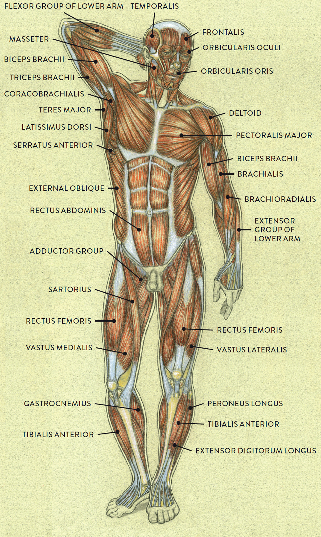
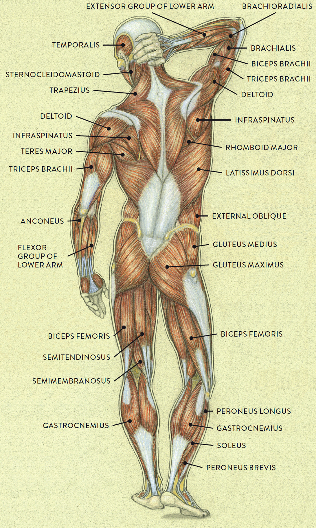
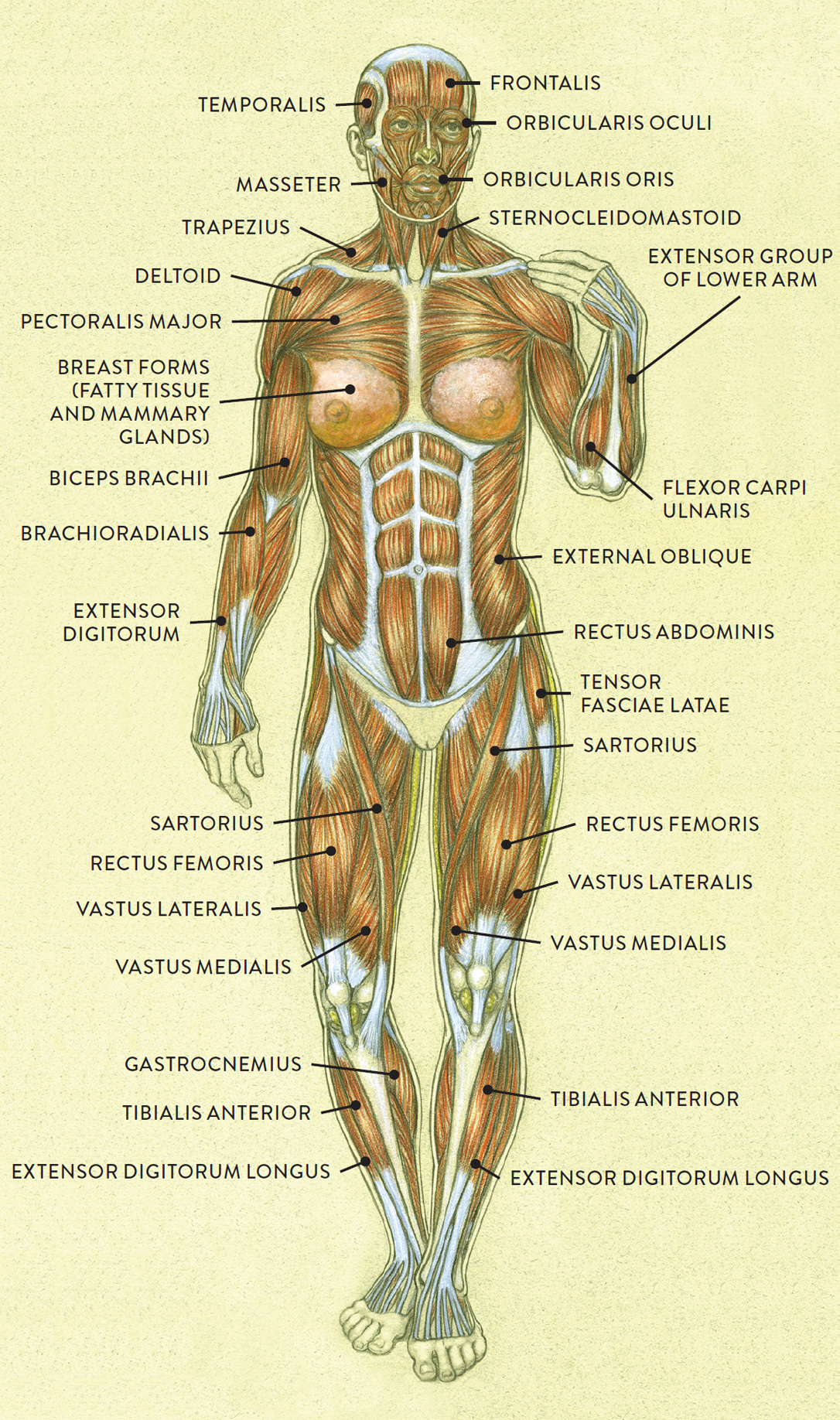
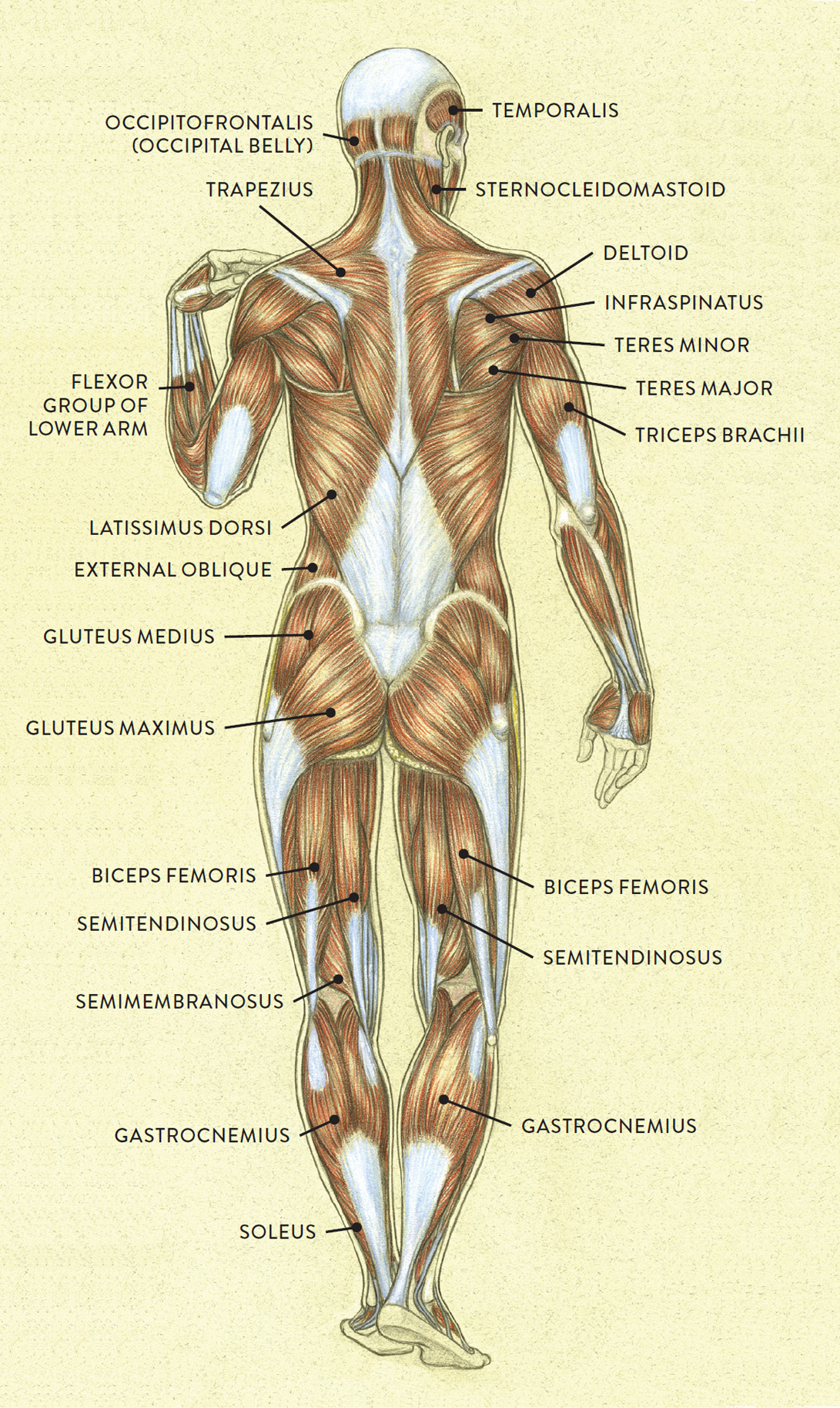
Let’s begin by looking at the entire muscular system. The following drawings show the male and female figures in both anterior and posterior views.
MUSCLES OF THE MALE FIGURE—ANTERIOR VIEW
MUSCLES OF THE MALE FIGURE—POSTERIOR VIEW
MUSCLES OF THE FEMALE FIGURE—ANTERIOR VIEW
MUSCLES OF THE FEMALE FIGURE—POSTERIOR VIEW
Muscles are situated within the body in two basic layers: the superficial muscle layer (also known as externus or superficialis) and the deep muscle layer (also called internus or profundus). Many figurative artists become familiar with the superficial layer when learning the basic anatomical forms of the body. With only a few exceptions, such as the sacrospinalis muscle of the back, the muscles of the deep layer do not influence the surface forms and usually are not visible. Some anatomy books refer to a middle layer, called the intermediate layer, located in the lower arm, foot, and torso.
One way to become familiar with muscles is to categorize them into groups wherever possible. Besides being identified by the layer to which they belong, muscles can be grouped in a number of other ways, including the following:
· By their function or action (e.g., flexor group, extensor group, adductor group)
· By their location in the body or by reference to other anatomical forms (e.g., gluteal group, abdominal group, pectoral group, scapula group, radial group, thenar group, peroneal group)
· By compartment, because muscles are separated into different compartments by deep fascia called intermuscular septa (e.g., anterior compartment, posterior compartment, medial compartment)
· By colloquial (common) names (e.g., thumb group, inner thigh group, upper thigh group, hamstring group)
Some muscles, such as the sartorius muscle of the upper leg, do not belong to any of these categories. These muscles assist in various movements while remaining independent of any group.
Skeletal Muscles
Beyond the groupings given above, muscles are classified as belonging to three basic types: cardiac muscle (pertaining to the heart), smooth muscles (usually affiliated with the tubular structures of the body, such as the arteries, colon, and bronchial tubes, as well as the iris of the eye), and skeletal muscles. As their name implies, the skeletal muscles attach to bones. It is the skeletal muscles that most interest artists because they are instrumental in creating bodily movement and because their shapes are often easy to see beneath the surface of the body.
The main function of skeletal muscles is to shorten, or contract, their overall shape to produce movement. To better understand how they accomplish this, we need to walk through muscles’ basic internal structure. Muscles are composed of a series of elongated muscle fibers (muscle cells) that are grouped together in muscle fiber bundles, or fascicles. Even though muscle fibers are parallel to each other, they can be short, long, circular, fan shaped, or obliquely positioned on a tendon.
SKELETAL MUSCLE ARCHITECTURE
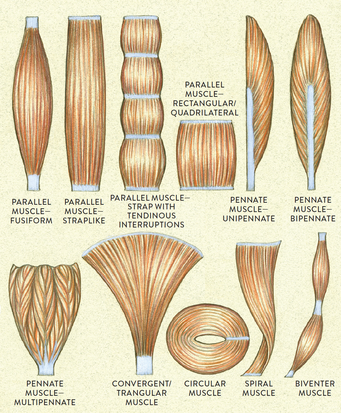
Arrangements of muscle fibers
Muscle Architecture
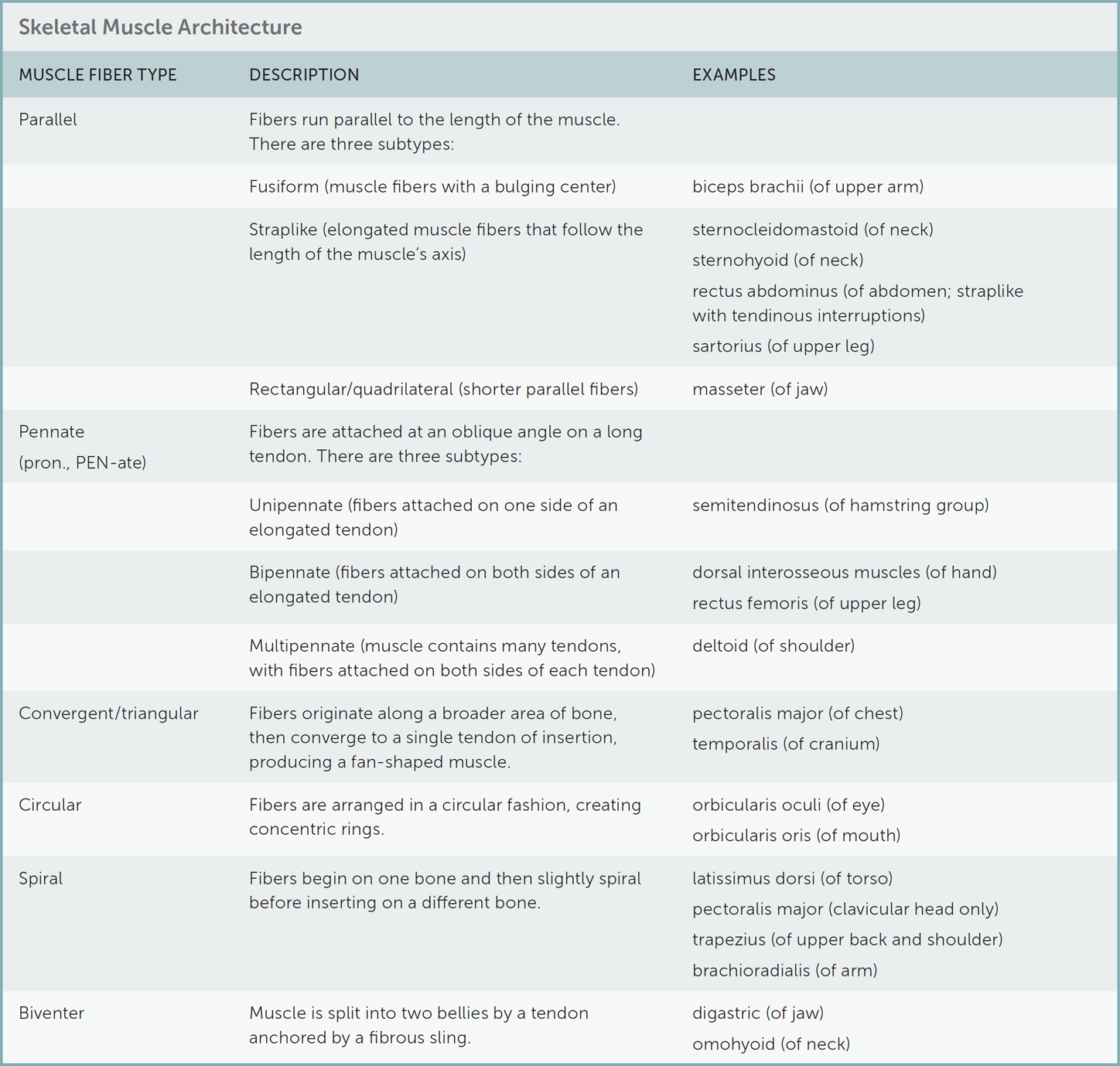
The varying lengths of muscle fibers affect the way muscles perform, with longer muscle fibers producing a greater range of movement while shorter fibers generate more power in movement. Muscle architecture is the term usually applied to the arrangement of muscle fibers. There are several muscle-architecture classifications: parallel, pennate, convergent/triangular, circular, spiral, and biventer. More information about each category, including examples of muscles belonging to each, appears in the following table.
Muscles’ Internal Structure
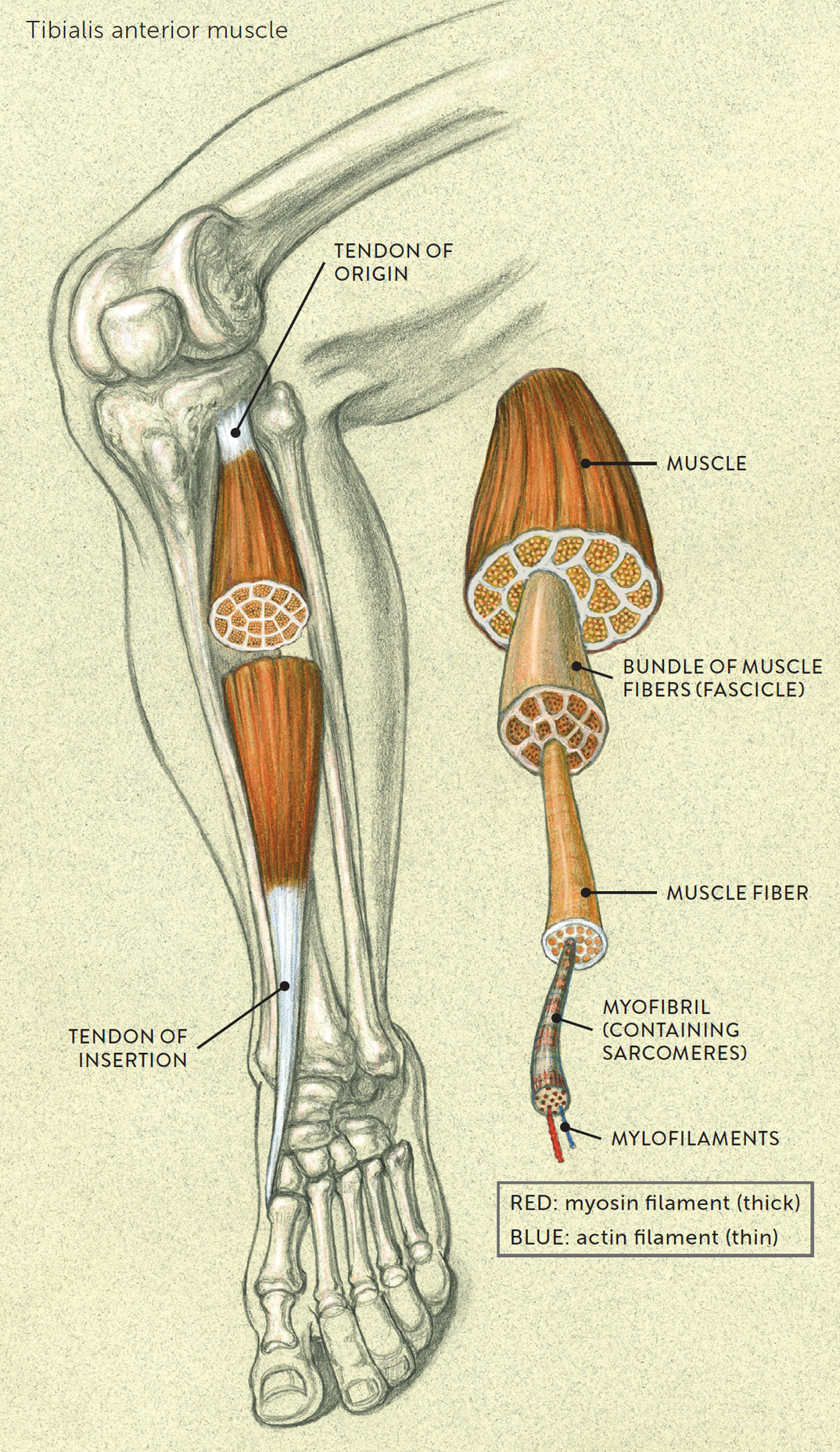
Within each individual muscle fiber are elongated, rodlike strands, called myofibrils, that run parallel to each other and extend to the entire length of the muscle fiber. Myofibrils, in turn, contain a series of smaller units called sarcomeres, which are positioned end to end inside the whole myofibril. And within each sarcomere are microscopic threads called myofilaments. The myofilaments are composed of contractile proteins of two different types—myosin and actin—and are specialized for contraction.
Contraction can be explained, somewhat simplistically, like this: When the sarcomere units receive a signal (an electrical impulse) from the central nervous system, the actin and myosin filaments slide along each other, an action referred to as the sliding filament mechanism/theory. This creates the dynamic force needed for the contraction of a muscle. When a muscle contracts, its fibers shorten toward the center of the muscle. This is called the line of pull, or pulling force, and when enough pulling force is exerted on the muscle attachments on bones, it lifts or pulls the bones, creating movement.
Muscle Attachments (Tendons)
In most cases, a muscle attaches to two different bones (though sometimes more), generating movement, called joint action, at the joint between the bones when it contracts. The places where the muscle attaches are called the origin and insertion sites, and these beginning and ending points are generally on different bones because if they were on the same bone, the muscle would simply lock in place when trying to contract. There are exceptions to this rule, particularly regarding the muscles of the face, but we’ll deal with those separately.
The muscles do not attach directly to bones; rather, the attachments are made via fibrous connective tissues called muscle tendons. The tendon at the muscle’s origin site is called the tendon of origin, or fixed attachment, because the bone to which it attaches stays more or less stationary, or fixed, during movement. The tendon at the muscle’s insertion site is referred to as the tendon of insertion, or mobile attachment, since it is connected to the bone that moves when the muscle is contracting. The following drawing, depicts the sartorius muscle of the upper leg, showing the tendon of origin on the upper end of the muscle and the tendon of insertion on the lower end.
ORIGIN AND INSERTION OF A MUSCLE
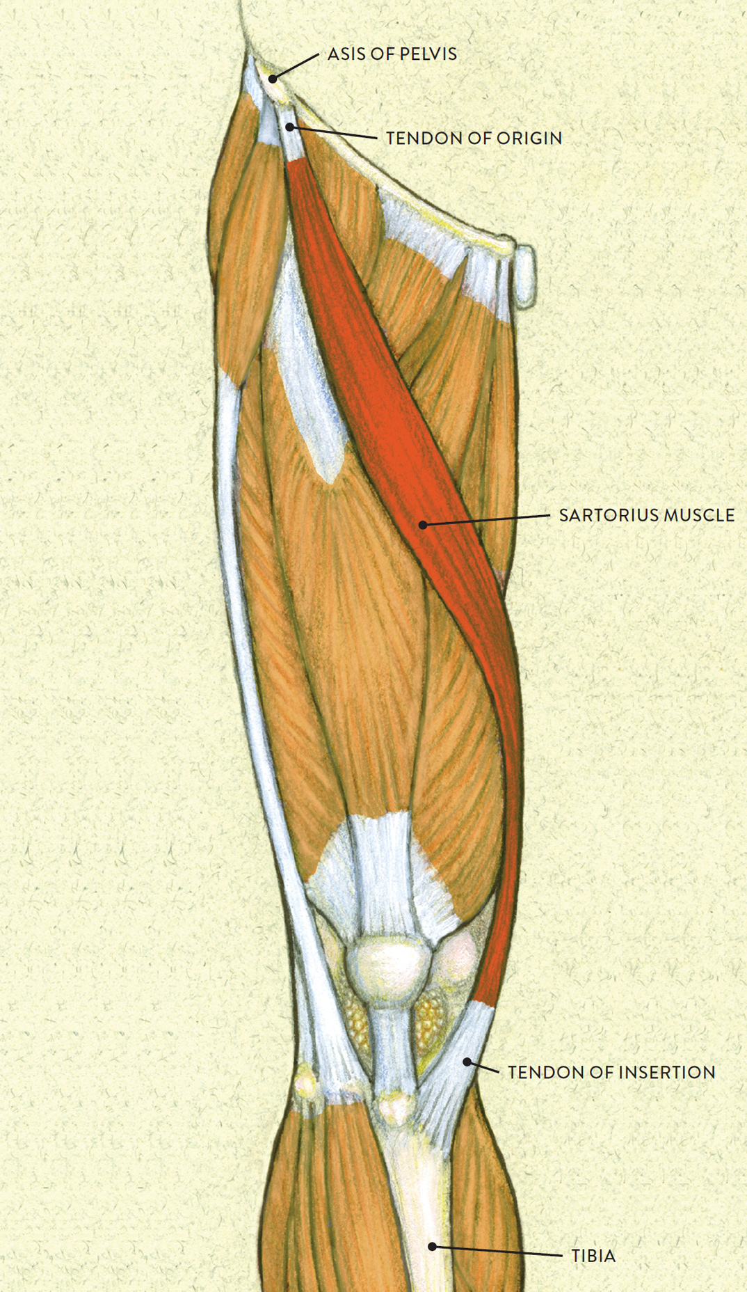
Right upper leg, anterior view
Patterns of skeletal muscle attachments are, however, somewhat varied: Some have multiple origins and a single insertion while others have a single origin and multiple insertions. In some cases, it is hard to decide which is the fixed attachment and which the mobile attachment because the muscle changes roles during various movements. Some anatomists have therefore abandoned the terms origin and insertion, replacing them with proximal attachment and distal attachment, which identify the attachments according to their placement on the body rather than their role in movement. In this book, however, I follow traditional practice, referring to muscle attachments as origins and insertions.
INTERNAL STRUCTURE OF A MUSCLE
How much you want to learn about muscle attachments is up to you, but this knowledge can be advantageous for an artist. Medical illustrators and forensic artists of course need to know the locations of attachments for superficial- and deep-layer muscles, but any artist whose intention is to create realistic (as oppose to stylized) figures, who draws from memory, or who works with figural movement will also find this information beneficial, because without this awareness, the muscular forms might appear inaccurate or possibly distorted. When certain tendons become prominent, then a fairly accurate depiction of how these tendons connect to bone is essential for the overall dynamics of that particular region of the body. An example is the tendon of insertion of the sternocleidomastoid (sternal portion) as it inserts into the upper part of the sternum; this tendon projects quite strongly when the head is rotated. If the tendon is erroneously placed in the wrong location, it might cause that part of the figure to look peculiar.
Any depiction of muscle attachments, including those presented here, should be used as an approximate reference. Human beings are physically diverse, and through many dissections anatomists have found that there are slight variations in the locations of muscle attachments. So figurative artists should know approximately where the two (or more) ends of a muscle attach rather than worrying about the attachments’ precise location.
As I alluded to earlier, most facial muscles work differently from the skeletal muscles of the rest of the body. Except for the muscles controlling the mandible (lower jaw), facial muscles do not move any bones when their fibers contract because, aside from the mandible, the cranium consists of fused bones. Instead, they move soft-tissue forms, creating facial expressions. Similarities and differences between skeletal and facial muscle attachments are summarized in the following table.
|
Skeletal versus Facial Muscle Attachments |
|
|
SKELETAL MUSCLES |
FACIAL MUSCLES |
|
Origin: Muscle attaches to a bone with a tendon. |
Origin: Muscle attaches to a bone with a tendon. |
|
Insertion: Same muscle attaches to a different bone with a different tendon. |
Insertion: Same muscle attaches into a soft-tissue structure such as skin, fascia, subcutaneous tissue, or another facial muscle. |
|
Action: When the muscle contracts, the second bone moves. |
Action: When the muscle contracts, the soft-tissue region moves, possibly creating facial movement. |
Tendon Landmarks
Tendons come in a variety of shapes, including cordlike forms, flat wide sheaths, and thin flat strips. Although the subcutaneous layer of adipose (fatty) tissue obscures many tendons, a few that are shaped like elongated cords do make occasional appearances on the surface form. This generally occurs when a tendon’s muscle is contracting, pulling the tendon close to the skin. Indicating these tendons in figural studies can create a sense of dynamic tension, but the trick is to avoid making the tendons too obvious. For example, when depicting a series of tendons, such as those appearing on the dorsal (back) side of the hand, treat each one slightly differently: One or two can be prominent, the others lightly suggested; otherwise, they might look like stiff spaghetti strands glued on the hand.
Cordlike fibrous structures are generally known as tendons, but broad flat sheathings of fibrous material are identified as aponeuroses (sing., aponeurosis). These wider sheets, which cover larger areas for muscle attachment, can be seen, for example, in the latissimus dorsi and external oblique muscles of the torso and abdominal regions. The following three drawings entitled Tendons and Aponeuroses—Surface Form Landmarks, depict the front torso and arms, back torso and arms, and three views of the legs, showing the basic locations of the tendons of the main superficial muscles. Key bony landmarks and triangular surface-form characteristics are also shown.
TENDONS AND APONEUROSES—SURFACE FORM LANDMARKS
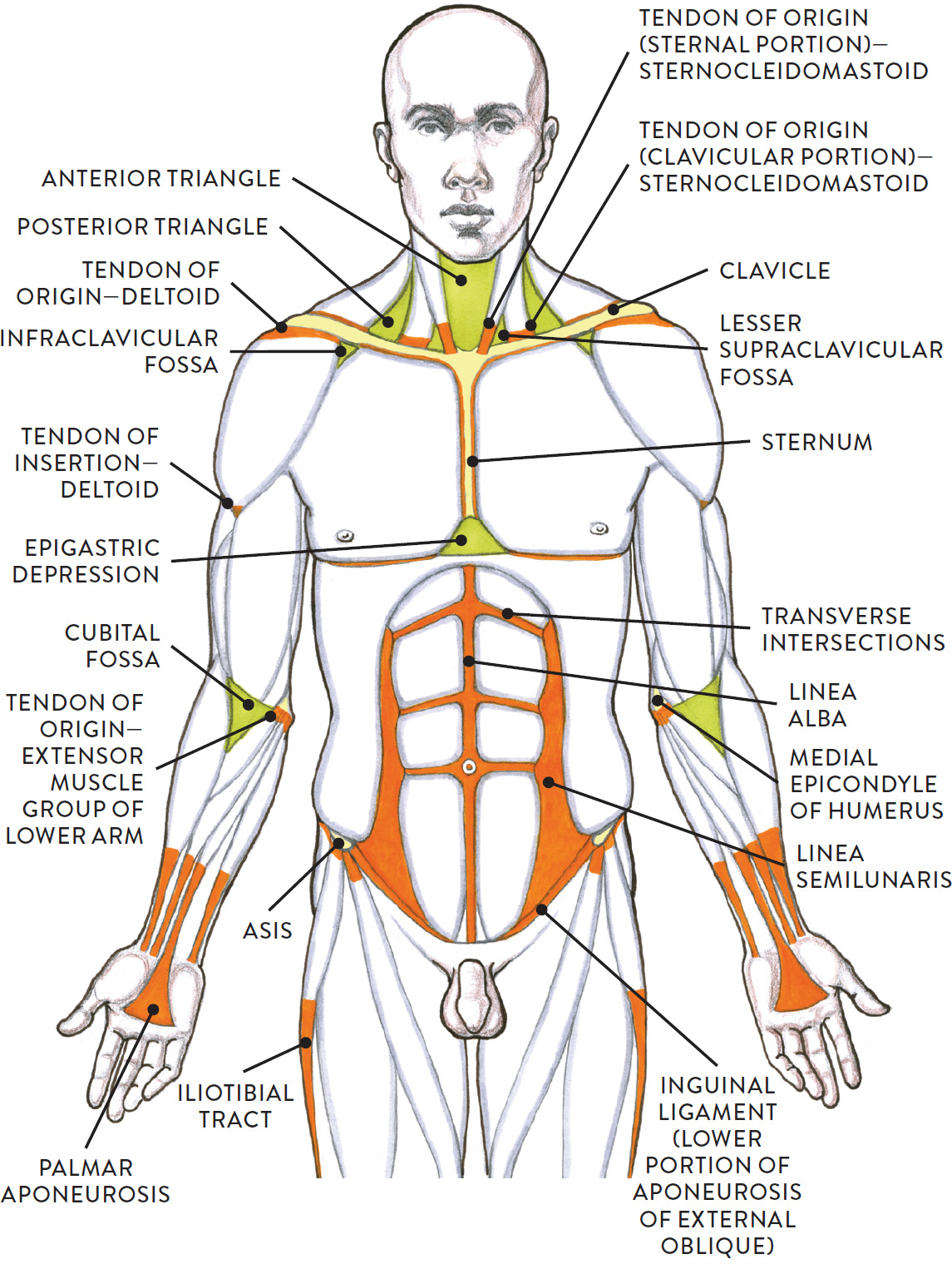
Torso and arms, anterior view
ORANGE: Tendons and aponeuroses
GREEN: Triangular depressions and projections on surface form
YELLOW: Bony landmarks
TENDONS AND APONEUROSES—SURFACE FORM LANDMARKS
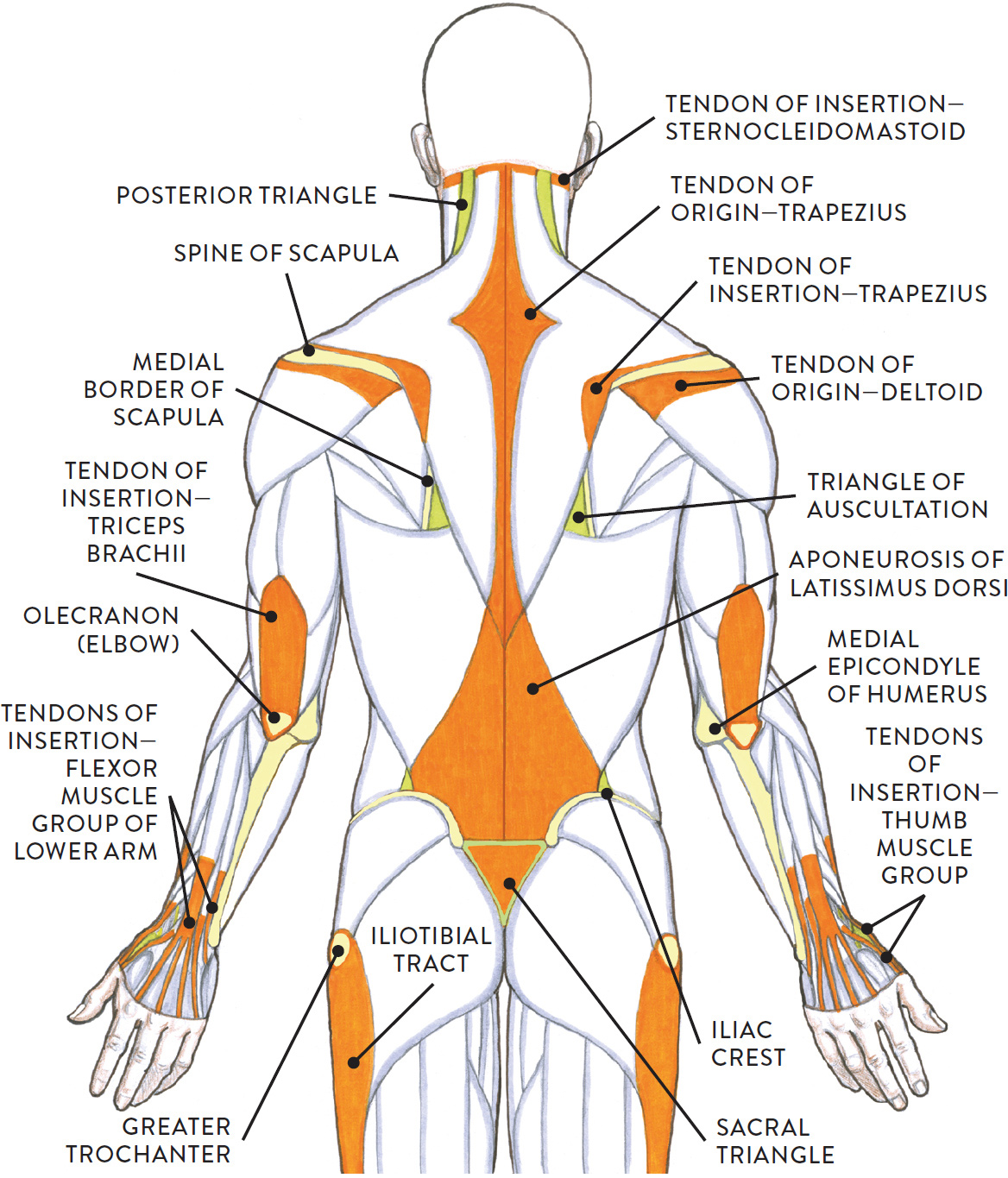
Torso and arms, posterior view
ORANGE: Tendons and aponeuroses
GREEN: Triangular depressions and projections on surface form
YELLOW: Bony landmarks
TENDONS AND APONEUROSES—SURFACE FORM LANDMARKS
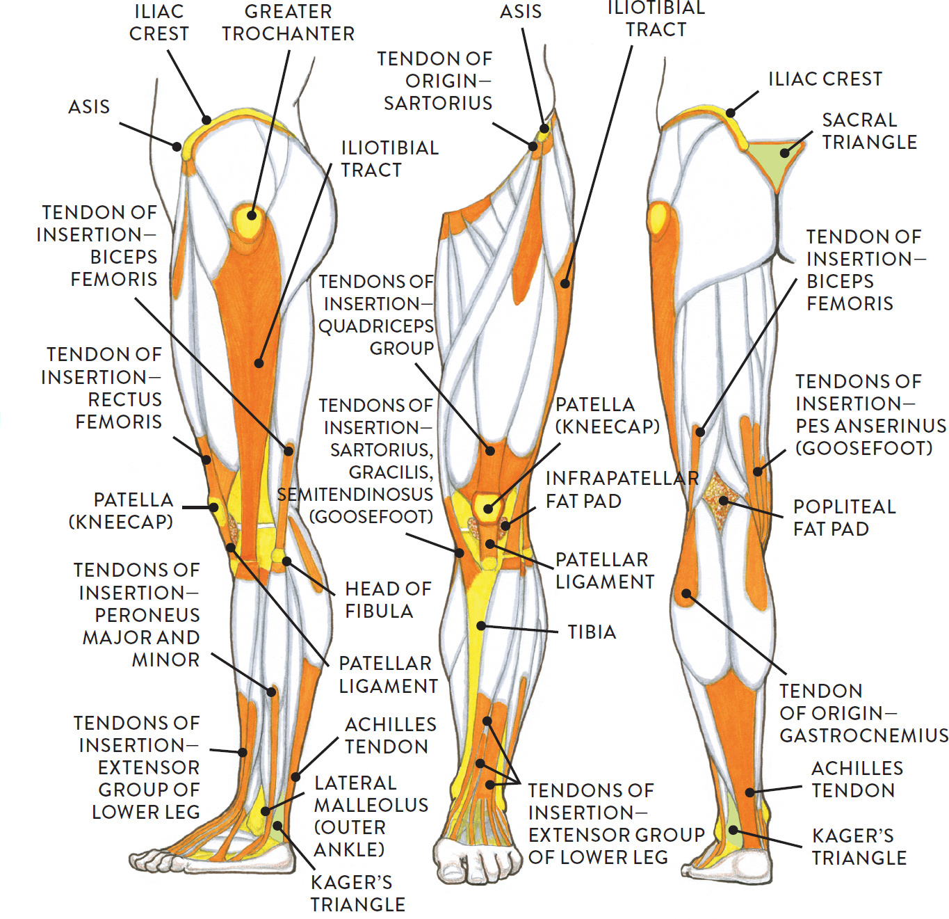
Upper and lower leg, three views
ORANGE: Tendons and aponeuroses
GREEN: Triangular depressions and projections on surface form
YELLOW: Bony landmarks
Tendons of the Sternocleidomastoid Muscle of the Neck
Tendons from each of the sternal portions of the sternocleidomastoid (SCM) muscle attach into the manubrium of the sternum. Between them is the suprasternal notch (pit of the neck). When the head turns sideways in a rotational movement, one of the tendons becomes quite prominent on the surface form, as can be seen in the following portrait study.
PORTRAIT STUDY OF CLAUDIO, WITH HEAD TURNED
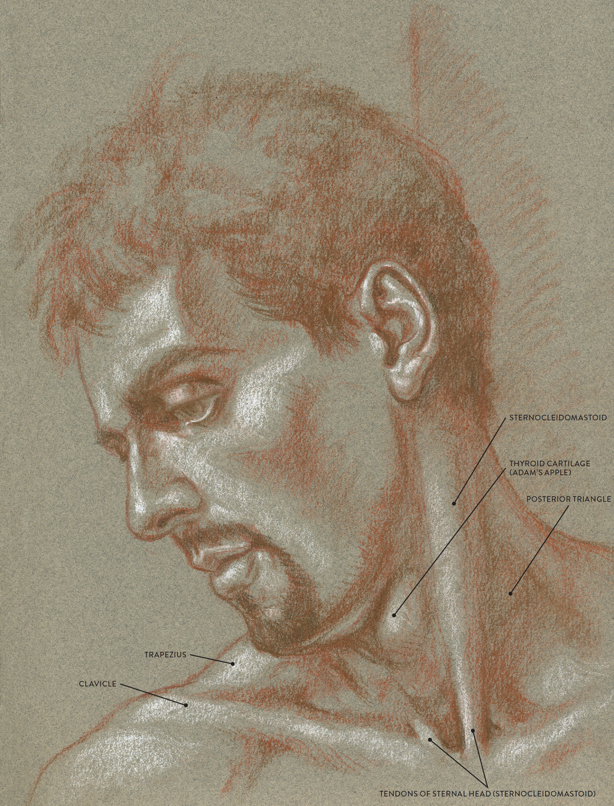
Sanguine and brown pastel pencils and white chalk on toned paper.
Tendons of the Dorsal Side of the Hand
The four tendons of the extensor digitorum muscle of the lower arm insert into the four fingers. They are most easily seen on the surface when the fingers are spread apart forcefully. Because the skin on the dorsal side (back) of the hand is very thin, these tendons sometimes appear, though subtly, when a hand is more relaxed, depending on the position of the hand and the way the light source is illuminating it. Again, when drawing a tendon, avoid emphasizing both sides of the tendon in heavy, dark lines, as this will give the tendon a flat look. One side should be emphasized in a soft, tonal line while the other side is indicated in a lighter value (or with white chalk if drawing on a toned paper surface) to achieve a more natural, organic look, as in the following life study. If there is tension in the tendons, then by all means accentuate them—but be careful not to overdo it.
STUDY OF A HAND, SHOWING TENDONS
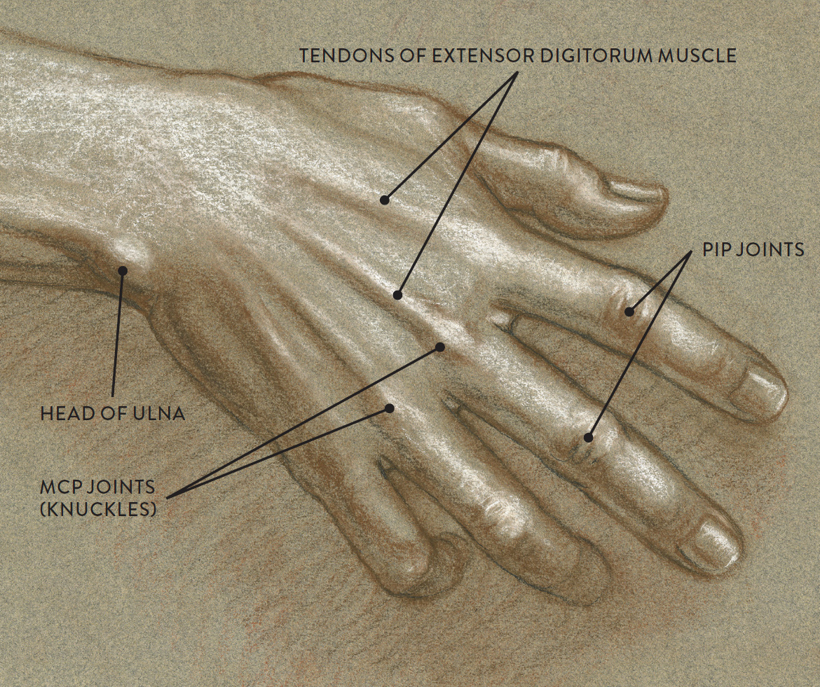
Sanguine and brown pastel pencils, charcoal, and white chalk on toned paper.
Tendons of the Anterior Region of the Lower Arm
The tendons of the various flexor muscles of the lower arm are usually seen on the surface when the hand clenches in a fist, as shown in the following life study. When the hand relaxes, the tendons become harder to detect.
STUDY OF A TIGHTLY CLENCHED FIST
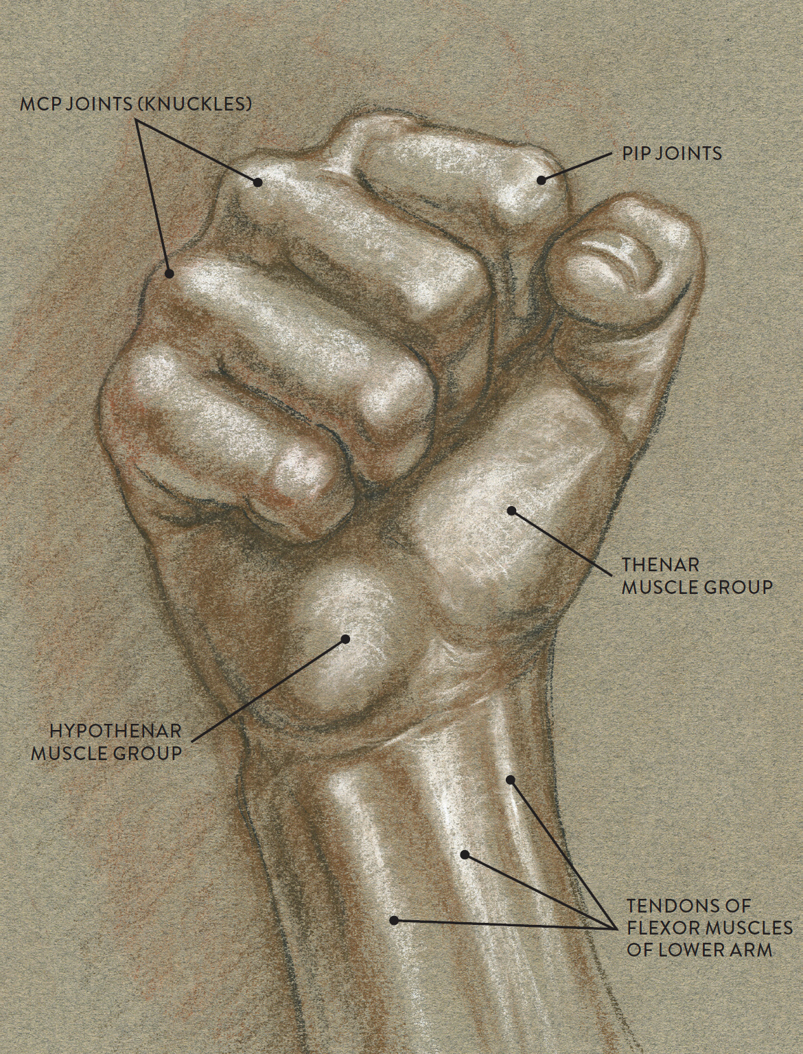
Sanguine and brown pastel pencils, charcoal, and white chalk on toned paper.
Tendons of the Hamstring Muscles
The tendons of the hamstring muscles attach on both sides of the popliteal fossa, located on the back of the knee. When the knee bends, these tendons become much more visible on the surface, as can be seen in the bent left leg in the following life study. The tendon of the biceps femoris muscle becomes especially prominent. This muscle is located on the outer side of the upper leg, and its tendon attaches into the head of the fibula bone, which is positioned on the outer side of the lower leg.
STUDY OF THE BACK OF THE LEGS
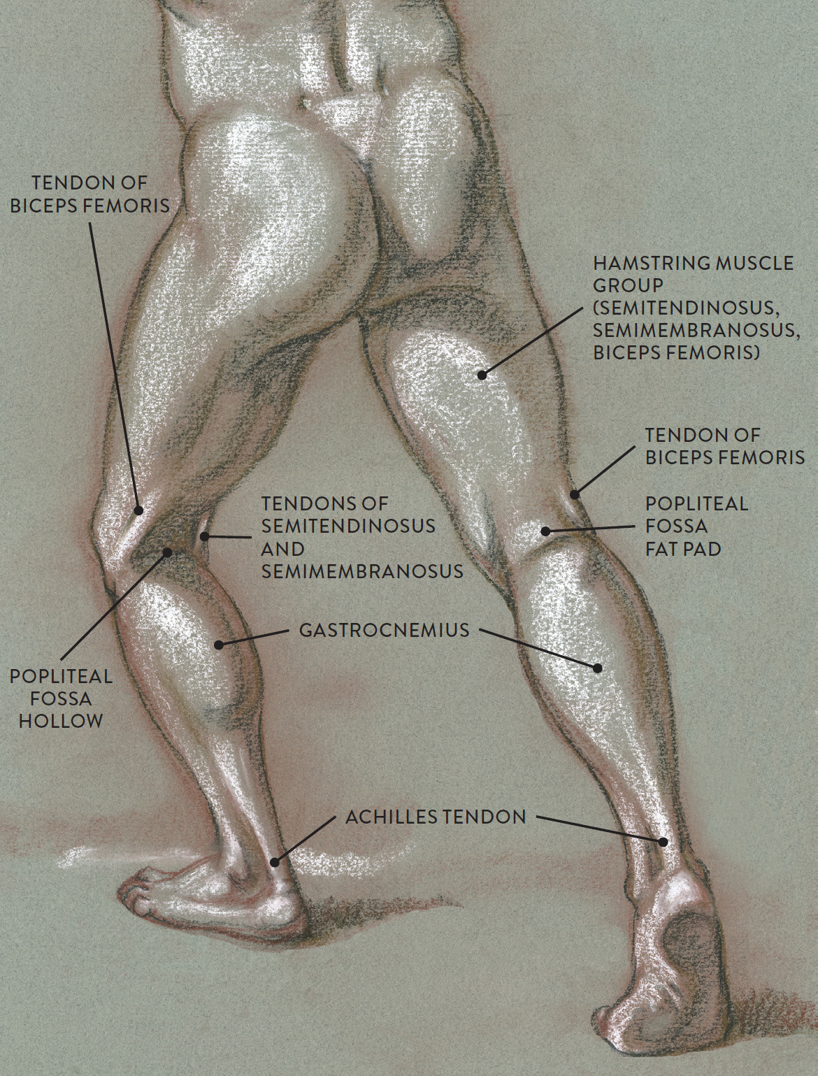
Charcoal pencil, sanguine and brown pastel pencils, and white chalk on toned paper.
The Achilles Tendon
The Achilles tendon—named for the warrior of Greek mythology who was killed by an arrow shot into his heel—is the tendon of the gastrocnemius and soleus muscles of the lower leg. It inserts into the heel bone (calcaneus) and appears on the surface form, sometimes quite prominently, as a thick, ropelike structure. The Achilles tendons are clearly visible in the following life study.
STUDY OF FEET
Graphite pencil, ballpoint pen, colored pencil, and white chalk on toned paper.
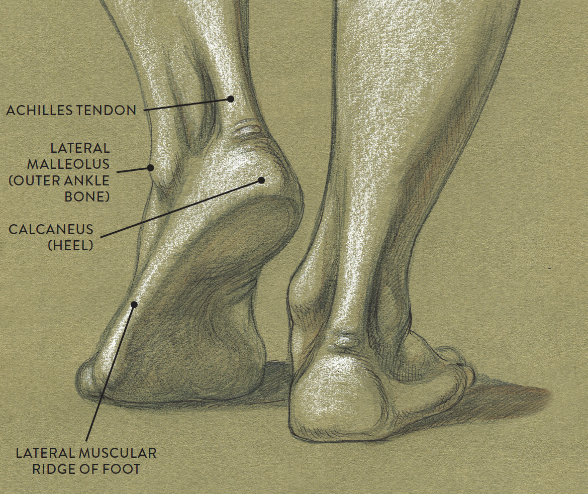
Tendons of the Dorsal Side of the Foot
The four tendons of the extensor digitorum longus muscle of the lower leg attach into each of the four lesser toes. When the toes are spread apart in a forceful manner, these tendons become very obvious on the surface form. A slender but prominent tendon from the extensor hallucis longus (long extender of the great toe) inserts directly into the great toe. This tendon becomes particularly noticeable when the large toe points upward.
A ropelike tendon on the lower part of the tibialis anterior muscle (shin muscle) of the lower leg can be seen only when the foot is in certain positions. Many artists confuse this muscle with the great toe’s tendon and draw a continuous line from the tibialis anterior muscle directly into the great toe. This is fine for gesture studies, but in more detailed renderings you should try to distinguish the subtle separation between the two tendons near the ankle. In the life study below, I’ve slightly exaggerated the tendons to show their placement on the surface more clearly.
STUDY OF A FOOT
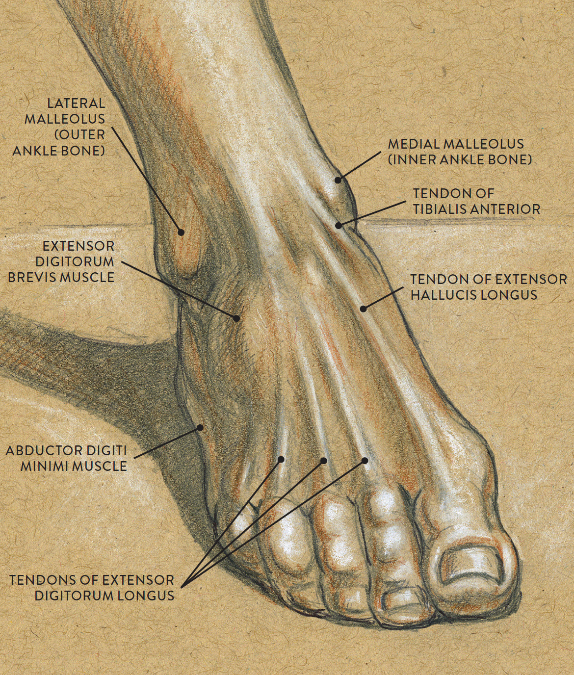
Graphite pencil, colored pencil, and white chalk on toned paper.
Muscle Contraction
Muscle contraction is also known as muscle action or muscle tension. Muscles can shorten their muscle fibers, lengthen them from a contracted state, or stabilize them at the same length. All these actions produce tension within the muscle fibers. When a muscle is not in any state of contraction, it is said to be relaxed, or in a “resting state.”
Muscle contractions can activate movement (initiating joint action), control the tempo of a movement (accelerating or slowing down joint action), or prevent unwanted movement by stabilizing a joint. The two basic categories of muscle contraction are called dynamic (isotonic) and static (isometric). Let’s look at each.
Dynamic Muscle Contraction
When a muscle changes length during a specific movement, either shortening its muscle fibers or lengthening them, this action is known as dynamic muscle contraction, or as isotonic contraction, dynamic muscle tension, or dynamic movement. There are two different types of dynamic muscle contraction: concentric and eccentric. Basically, concentric contractions shorten the muscle fibers and eccentric contractions lengthen them. For many years, these opposing actions were commonly referred to as squash and stretch in the animation industry, but there are other names for the actions, including stretching and compressing and extension and contraction.
DYNAMIC MUSCLE CONTRACTION
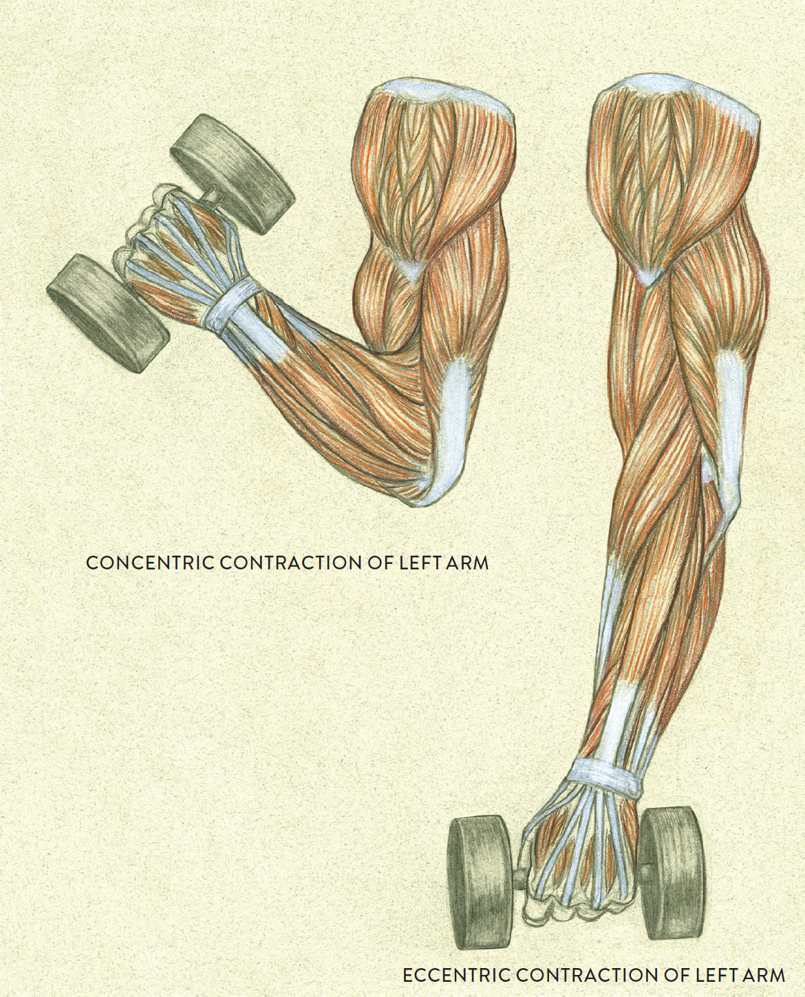
Concentric and eccentric contractions
When a muscle is in a state of concentric contraction, the muscle fibers shorten toward their centers, and, in the process, pull a bone in a certain direction, causing movement at a joint. This type of action usually occurs in the “up phase” of a movement, as when lifting a barbell.
When a muscle is in a state of eccentric contraction, the muscle fibers lengthen from the contracted state and the muscle is returned to its resting length. This type of action usually occurs in the “down phase” of a movement, as when lowering a barbell.
Don’t confuse eccentric contraction with the state of rest, however. During eccentric contraction, the muscle fibers lengthen from a contracted state in a controlled manner, slowing down the movement against the influence of gravity. This smoothing out of a movement is known as the “braking force.” For example, when lifting a weight, the biceps brachii and brachialis muscles shorten their fibers (concentric contraction) to lift the forearm and hand holding the weight in an upward direction (the up phase). Then, when the weight is being lowered (the down phase), the biceps and brachialis lengthen their muscle fibers but in a controlled way that resists gravity and thus prevents the forearm from slamming down. Even though the muscle fibers are lengthening, there is tension within the muscle. The drawings on this page illustrate concentric/eccentric phases of dynamic muscle contraction.
When depicting any active or semi-active pose, you should try to locate any muscles that are in a state of compression or stretching. Visual clues include
· compact shape of a muscle
· one muscle pressing against another muscle
· stretched muscles
· a tendon protruding close to the surface due to tension within its muscle
I made the study at right from the ancient marble sculpture called Laocoön and His Sons (or The Laocoön Group), housed in the Vatican Museum, which depicts a man and his two sons writhing in agony as they are attacked by serpents. The focus of my study is the central figure, whose anguished, twisting action exemplifies the energetic dynamics of stretching and compression. I always recommend drawing directly from figurative sculpture, whether ancient or contemporary, whenever possible. The three-dimensional anatomical forms of stone or bronze statues appear so much more clearly than they do in photographs of sculptures. Plus, you can usually walk around the sculpture and draw it from different viewpoints.
STUDY OF CENTRAL FIGURE OF LAOCOÖN AND HIS SONS
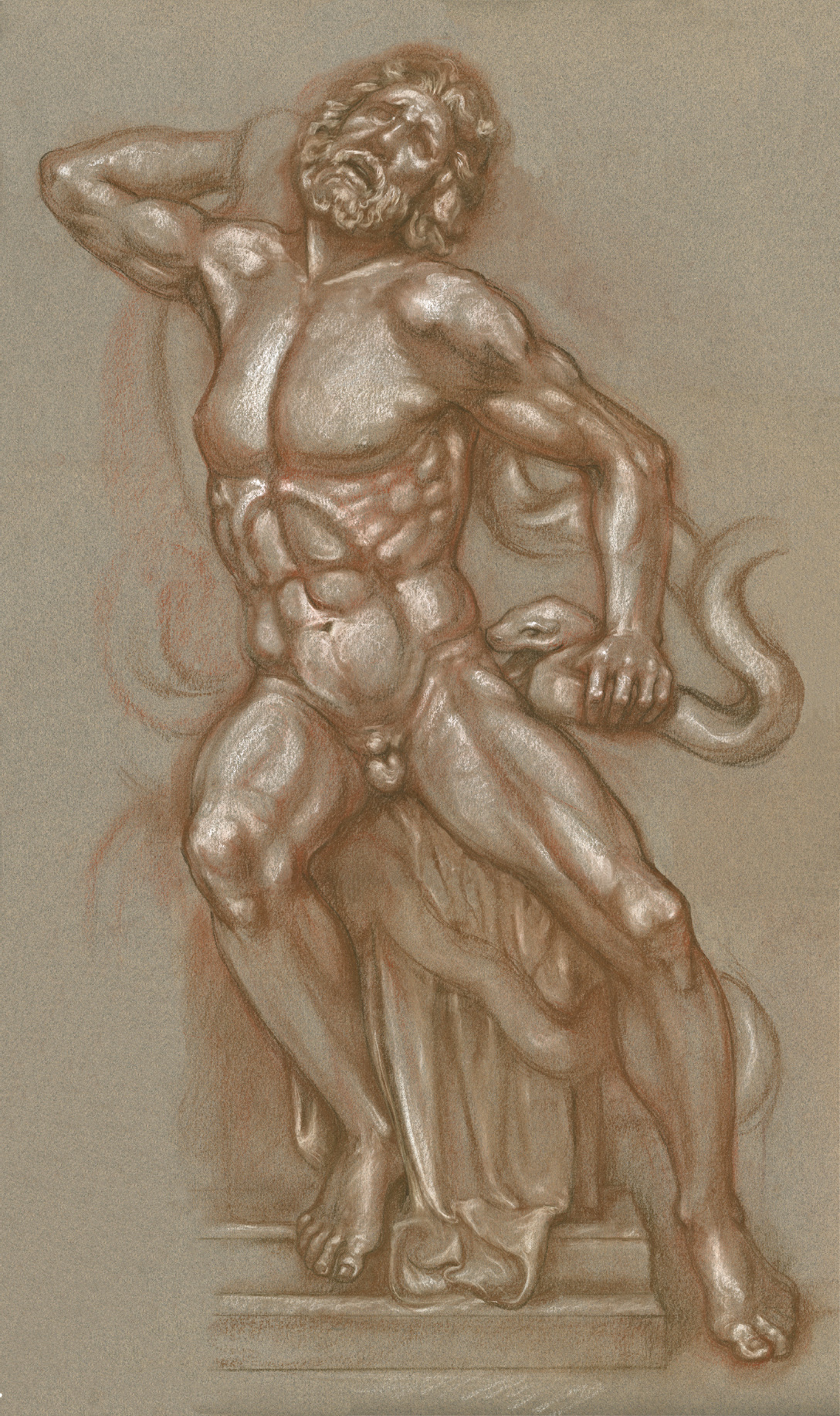
Sanguine and brown pastel pencils and white chalk on toned paper.
Static Muscle Contraction
In static contraction—also called isometric contraction—a muscle increases tension within its muscle fibers but does not change its length, thereby remaining stationary. No movement occurs at any joint, and in fact, this type of contraction stops movement altogether. Static contraction prepares muscles for possible action, as when a sprinter adopts a stationary position before taking off in a race. It is essential for maintaining posture (otherwise, gravitational forces would pull us down) and is activated when holding heavy objects stationary, as shown in next drawing. It also occurs when a muscle needs to stabilize a joint when movement is not wanted.
STATIC MUSCLE CONTRACTION
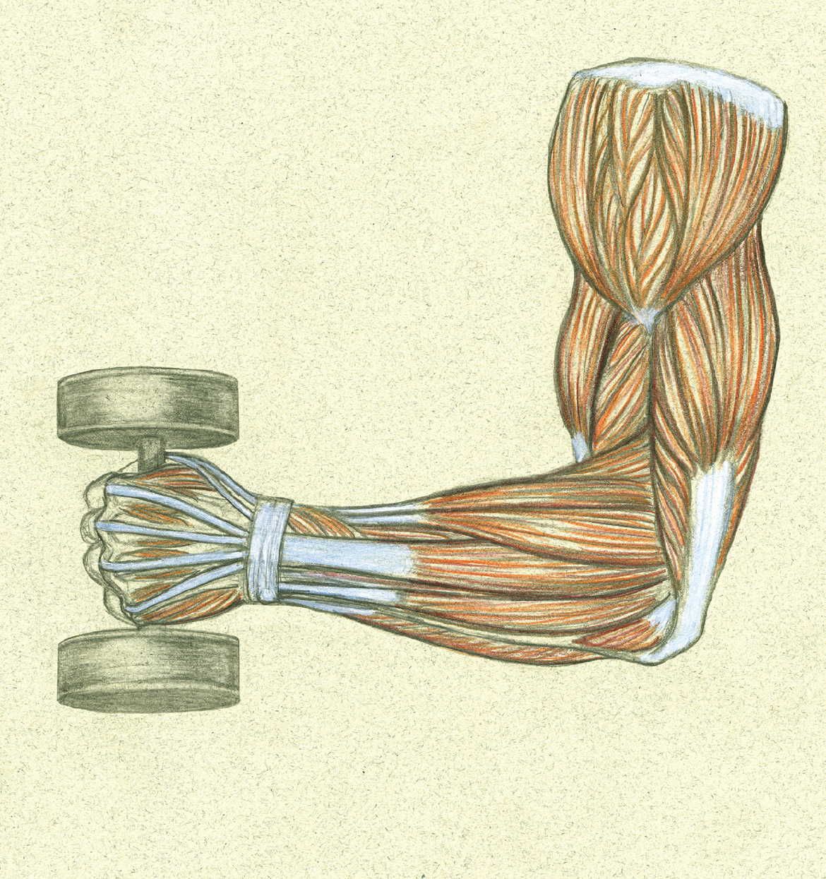
STATIC CONTRACTION OF LEFT ARM HOLDING WEIGHT STATIONARY
Left arm, lateral view
No matter how relaxed a person may look when standing or sitting, his or her muscles are still contracting to some degree to maintain the position and to prevent the person from being pulled down by gravity. Artist’s models have to maintain each pose for a length of time without moving or shifting their weight, so their bodies are always in a state of static contraction while posing. They can usually hold difficult action poses for a short time, but for longer poses they need to make sure their weight is balanced. The mark of a good model is being able to do a long pose and make it look interesting—dynamic or lyrical, not symmetrical or stiff—and to hold that pose (usually for a twenty-minute interval) without moving or twitching. In interpreting the pose, the artist will try to convey the tension or relaxation of the various anatomical forms, as I did in the following life study.
STUDY OF A MUSCULAR MALE FIGURE SITTING
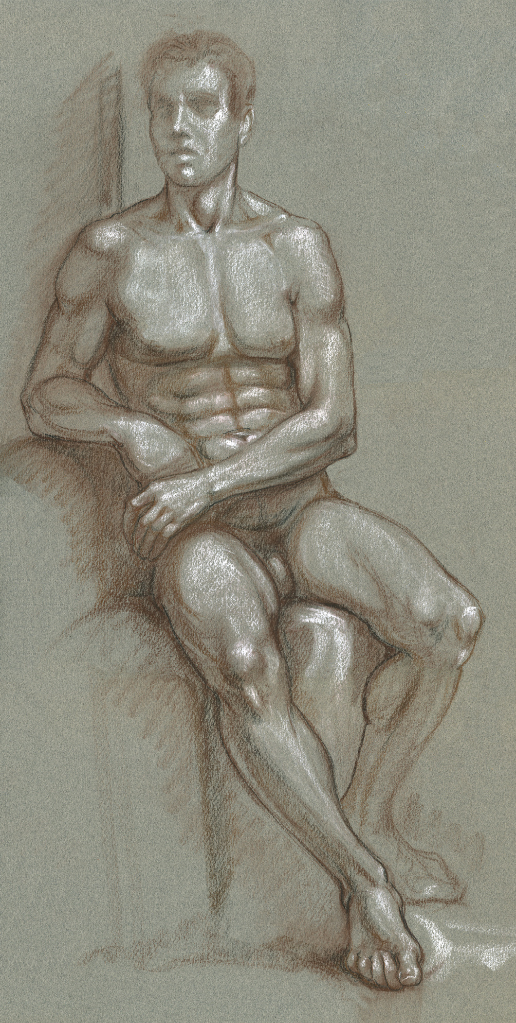
Colored pastel pencil, charcoal pencil, and white chalk on toned paper.
Muscles’ Differing Roles
When a particular movement occurs at a joint, several muscles in the vicinity of the joint participate. Although the main function of every muscle is to contract its muscle fibers, a muscle can play different roles at different times during a movement or series of movements. Generally speaking, there are four different roles muscles can play: agonist (prime mover), antagonist, synergist (assistor), and stabilizer (fixator).
Agonist (Prime Mover) and Antagonist
The muscle that is mainly responsible for activating a bone or body part for a specified movement is known as the agonist, or prime mover. When the agonist muscle contracts, it becomes a more compact version of itself. As this action is occurring, another muscle—the antagonist—has to act in opposition. This muscle, usually positioned on the side of the bone or body part opposite from the agonist, has to stretch its muscle fibers to yield to the agonist muscle’s contraction. This relationship can be clearly seen in the flexion and extension of the lower arm at the elbow joint, shown in the following drawing.
AGONIST (PRIME MOVER) AND ANTAGONIST MUSCLES
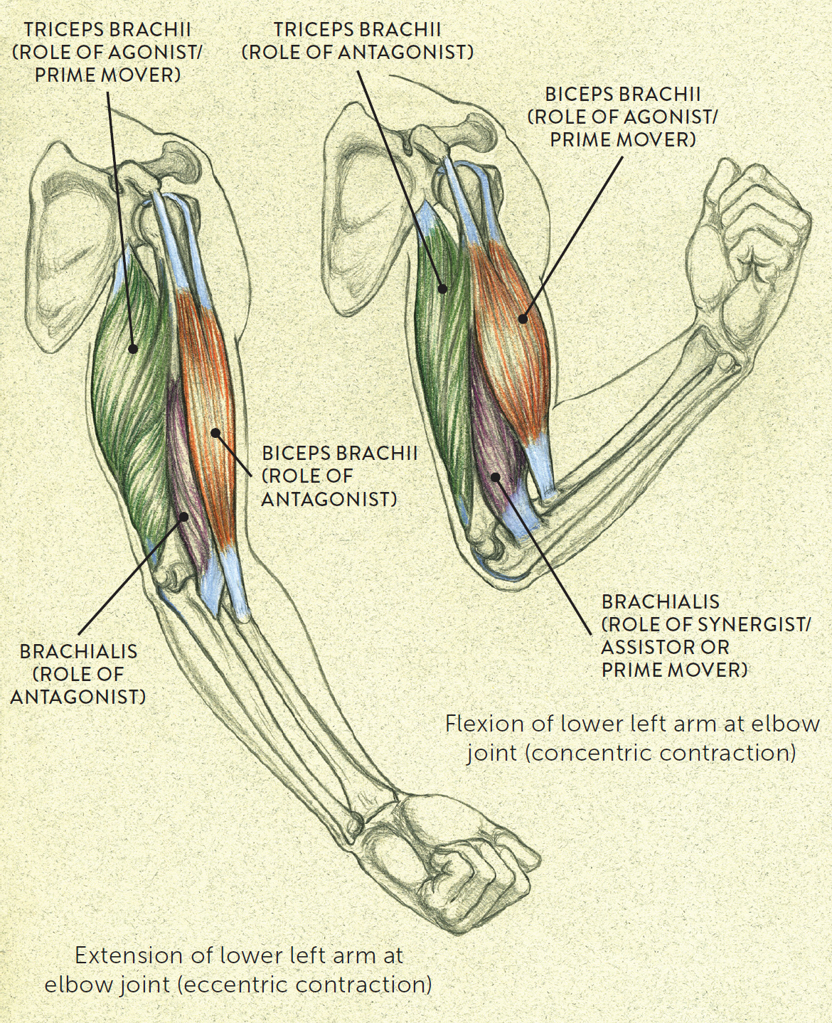
Upper and lower left arm, anterior view of scapula with arm moving sideways of torso
What happens is this: When the biceps brachii (located on the anterior portion of the upper arm) contracts its muscle fibers, it lifts up the lower arm, becoming a bulging shape. In this action, the biceps brachii is considered the agonist or prime mover (with the brachialis and brachioradialis acting as synergists, or assistors). (Some experts think that the brachialis, along with the biceps, is also a prime mover when it lifts the lower arm.) The antagonist muscle in this action is the triceps brachii, which is positioned on the posterior region of the upper arm. While the biceps is contracting, the triceps stretches its muscle fibers so that the elbow joint and lower arm can move freely, without interference. When the forearm is lowered, the biceps and triceps reverse their actions, switching roles. The triceps, which now contracts its muscle fibers, becomes the prime mover in lowering the forearm, while the biceps is now the antagonist, stretching its muscle fibers. (If only the lower arm is being moved, the humerus and scapula bones remain stationary, with the shoulder joint stabilized by the rhomboids, the trapezius, and the rotator cuff muscles of the scapula.)
Synergists and Stabilizers
As mentioned, a muscle usually does not move a bone or other body structure all by itself; other muscles assist in the process. These synergist muscles, or assistor muscles, provide additional pull near the prime mover’s tendon of insertion. They also help prevent any unwanted actions that could occur during a particular movement.
While a particular movement occurs, other muscles may act as stabilizers, or fixators, holding a bone firmly in place—usually the bone where the prime mover muscle originates. This prevents any unwanted movement from occurring so that the agonist and synergists can act more efficiently. When stabilizer muscles hold a bone stationary, their muscle fibers contract but do not shorten—the type of contraction called static or isometric contraction.
The Influence of Force on Anatomical Forms
The term force refers to energy that produces, modifies, or restrains movement of bodily components. There are two basic types: internal force, created within the body, and external force, produced outside the body. Internal force results from muscle contractions. When the nervous system sends a message to a muscle, telling it to contract, a tension is generated, creating movement at a joint. But it’s not just the muscle and bone that are affected. Soft-tissue anatomical forms such as tendons, ligaments, the subcutaneous layer, and the skin are also influenced by internal force and may change shape in any of the following ways, also schematized in the following drawing:
· Compression of forms, which occurs during the contraction of a muscle, making it shorter or compressed, or when a form is pressing against an object
· Stretching of forms, or tension, which occurs when a muscle stretches or when forms are pulling in opposite directions
· Bending of forms, which occurs when two forms press against each other
· Twisting or spiraling of forms, also called torsion or torque, which usually occurs when one structure twists in one direction while another twists in a different direction
INFLUENCE OF FORCE ON ANATOMICAL FORMS
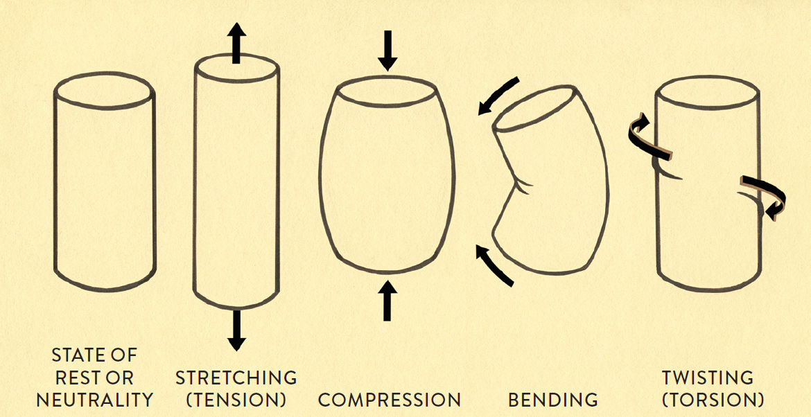
Black arrows indicate the direction of the force influencing the anatomical form.
Combinations of these actions (called combined loading) can also occur, as when a figure or bodily structure twists while bending or stretches while twisting. There are countless variations, depending on the type of action taking place. When you draw the figure in an active pose, you’ll observe that many forms are influenced by the dynamics of force, and you can interpret that dynamism in your own unique way, infusing the forms and lines of the body with vitality and energy.
The gesture drawing called Study of Male Figure in Dynamic Twisting Movement, conveys the tremendous amount of energy the model is exhibiting in this pose. The twisting action at the waist region shows both tension and compression, and the swinging of the hair contributes to the figure’s overall rhythm. The gesture drawing Study of a Female Figure Bending, shows the anatomical forms stretching and compressing throughout.
STUDY OF MALE FIGURE IN DYNAMIC TWISTING MOVEMENT
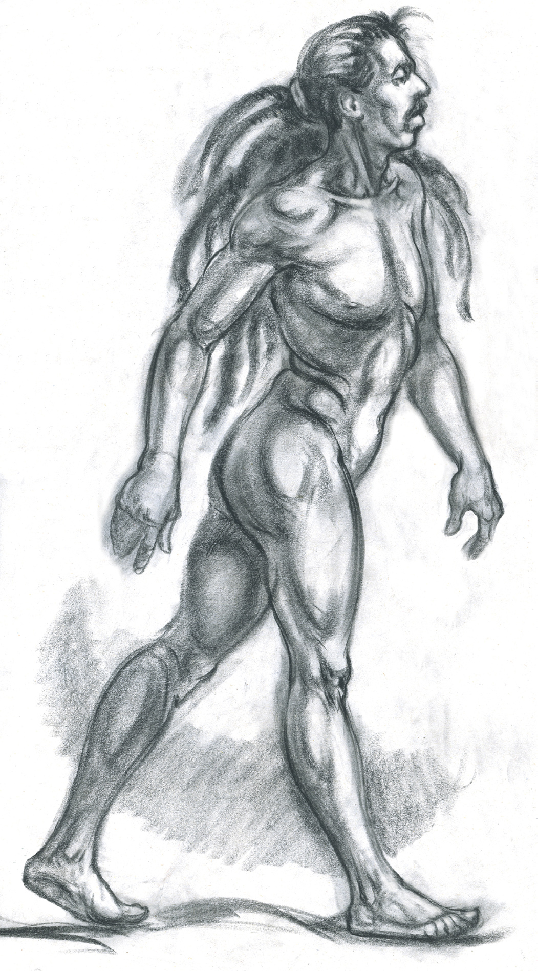
Black Conté crayon on newsprint.
STUDY OF A FEMALE FIGURE BENDING
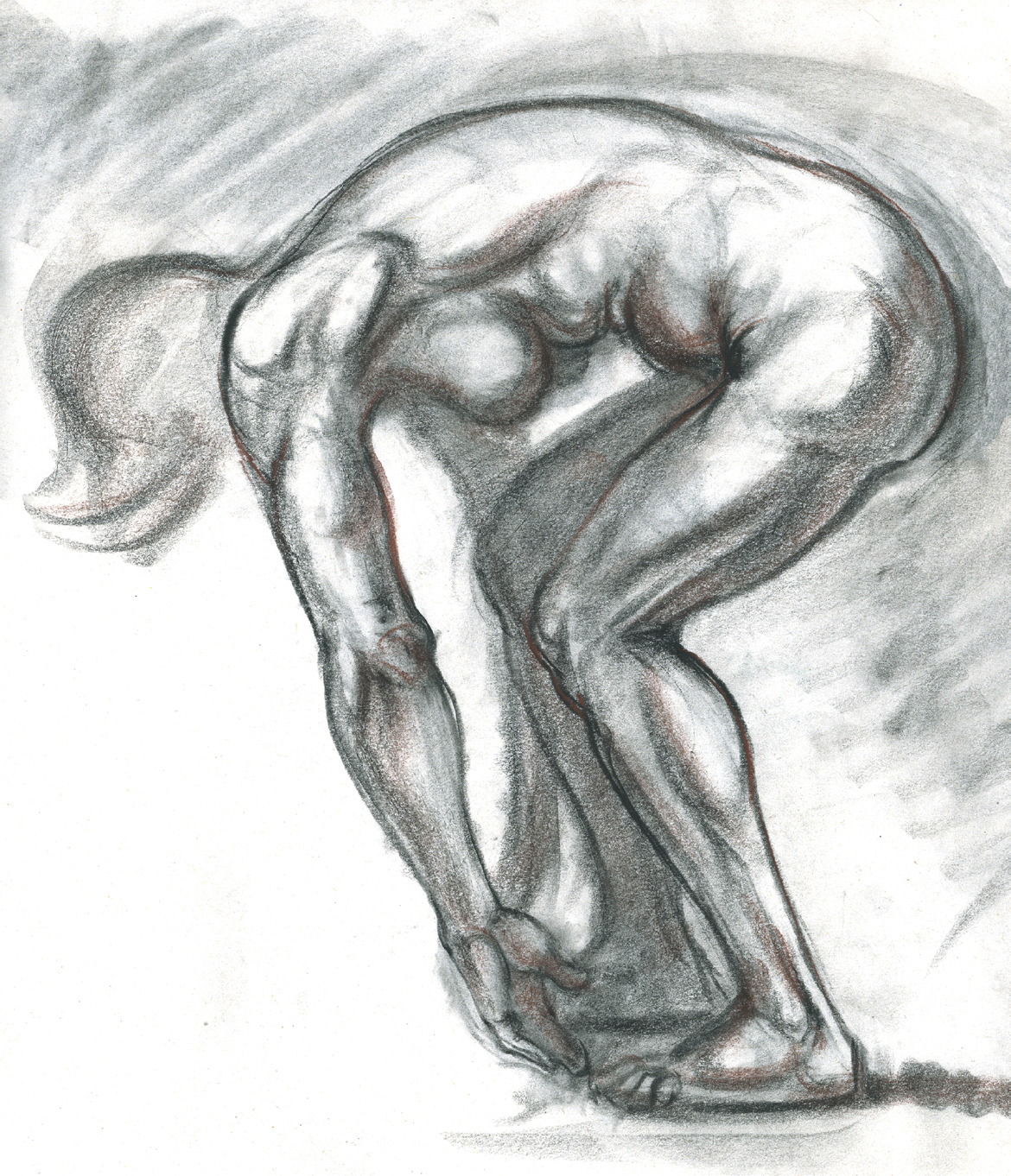
Black and brown Conté crayon on newsprint.
Of the external forces affecting the body, gravity may be the most important, because it is always at work, pulling everything—including human bodies—toward the center of the earth. Certain muscles contract without moving any joints (isometric contraction) to counter the force of gravity and help hold the body upright. But there are other external forces that can strongly affect the body, making it difficult to maintain equilibrium. These include forces of nature (a turbulent wind or a large wave), physical impact with external objects, and combat with another body (wrestling, boxing).
When the body is performing an action requiring forceful exertion, such as pushing a heavy cart or lifting a heavy box, the external impact is channeled as tension, which is also created internally within the muscles. So external and internal forces can affect the human body simultaneously—and, in fact, they do so almost constantly. Illustrators, storyboard artists, comic book artists, and animators (traditional and digital) all depict figures in various forms of movement being affected by external and internal forces of every kind.
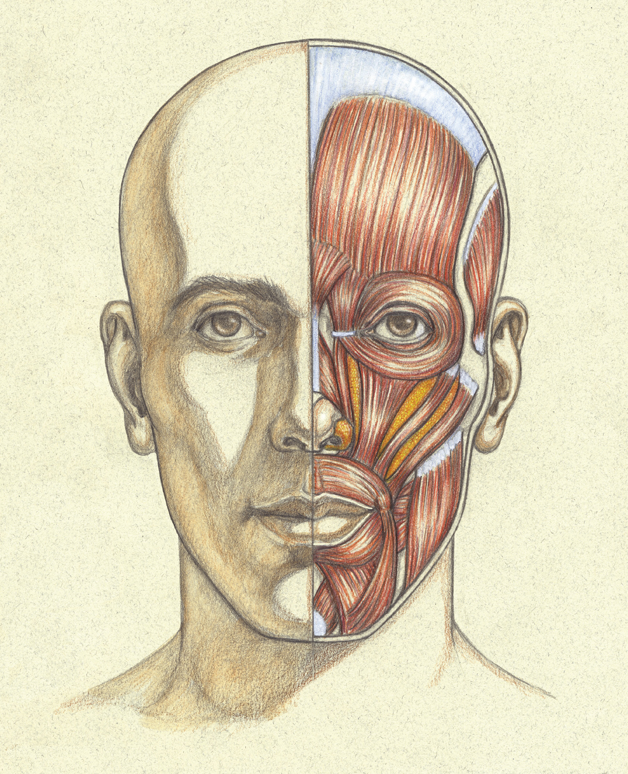
HEAD STUDY—HALF FACE/HALF MUSCLES
Graphite pencil and colored pencil on lightly toned paper.