THE LIVING WORLD
Unit Six. Animal Life
29. The Senses
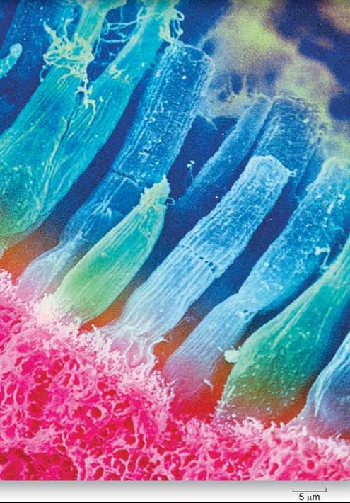
Sensory neurons receive input from different kinds of sensory receptor cells, such as the rods and cones pictured above that are found in the vertebrate eye. All input from sensory neurons to the central nervous system arrives in the same form, as action potentials propagated by afferent (inward-conducting) sensory neurons. Different sensory neurons project to different brain regions, and so are associated with different sensory modalities. The intensity of the sensation depends on the frequency of action potentials conducted by the sensory neuron. A sunset, a symphony, and searing pain are distinguished by the brain only in terms of the identity of the sensory neuron carrying the action potentials and the frequency of these impulses. Thus, if the auditory nerve is artificially stimulated, the brain perceives the stimulation as sound. But if the optic nerve is artificially stimulated in exactly the same manner and degree, the brain perceives a flash of light.
29.1. Processing Sensory Information
Did you ever wonder what it would be like not to know anything about what is going on around you? Imagine if you couldn’t hear, or see, or feel, or smell. After a while, a human goes mad if completely deprived of sensory input. The senses are the bridge to experience and perceive the way the body relates to everything around it.
Sensory Receptors
The sensory nervous system tells the central nervous system what is happening. Sensory neurons carry impulses to the CNS from more than a dozen different types of sensory cells that detect changes outside and inside the body. Called sensory receptors, these specialized sensory cells detect many different things, including changes in blood pressure, strain on ligaments, and smells in the air. Particularly complex sensory receptors, made up of many cell and tissue types, are called sensory organs. The eyes and ears (figure 29.1) are sensory organs, and so are the taste buds in your mouth.
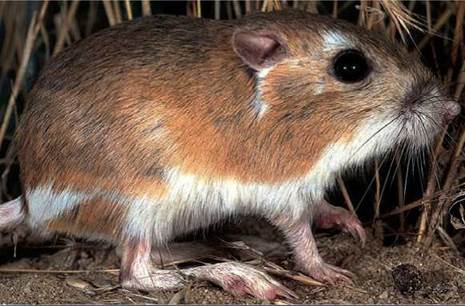
Figure 29.1. Kangaroo rats have specialized ears.
The ears of kangaroo rats (Dipodomys) are adapted to nocturnal life and allow them to hear the low-frequency sounds of their predators, such as an owl's wingbeats or a sidewinder rattlesnake's scales rubbing against the ground. Also, the ears seem to be adapted to the poor sound-carrying quality of dry, desert air.
How does the brain know whether an incoming nerve impulse is light, sound, or pain? This information is built into the “wiring”—into which neurons interact while passing the information to the CNS and into the location in the brain where the information is sent. The brain “knows” it is responding to light because the message from a sensory neuron is wired to light receptor cells. That is why when you press your fingertips gently against the corners of your eyes, you “see stars”—the brain treats any impulse from the eyes as light, even though the eye received no light.
The Path of Sensory Information
The path of sensory information to the CNS is a simple one, composed of three stages:
1. Stimulation. A stimulus impinges on a sensory receptor.
2. Transduction. Sensory receptors change the stimulus into an electrical potential by initiating the opening or closing of ion channels in a sensory neuron.
3. Transmission. The sensory neuron conducts a nerve impulse along an afferent pathway to the CNS.
All sensory receptors are able to initiate nerve impulses by opening or closing stimulus-gated channels within sensory neuron membranes. Except for visual photoreceptors, these channels are sodium ion channels that depolarize the membrane and so start an electrical signal. If the stimulus is large enough, the depolarization will trigger an action potential. The greater the sensory stimulus, the greater the depolarization of the sensory receptor and the higher the frequency of action potentials. The channels are opened by chemical or mechanical stimulation, often a disturbance such as touch, heat, or cold. The receptors differ from one another in the nature of the environmental input that triggers the opening of the channel. The body contains many sorts of receptors, each sensitive to a different aspect of the body’s condition or to a different quality of the external environment.
Exteroceptors are receptors that sense stimuli that arise in the external environment. Almost all of a vertebrate’s exterior senses evolved in water before vertebrates invaded the land. Consequently, many senses of terrestrial vertebrates emphasize stimuli that travel well in water, using receptors that have been retained in the transition from the sea to the land. Hearing, for example, converts an airborne stimulus into a waterborne one, using receptors similar to those that originally evolved in aquatic animals. A few vertebrate sensory systems that function well in the water, such as the electric organs of fish, cannot function well in the air and are not found among terrestrial vertebrates. On the other hand, some land dwellers have sensory systems, such as infrared receptors, that could not function in the sea. Still other organisms seem to be able to detect earth’s magnetic field, as we will discuss later on in this chapter.
Interoceptors sense stimuli that arise from within the body. These internal receptors detect stimuli related to muscle length and tension, limb position, pain, blood chemistry, blood volume and pressure, and body temperature. Many of these receptors are simpler than those that monitor the external environment and are believed to bear a closer resemblance to primitive sensory receptors.
Sensing the Internal Environment
Sensory receptors inside the body inform the CNS about the condition of the body. Much of this information passes to a coordinating center in the brain, the hypothalamus. This part of the brain is responsible for maintaining the body’s homeostasis— that is, keeping the body’s internal environment constant. The vertebrate body uses a variety of different sensory receptors to respond to different aspects of its internal environment.
Temperature change. Two kinds of nerve endings in the skin are sensitive to changes in temperature—one stimulated by cold, the other by warmth. By comparing information from the two, the CNS can learn what the temperature is and if it is changing.
Blood chemistry. Receptors in the walls of arteries sense CO2 levels in the blood. The brain uses this information to regulate the body’s respiration rate, increasing it when CO2 levels rise above normal.
Pain. Damage to tissue is detected by special nerve endings within tissues, usually near the surface, where damage is most likely to occur. When these nerve endings are physically damaged or deformed, the CNS responds by reflexively withdrawing the body segment and often by changing heartbeat and blood pressure as well.
Muscle contraction. Buried deep within muscles are sensory receptors called stretch receptors (see also pages 600-601). In each, the end of a sensory neuron is wrapped around a muscle fiber, like the receptor shown in figure 29.2. When the muscle is stretched, the fiber elongates, stretching the spiral nerve ending (like stretching a spring) and causing repeated nerve impulses to be sent to the brain. From these signals the brain can determine the rate of change of muscle length at any given moment. The CNS uses this information to control movements that require the combined action of several muscles, such as those that carry out breathing or locomotion.
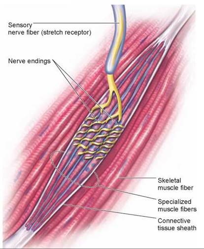
Figure 29.2. A stretch receptor embedded within skeletal muscle.
Stretching the muscle elongates the specialized muscle fibers, which deforms the nerve endings, causing them to send a nerve impulse out along the nerve fiber.
Blood pressure. Blood pressure is sensed by neurons called baroreceptors with highly branched nerve endings within the walls of major arteries. When blood pressure increases, the stretching of the arterial wall, like the expansion of the artery in figure 29.3, causes the sensory neuron to increase the rate at which it sends nerve impulses to the CNS. When the wall of the artery is not stretched, the rate of firing of the sensory neuron goes down. Thus, the frequency of impulses provides the CNS with a continuous measure of blood pressure.
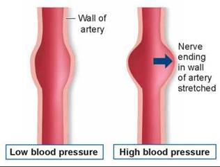
Figure 29.3. How a baroreceptor works.
A network of nerve endings covers a region where the wall of the artery is thin. High blood pressure causes the wall to balloon out there, stretching the nerve endings and causing them to fire impulses.
Touch. Touch is sensed by pressure receptors buried below the surface of the skin. There are a variety of different types, some specialized to detect rapid changes in pressure, others to measure the duration and extent to which pressure is applied, and still others sensitive to vibrations.
Key Learning Outcome 29.1. Sensory receptors initiate nerve impulses in response to stimulation. All sensory nerve impulses are the same, differing only in the stimulus that fires them and their destination in the brain. A variety of different sensory receptors inform the hypothalamus about different aspects of the body's internal environment, enabling it to maintain the body's homeostasis.