5 Steps to a 5: AP Biology - Mark Anestis 2021
STEP 4 Review the Knowledge You Need to Score High
CHAPTER 8 Cell Communication and Cell Cycle
Exam Weight: 10—15%
IN THIS CHAPTER
Summary: This chapter teaches you what you need to know about cell communication, signal transduction pathway, feedback mechanisms, the cell cycle, mitosis, and cell cycle regulation.

Key Ideas
![]() Cells communicate by cell-to-cell contact or via chemical signals over distance in an organism.
Cells communicate by cell-to-cell contact or via chemical signals over distance in an organism.
![]() Signal transduction pathway starts with a signal followed by a transduction of the signal resulting in a cellular response.
Signal transduction pathway starts with a signal followed by a transduction of the signal resulting in a cellular response.
![]() Feedback mechanisms are used to maintain homeostasis.
Feedback mechanisms are used to maintain homeostasis.
![]() There are four main stages in the cell cycle: G1, S, G2, and M.
There are four main stages in the cell cycle: G1, S, G2, and M.
![]() The stages of mitosis are prophase, metaphase, anaphase, telophase, and cytokinesis.
The stages of mitosis are prophase, metaphase, anaphase, telophase, and cytokinesis.
![]() Examples of cell division control mechanisms are growth factors, checkpoints, density-dependent inhibition, and cyclins/protein kinases.
Examples of cell division control mechanisms are growth factors, checkpoints, density-dependent inhibition, and cyclins/protein kinases.
Introduction
Cells use energy in many ways: for maintenance of homeostasis through feedback mechanisms, for communication with other cells, and for replication. In this chapter, we will discuss how cells communicate with one another via cell-to-cell contact or chemical signals with signal transduction pathways. Next, we will discuss how cells respond to their changing environments via negative and positive feedback pathways. After dissecting the cell cycle and the phases of mitosis, we will touch on cell cycle regulation.
Cell Communication
The ability of a cell to communicate with its environment and with itself is key to maintaining homeostasis and surviving. This survival depends on receiving and processing information from outside the cell—information about changing environmental conditionals, availability of nutrients, or any other information vital to surviving. Cells have developed a variety of signaling mechanisms to accomplish the transmission of important biological information. For example, the presence of receptors that allow ion currents to flow in response to photons, which translate light into chemical messengers inside the retina of the eye.
IST-3
Cells communicate by generating, transmitting, receiving, and responding to chemical signals.
Cell signaling involves a ligand, a signaling molecule, and a receptor protein to which the ligand binds. The receptor can be located on the cell membrane for hydrophilic ligands that can’t cross the membrane, or it can be located inside the cell for hydrophobic ligands that are small enough to cross the membrane.
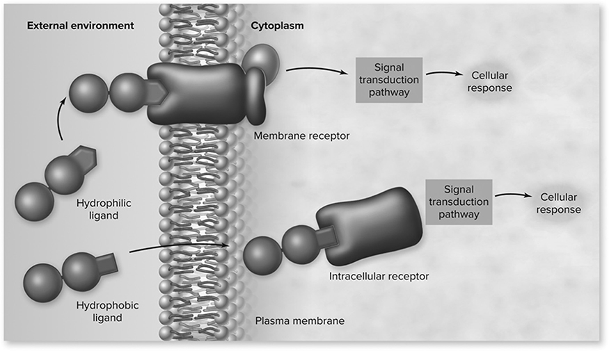
Figure 8.1 Overview of cell signaling. Cell signaling involves a signal molecule called a ligand, a receptor, and a signal transduction pathway that produces a cellular response. The location of the receptor can either be intracellular, for hydrophobic ligands that can cross the membrane, or in the plasma membrane, for hydrophilic ligands that cannot cross the membrane. (Reproduced with permission from Raven P, Johnson G, Mason K, Losos J, Duncan T; Biology, 12th ed. New York: McGraw Hill; 2020)
Signaling
Cells communicate in a variety of ways, depending on the distance between the cells communicating.
1. Direct cell-to-cell signaling involves the direct physical contact between cells during communication. Gap junctions in animals and plasmodesmata in plants are tiny channels that directly connect to neighboring cells, which allow the cells to transfer signaling molecules that transmit their current state of homeostasis with one another.
2. Paracrine signaling involves cells close to one another but not in direct contact. A cell releases a short-lived signal into a localized area that will induce changes in a nearby cell. Paracrine signals can diffuse only over relatively short distances.
3. Endocrine signaling involves cells far apart in which a longer-lasting signal, called a hormone, is released into the extracellular fluid and travels widely throughout the organisms to target cells. Protein hormones are large molecules that must bind to receptors on the cell membrane. Steroid hormones are lipid-soluble molecules that are able to pass through the cell membrane and attach to an intracellular receptor. One example of endocrine signaling involves the release of human growth hormone (HGH) from the pituitary gland into the bloodstream, which targets bone and muscle cells to trigger growth (See Figure 8.2 a-d).
4. Synaptic signaling involves a specialized nerve cell, a neuron, and its target cell. This association is called a chemical synapse and involves the release of neurotransmitters from the neuron into the synaptic gap to target the target cell.
5. Autocrine signaling occurs when a cell sends a signal to itself by secreting something that in turn binds to specific receptors on its own membrane. This plays an important role in cell development and immune system.
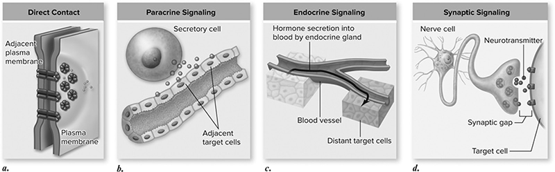
Figure 8.2 Four kinds of cell signaling. Cells communicate in several ways. a. Two cells in direct contact with each other may send signals across gap junctions. b. In paracrine signaling, secretions from one cell have an effect only on cells in the immediate area. c. In endocrine signaling, hormones are released into the organism’s circulatory system, which carries them to the target cells. d. Chemical synapse signaling involves transmission of signal molecules, called neurotransmitters, from a neuron over a small synaptic gap to the target cell. (Reproduced with permission from Raven P, Johnson G, Mason K, Losos J, Duncan T; Biology, 12th ed. New York: McGraw Hill; 2020)
Signal Transduction Pathway
When a ligand binds to a receptor on a cell, the work has just begun for the cell. The cell relays the message through a series of reactions to elicit a cellular response known as the signal transduction pathway (Figure 8.1. The binding of the ligand to the receptor generally causes the receptor to change shape, resulting in an activation of an enzyme or binding of other molecules. This starts a signaling cascade that can amplify the signal through a series of reactions that leads to a cellular response, resulting in a change to a cell’s behavior or characteristics.
Phosphorylation
The signal transduction pathway may require activating or inactivating proteins via the addition of a phosphate group in a process called phosphorylation.
The phosphorylation of proteins (addition of phosphate groups) is catalyzed by enzymes called kinases. There are many different types of kinases that target different proteins in the cell. The dephosphorylation of proteins (removal of phosphate groups) is catalyzed by enzymes called phosphatases. Many proteins are activated when phosphorylated and deactivated when dephosphorylated, which creates a pretty nifty way for cells to turn on and off various important cellular pathways (See Figure 8.3).
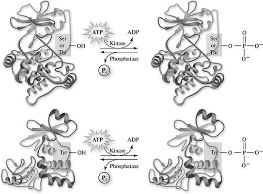
Figure 8.3 Phosphorylation of proteins. Many proteins are controlled by their phosphorylation state—that is, they are activated by phosphorylation and deactivated by dephosphorylation or the reverse. The enzymes that add phosphate groups are called kinases. These form two classes depending on the amino acid the phosphate is added to, either serine— threonine kinases or tyrosine kinases. The action of kinases is reversed by protein phosphatase enzymes. (Reproduced with permission from Raven P, Johnson G, Mason K, Losos J, Duncan T; Biology, 12th ed. New York: McGraw Hill; 2020)
Secondary Messengers
While proteins are a main component of most signal transduction pathways, many other molecules play important roles in the process as secondary messengers, which are small, nonprotein molecules that pass messages along.
1. Calcium (Ca2+): Calcium is widely used by cells as a secondary messenger. Some proteins have binding sites for Ca2+ and when calcium binds to the protein, the shape changes, leading to a change in function. One such example is the use of Ca2+ in muscle to start muscle contraction after receiving a signal from a neuron.
2. Cyclic AMP (cAMP): Cyclic adenosine monophosphate (cAMP) is involved in many signal cascade pathways. Protein hormones activate cAMP though a multistep process that begins with protein-hormone activation of relay proteins such as G-proteins. These proteins are able to directly activate a compound known as adenyl cyclase, which in turn produces cAMP (See Figure 8.4).
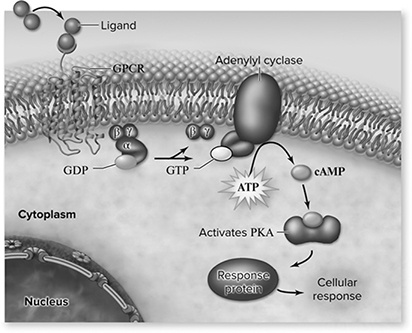
Figure 8.4 cAMP signaling pathway. Extracellular signal binds to a GPCR, activating a G protein. The G protein then activates the effector protein adenylyl cyclase, which catalyzes the conversion of ATP to cAMP. The cAMP then activates protein kinase A (PKA), which phosphorylates target proteins to cause a cellular response. (Reproduced with permission from Raven P, Johnson G, Mason K, Losos J, Duncan T; Biology, 12th ed. New York: McGraw Hill; 2020)
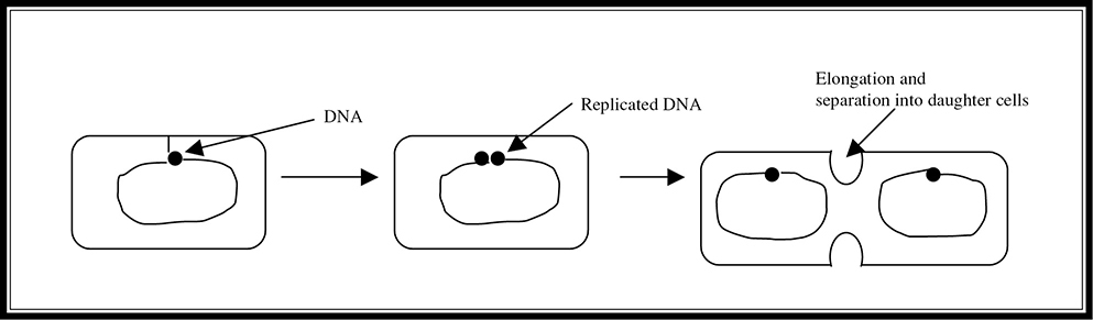
Figure 8.5 Binary fission.
Cell Division in Prokaryotes
Prokaryotes are simple single-celled organisms without a nucleus. Their genetic material is arranged in a single circular chromosome of DNA, which is anchored to the cell membrane. As in eukaryotes, the genetic material of prokaryotes is duplicated before division. However, instead of entering into a complex cycle for cell division, prokaryotes simply elongate until they are double their original size. At this point, the cell pinches in and separates into two identical daughter cells in a process known as binary fission (Figure 8.5.
The Cell Cycle
Eukaryotic cell reproduction is a bit more complicated. The cell cycle functions as the daily planner of growth and development for the eukaryotic cell. It tells the cell when and in what order it is going to do things, and consists of all the necessary steps required for the reproduction of a cell. It begins after the creation of the cell and concludes with the formation of two daughter cells through cell division. It then begins again for the two daughter cells that have just been formed. There are four main stages to the cell cycle, and they occur in the following sequence: phases G1, S, G2, and M (Figure 8.6. Phases G1 and G2 are growth stages; S is the part of the cell cycle during which the DNA is duplicated; and the M phase stands for mitosis, the cell division phase.
IST-1
Heritable information provides for continuity.
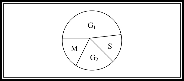
Figure 8.6 Pie chart showing the four main stages of the cell cycle.
Stages of the Cell Cycle
G1 phase. During the first growth phase of the cell cycle, the cell prepares itself for the synthesis stage of the cycle, making sure that it has all the necessary raw materials for DNA synthesis.
S phase. The DNA is copied so that each daughter cell has a complete set of chromosomes at the conclusion of the cell cycle.
G2 phase. During the second growth phase of the cycle, the cell prepares itself for mitosis (for producing body cells) and/or meiosis (for producing gametes), making sure that it has the raw materials necessary for the physical separation and formation of daughter cells.
M phase. Mitosis is the stage during which the cell separates into two new cells.
The first three stages of the cycle (G1, S, and G2) make up the portion of the cell cycle known as interphase. A cell spends approximately 90 percent of its cycle in this phase. The other 10 percent is spent in the final stage, mitosis.
The amount of time that a cell requires to complete a cycle varies by cell type. Some cells complete a full cycle in hours, while others can take days to finish. The rapidity with which cells replicate also varies. Skin cells are continually zipping along through the cell cycle, whereas nerve cells do not replicate—once they are damaged, they are lost for good. This is one reason why the death of nerve cells is such a problem—these cells cannot be repaired or regenerated through mitotic replication.
Mitosis
During mitosis, the fourth stage of the cell cycle, the cell actually takes the second copy of DNA made during the S phase and divides it equally between two cells. Single-cell eukaryotes undergo mitosis for the purpose of asexual reproduction. More complex multi-cellular eukaryotes use mitosis for other processes as well, such as growth and repair.
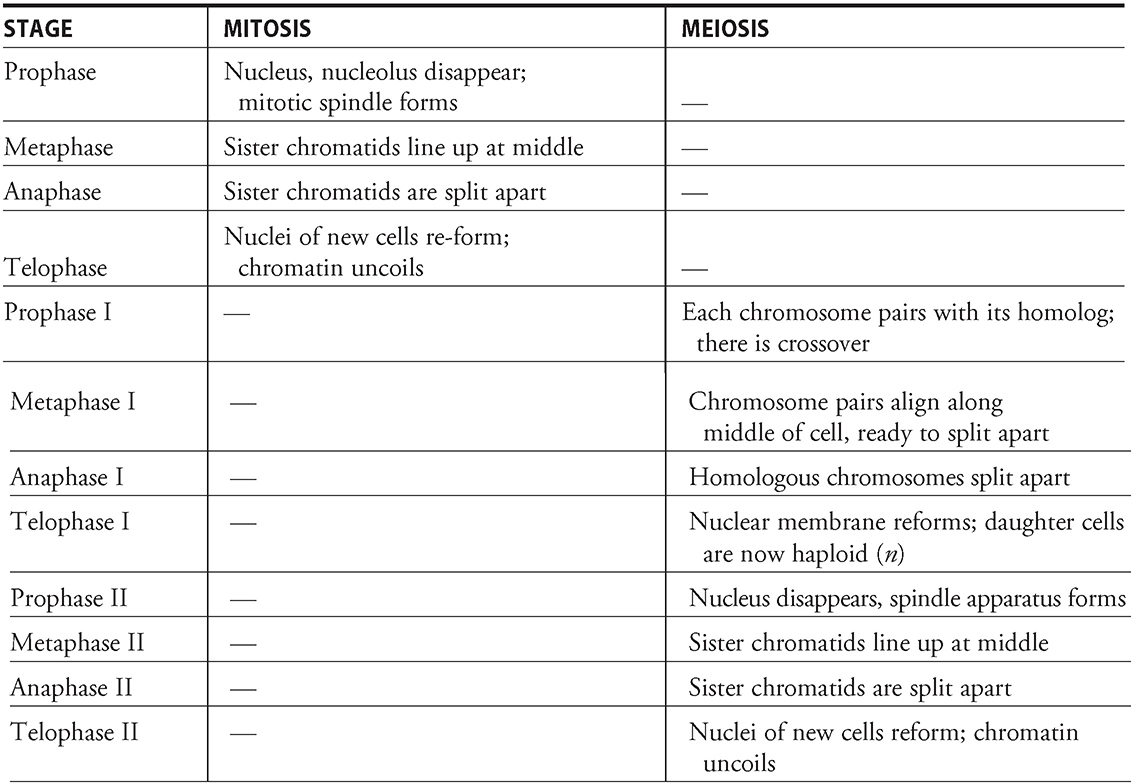
Mitosis consists of four major stages: prophase, metaphase, anaphase, and telo-phase. These stages are immediately followed by cytokinesis—the physical separation of the newly formed daughter cells. During interphase, chromosomes are invisible. The chromatin—the raw material that gives rise to the chromosomes—is long and thin during this phase. When the chromatin condenses to the point where the chromosome becomes visible through a microscope, the cell is said to have begun mitosis. The AP Biology exam is not going to ask you detailed questions about the different stages of mitosis; just have a general understanding of what happens during each step.
Mitosis
Prophase. Nucleus and nucleolus disappear; chromosomes appear as two identical, connected sister chromatids; mitotic spindle (made of microtubules) begins to form; centrioles move to opposite poles of the cell (plant cells do not have centrioles).
Metaphase. For metaphase, think middle. The sister chromatids line up along the middle of the cell, ready to split apart.
Anaphase. For anaphase, think apart. The split sister chromatids move via the microtubules to the opposing poles of the cell—the chromosomes are pulled to opposite poles by the spindle apparatus. After anaphase, each pole of the cell has a complete set of chromosomes.
Telophase. The nuclei for the newly split cells form; the nucleoli reappear, and the chromatin uncoils.
Cytokinesis. Newly formed daughter cells split apart. Animal cells are split by the formation of a cleavage furrow, and plant cells split by the formation of a cell plate (See Figure 8.7).
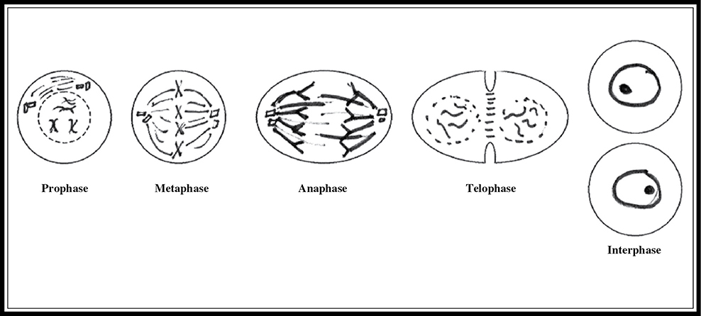
Figure 8.7 Pictorial representation of the stages of mitosis.
Here are the definitions for words you may need to know:
Cell plate: plant cell structure, constructed in the Golgi apparatus, composed of vesicles that fuse together along the middle of the cell, completing the separation process.
Cleavage furrow: groove formed (in animal cells) between the two daughter cells that pinches together to complete the separation of the two cells after mitosis.
Cytokinesis: the actual splitting of the newly formed daughter cells that completes each trip around the cell cycle—some consider it part of mitosis; others regard it as the step immediately following mitosis.
Mitotic spindle: apparatus constructed from microtubules that assists the cell in the physical separation of the chromosomes during mitosis.
Control of Cell Division
Control of the cell cycle is important to normal cell growth. There are various ways in which the cell controls the process of cell division:
1. Checkpoints. There are checkpoints throughout the cell cycle where the cell verifies that there are enough nutrients and raw materials to progress to the next stage of the cycle. The G1 checkpoint, for example, makes sure that the cell has enough raw materials to progress to and successfully complete the S phase.
Sam (12th grader): “Control mechanisms are an important theme for this test. Be able to write about them.”
2. Density-dependent inhibition. When a certain density of cells is reached, growth of the cells will slow or stop because there are not enough raw materials for the growth and survival of more cells. Cells that are halted by this inhibition enter a quiescent phase of the cell cycle known as G0. Cancer cells can lose this inhibition and grow out of control.
3. Growth factors. Some cells will not divide if certain factors are absent. Growth factors, as their name indicates, assist in the growth of structures.
4. Cyclins and protein kinases. Cyclin is a protein that accumulates during G1, S, and G2 of the cell cycle. A protein kinase is a protein that controls other proteins through the addition of phosphate groups. Cyclin-dependent kinase (CDK) is present at all times throughout the cell cycle and binds with cyclin to form a complex known as MPF (maturation or mitosis promoting factor). Early in the cell cycle, because the cyclin concentration is low, the concentration of MPF is also low. As the concentration of cyclin reaches a certain threshold level, enough MPF is formed to push the cell into mitosis. As mitosis proceeds, the level of cyclin declines, decreasing the amount of MPF present and pulling the cell out of mitosis.
Apoptosis
Cells have the ability to undergo programmed cell death known as apoptosis, which is key to maintaining a proper balance of cells in the human body. Apoptosis removes cells during development, which is vital to the elimination of cancerous and virus-infected cells. If a cancerous cell is able to escape apoptosis, it will continue to divide and eventually create a tumor.
Feedback
How is the hormone secretion process of the body regulated? The two main types of regulation with which you should be familiar are negative feedback and positive feedback. Negative feedback occurs when a hormone acts to directly or indirectly inhibit further secretion of the hormone of interest. A good example of negative feedback involves insulin, which is secreted by the pancreas. When the blood glucose gets too high, the pancreas is stimulated to produce insulin, which causes cells to use more glucose. As a result of this activity, the blood glucose level declines, halting the production of insulin by the pancreas. Positive feedback occurs when a hormone acts to directly or indirectly cause increased secretion of the hormone. An example of this feedback mechanism is the LH surge that occurs prior to ovulation in females. Estrogen is released as a result of the action of FSH, and travels to the anterior pituitary to stimulate the release of LH, which acts on the ovaries to stimulate further secretion of estrogen.
ENE-3
Timing and coordination of biological mechanisms involved in growth, reproduction, and homeostasis depend on organisms responding to environmental cues.
Homeostasis
Homeostasis is the maintenance of balance. Hormones can work antagonistically to maintain homeostasis in the body. Two examples we will talk about are insulin/glucagon and calcitonin/PTH:
1. Insulin/glucagon. Both are hormones of the pancreas and have opposing effects on blood glucose. Let’s say that you eat a nice sugary snack that pushes the blood glucose above its desired level. This results in the release of insulin from the pancreas to stimulate the uptake of glucose from the blood to the liver to be stored as glycogen. It also causes other cells of the body to take up glucose to be used for energy. Sometimes if you go a long time between meals, your blood glucose can dip below the desired level. This sets glucagon into action and causes its release from the pancreas. Glucagon acts on the liver to stimulate the removal of glycogen from storage to produce glucose to pump into the bloodstream. When the glucose level gets back to the appropriate level, glucagon release ceases. This back-and-forth dance works to keep the glucose concentration in our bodies relatively stable over time.
NYC teacher: “This could make a nice subquestion to an essay. Understand these relationships.”
2. Calcitonin/PTH. Like glucose, the body has a desired blood calcium (Ca2+) level it tries to maintain. If it drops below this level, PTH is released by the parathyroid gland and works to increase the amount of Ca2+ in circulation in three major ways: it (a) releases Ca2+ from bones, (b) increases absorption of Ca2+ by the intestines, and (c) increases reabsorption of Ca2+ by the kidneys. If the blood Ca2+ level gets too high, the thyroid gland releases calcitonin, which pretty much performs the three opposite responses to PTH’s work: it (a) puts Ca2+ into bone, (b) decreases absorption of Ca2+ by the intestines, and (c) decreases reabsorption of Ca2+ by the kidneys.
ENE-3
Timing and coordination of biological mechanisms involved in growth, reproduction, and homeostasis depend on organisms responding to environmental cues.
![]() Review Questions
Review Questions
1. Which of the following plant types has the gametophyte as its prominent generation?
A. Angiosperms
B. Bryophytes
C. Conifers
D. Gymnosperms
2. During which phase of the cell cycle does crossing over occur?
A. Metaphase of mitosis
B. Metaphase I of meiosis
C. Prophase I of meiosis
D. Prophase of mitosis
For questions 3—6, please use the following answer choices:
A. Prophase
B. Metaphase
C. Anaphase
D. Cytokinesis
3. During this phase, the split sister chromatids, now considered to be chromosomes, are moved to the opposite poles of the cell.
4. During this phase, the nucleus deteriorates, and the mitotic spindle begins to form.
5. During this phase, the two daughter cells are actually split apart.
6. During this phase, the sister chromatids line up along the equator of the cell, preparing to split.
7. Which of the following organisms is diploid (2n) only as a zygote and is haploid for every other part of its life cycle?
A. Humans
B. Bryophytes
C. Fungi
D. Bacteria
8. Which of the following statements is true about a human meiotic cell after it has completed meiosis I?
A. It is diploid (2n).
B. It is haploid (n).
C. It has divided into four daughter cells.
D. It proceeds directly to meiosis II without an intervening intermission.
9. Which of the following is not true about cyclin-dependent kinase (CDK)?
A. It is present only during the M phase of the cell cycle.
B. When enough of it is combined with cyclin, the MPF (mitosis promoting factor) formed initiates mitosis.
C. It is a protein that controls other proteins using phosphate groups.
D. It is present at all times during the cell cycle.
10. Which of the following statements about meiosis and/or mitosis is incorrect?
A. Mitosis results in two diploid daughter cells.
B. Meiosis in humans occurs only in gonad cells.
C. Homologous chromosomes line up along the metaphase plate during mitosis.
D. Crossover occurs during prophase I of meiosis.
![]() Answers and Explanations
Answers and Explanations
1. B—Bryophytes, or mosses, are the plant type that has the gametophyte (haploid) as its dominant generation. The others in this question have the sporophyte (diploid) as their dominant generation.
2. C—Crossover occurs in humans only in prophase I. Prophase I is a major source of variation in the production of offspring.
3. C
4. A
5. D
6. B
7. C—The life cycle for fungi is different from that of humans. Fungi exist as haploid organisms, and the only time they exist in diploid form is as a zygote. Like humans, the gametes for fungi are haploid (n) and combine to form a diploid zygote. Unlike in humans, the fungus zygote divides by meiosis to form a haploid organism.
8. B—Human cells start with 46 chromosomes arranged in 23 pairs of homologous chromosomes. At this time, they are 2n because they have two copies of each chromosome. After the S phase of the cell cycle, the DNA has been doubled in preparation for cell division. The first stage of meiosis pulls apart the homologous pairs of chromosomes. This means that after meiosis I, the cells are n, or haploid—they no longer consist of two full sets of chromosomes.
9. A—CDK is present at all times during the cell cycle. It combines with a protein called cyclin, which accumulates during interphase of the cell cycle, to form MPF. When enough MPF is formed, the cell is pushed to begin mitosis. As mitosis continues, cyclin is degraded, and when the concentration of MPF drops below a level sufficient to maintain mitotic division, mitosis grinds to a halt until the threshold is reached again next time around the cycle.
10. C—Answer choices A, B, and D, are all correct. C is incorrect because homologous pairs of chromosomes pair together only during meiosis. During mitosis, the sister chromatid pairs align along the metaphase plate, separate from the homologous counterpart.
![]() Rapid Review
Rapid Review
You should be familiar with the following terms:
Binary fission: prokaryotic cell division; double the DNA, double the size, then split apart.
Cell cycle: G1 → S → G2 → M should be familiar with the following growth1 → synthesis → growth2 → mitosis → etc.
Interphase: G1 + S + G2 = 90 percent of the cell cycle.
Cytokinesis: physical separation of newly formed daughter cells of cell division.
Cell division control mechanisms:
1. Growth factors: factors that when present, promote growth, and when absent, impede growth.
2. Checkpoints: a cell stops growing to make sure it has the nutrients and raw materials to proceed.
3. Density-dependent inhibition: cell stops growing when certain density is reached—runs out of food!!!
4. Cyclins and protein kinases: cyclin combines with CDK to form a structure known as MPF that pushes cell into mitosis when enough is present.
Haploid (n): one copy of each chromosome.
Diploid (2n): two copies of each chromosome.
Homologous chromosomes: chromosomes that are similar in shape, size, and function.
Spermatogenesis: the process of male gamete formation (four sperm from one cell).
Oogenesis: the process of female gamete formation (one ovum from each cell).
Life cycles: Sequence of events that make up the reproductive cycle of an organism.
✵ Human: zygote (2n) → multicellular organism (2n) → gametes (n) → zygote (2n)
✵ Fungi: zygote (2n) → multicellular organism (n) → gametes (n) → zygote (2n)
✵ Plants: zygote (2n) → sporophyte (2n) → spores (n) → gametophyte (n) → gametes (n) → zygote (2n)
Sources of variation: crossover, 2n possible gametes that can be formed, random pairing of gametes.