5 Steps to a 5: AP Biology - Mark Anestis 2021
STEP 4 Review the Knowledge You Need to Score High
CHAPTER 6 Cell Structure and Function
Exam Weight: 10—13%
IN THIS CHAPTER
Summary: This chapter discusses the different types of cells (eukaryotic and prokaryotic) and the important organelles, structures, and transport mechanisms that power these cells.

Key Ideas
![]() Prokaryotic cells are simple cells with no nuclei or organelles.
Prokaryotic cells are simple cells with no nuclei or organelles.
![]() Animal cells do not contain cell walls or chloroplasts and have small vacuoles.
Animal cells do not contain cell walls or chloroplasts and have small vacuoles.
![]() Plant cells do not have centrioles.
Plant cells do not have centrioles.
![]() The fluid mosaic model states that a cell membrane consists of a phospholipid bilayer with proteins of various lengths and sizes interspersed with cholesterol among the phospholipids.
The fluid mosaic model states that a cell membrane consists of a phospholipid bilayer with proteins of various lengths and sizes interspersed with cholesterol among the phospholipids.
![]() Passive transport is the movement of a particle across a selectively permeable membrane down its concentration gradient (e.g., diffusion, osmosis).
Passive transport is the movement of a particle across a selectively permeable membrane down its concentration gradient (e.g., diffusion, osmosis).
![]() Active transport is the movement of a particle across a selectively permeable membrane against its concentration gradient (e.g., sodium-potassium pump).
Active transport is the movement of a particle across a selectively permeable membrane against its concentration gradient (e.g., sodium-potassium pump).
![]() A cell’s size affects its ability to obtain the necessary resources and eliminate wastes.
A cell’s size affects its ability to obtain the necessary resources and eliminate wastes.
![]() The endosymbiotic theory states that eukaryotic cells originated from a symbiotic partnership of prokaryotic cells.
The endosymbiotic theory states that eukaryotic cells originated from a symbiotic partnership of prokaryotic cells.
![]() Water potential is a force that drives water to move from areas of high water potential to areas of low water potential.
Water potential is a force that drives water to move from areas of high water potential to areas of low water potential.
Introduction
A cell is defined as a small room, sometimes a prison room, usually designed for only one person (but usually housing two or more inmates, except for solitary-confinement cells). It is a place for rehabilitation—whoops! We’re looking at the wrong notes here. Sorry, let’s start again. A cell is the basic unit of life (that’s more like it), discovered in the seventeenth century by Robert Hooke. There are two major divisions of cells: prokaryotic and eukaryotic. This chapter starts with a discussion of these two cell types, followed by an examination of the organelles found in cells. We conclude with a look at the fluid mosaic model of the cell membrane and a discussion of the different types of cell transport: diffusion, facilitated diffusion, osmosis, active transport, endocytosis, and exocytosis.
Types of Cells
The prokaryotic cell is a simple cell. It has no nucleus, and no membrane-bound organelles. The genetic material of a prokaryotic cell is found in a region of the cell known as the nucleoid. Bacteria are a fine example of prokaryotic cells and divide by a process known as binary fission; they duplicate their genetic material, divide in half, and produce two identical daughter cells. Prokaryotic cells are found only in the kingdom Monera (bacteria group).
The eukaryotic cell is much more complex. It contains a nucleus, which functions as the control center of the cell, directing DNA replication, transcription, and cell growth. Eukaryotic organisms may be unicellular or multicellular. One of the key features of eukaryotic cells is the presence of membrane-bound organelles, each with its own duties. Two prominent members of the “Eukaryote Club” are animal and plant cells; the differences between these types of cells are discussed in the next section.
Steve (12th grade): “Five questions on my test dealt with organelle function—know them.”
Endosymbiotic Theory
The endosymbiotic theory states that eukaryotic cells originated from a symbiotic partnership of prokaryotic cells. This theory focuses on the origin of mitochondria and chloroplasts from aerobic heterotrophic and photosynthetic prokaryotes, respectively.
SYI-1
Living systems are organized in a hierarchy of structural levels that interact.
Bill (11th grade): “Important concept to know.”
We can see why scientists examining these two organelles would think that they may have originated from prokaryotes. They share many characteristics: (1) they are the same size as eubacteria, (2) they also reproduce in the same way as prokaryotes (binary fission), and (3), if their ribosomes are sliced open and studied, they are found to more closely resemble those of a prokaryote than those of a eukaryote. They are prokaryotic groupies living in a eukaryotic world.
The eukaryotic organism that scientists believe most closely resembles prokaryotes is the archezoa, which does not have mitochondria. One phylum grouped with the archezoa is the diplomonads. A good example of a diplomonad you should remember is Giardia—an infectious agent you would do well to avoid. Giardia is a parasitic organism that takes hold in your intestines and essentially denies your body the ability to absorb any fat. This infection makes for very uncomfortable and unpleasant GI (gastrointestinal) issues and usually results from the ingestion of contaminated water.
Organelles
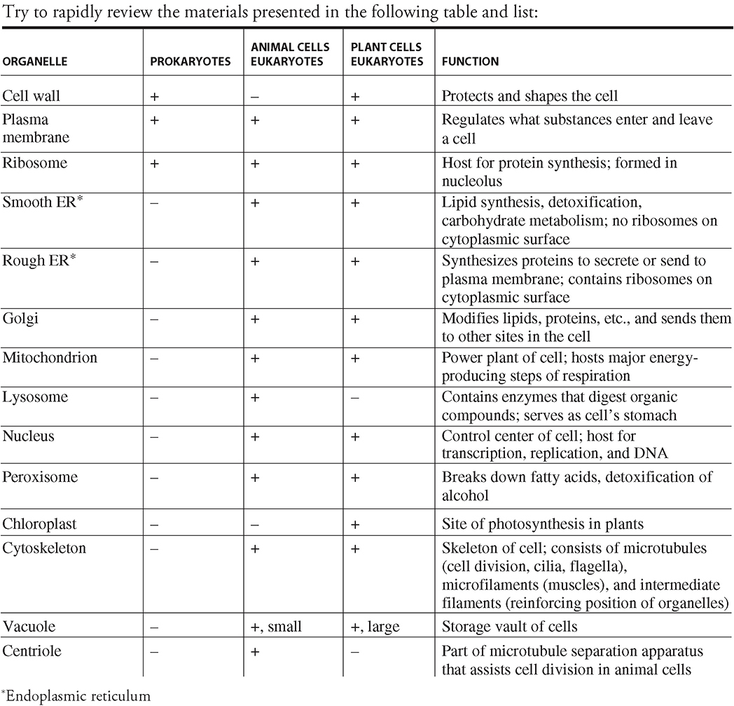
You should familiarize yourselves with approximately a dozen organelles and cell structures before taking the AP Biology exam:
SYI-1
Living systems are organized in a hierarchy of structural levels that interact.
Prokaryotic Organelles
You should be familiar with the following structures:
Plasma membrane. This is a selective barrier around a cell composed of a double layer of phospholipids. Part of this selectivity is due to the many proteins that either rest on the exterior of the membrane or are embedded in the membrane of the cell. Each membrane has a different combination of lipids, proteins, and carbohydrates that provide it with its unique characteristics.
Cell wall. This is a wall or barrier that functions to shape and protect cells. This is present in all prokaryotes.
Ribosomes. These function as the host organelle for protein synthesis in the cell. They are found in the cytoplasm of cells and are composed of a large unit and a small subunit.
Eukaryotic Organelles
You should be familiar with the following structures:
Ribosomes. As in prokaryotes, eukaryotic ribosomes serve as the host organelles for protein synthesis. Eukaryotes have bound ribosomes, which are attached to endoplasmic reticula and form proteins that tend to be exported from the cell or sent to the membrane. There are also free ribosomes, which exist freely in the cytoplasm and produce proteins that remain in the cytoplasm of the cell. Eukaryotic ribosomes are built in a structure called the nucleolus.
Smooth endoplasmic reticulum. This is a membrane-bound organelle involved in lipid synthesis, detoxification, and carbohydrate metabolism. Liver cells contain a lot of smooth endoplasmic reticulum (SER) because they host a lot of carbohydrate metabolism (glycolysis). It is given the name “smooth” endoplasmic reticulum because there are no ribosomes on its cytoplasmic surface. The liver contains much SER for another reason—it is the site of alcohol detoxification.
Rough endoplasmic reticulum. This membrane-bound organelle is termed “rough” because of the presence of ribosomes on the cytoplasmic surface of the cell. The proteins produced by this organelle are often secreted by the cell and carried by vesicles to the Golgi apparatus for further modification.
Golgi apparatus. Proteins, lipids, and other macromolecules are sent to the Golgi to be modified by the addition of sugars and other molecules to form glycoproteins. The products are then sent in vesicles (escape pods that bud off the edge of the Golgi) to other parts of the cell, directed by the particular changes made by the Golgi. We think of the Golgi apparatus as the post office of the cell—packages are dropped off by customers, and the Golgi adds the appropriate postage and zip code to make sure that the packages reach proper destinations in the cell.
Mitochondria. These are double-membraned organelles that specialize in the production of ATP. The innermost portion of the mitochondrion is called the matrix, and the folds created by the inner of the two membranes are called cristae. The mitochondria are the host organelles for the Krebs cycle (matrix) and oxidative phosphorylation (cristae) of respiration, which we discuss in Chapter 7. We think of the mitochondria as the power plants of the cell.
Lysosome. This is a membrane-bound organelle that specializes in digestion. It contains enzymes that break down (hydrolyze) proteins, lipids, nucleic acids, and carbohydrates. This organelle is the stomach of the cell. Absence of a particular lysosomal hydrolytic enzyme can lead to a variety of diseases known as storage diseases. An example of this is Tay-Sachs disease (discussed in Chapter 9), in which an enzyme used to digest lipids is absent, leading to excessive accumulation of lipids in the brain. Lysosomes are often referred to as “suicide sacs” of the cell. Cells that are no longer needed are often destroyed in these sacs. An example of this process involves the cells of the tail of a tadpole, which are digested as a tadpole changes into a frog.
Nucleus. This is the control center of the cell. In eukaryotic cells, this is the storage site of genetic material (DNA). It is the site of replication, transcription, and posttranscriptional modification of RNA. It also contains the nucleolus, the site of ribosome synthesis.
Vacuole. This is a storage organelle that acts as a vault. Vacuoles are quite large in plant cells but small in animal cells.
Peroxisomes. These are organelles containing enzymes that produce hydrogen peroxide as a by-product while performing various functions, such as breakdown of fatty acids and detoxification of alcohol in the liver. Peroxisomes also contain an enzyme that converts the toxic hydrogen peroxide by-product of these reactions into cell-friendly water.
Chloroplast. This is the site of photosynthesis and energy production in plant cells. Chloroplasts contain many pigments, which provide leaves with their color. Chloroplasts are divided into an inner portion and an outer portion. The inner fluid portion is called the stroma, which is surrounded by two outer membranes. Winding through the stroma is an inner membrane called the thylakoid membrane system, where the light-dependent reactions of photosynthesis occur. The light-independent (dark) reactions occur in the stroma.
Cytoskeleton. The skeleton of cells consists of three types of fibers that provide support, shape, and mobility to cells: microtubules, microfilaments, and intermediate filaments. Microtubules are constructed from tubulin and have a lead role in the separation of cells during cell division. Microtubules are also important components of cilia and flagella, which are structures that aid the movement of particles. Microfilaments, constructed from actin, play a big part in muscular contraction. Intermediate filaments are constructed from a class of proteins called keratins and are thought to function as reinforcement for the shape and position of organelles in the cell.

Remember me!
Of the structures listed above, animal cells contain all except cell walls and chloroplasts, and their vacuoles are small. Plant cells contain all the structures listed above, and their vacuoles are large. Animal cells have centrioles (cell division structure); plant cells do not.
As a cell grows in size, its internal volume increases, and its cell membrane (surface area) expands to respond to the growth of the cell. However, as the volume of the cell increases, the cell membrane (surface area) does not keep up. This results in the cell not having enough surface area to pass materials produced by the increasing volume of the cell. So the cell must stop growing in order to survive (See Figure 6.1).
Cell Size
ENE-1
The highly complex organization of living systems requires constant input of energy and the exchange of macromolecules.
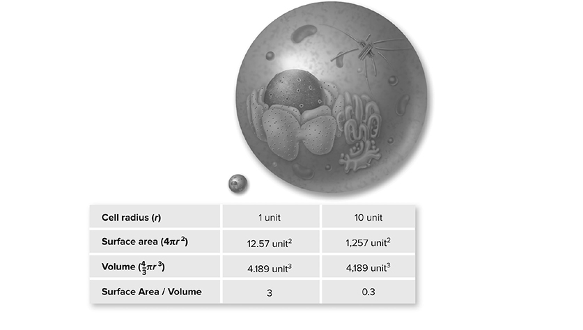
Figure 6.1 Surface area-to-volume ratio. As a cell gets larger, its volume increases at a faster rate than its surface area. If the cell radius increases by 10 times, the surface area increases by 100 times, but the volume increases by 1000 times. A cell’s surface area must be large enough to meet the metabolic needs of its volume. (Reproduced with permission from Raven P, Johnson G, Mason K, Losos J, Duncan T; Biology, 12th ed. New York: McGraw Hill; 2020)
✵ As the surface-area-to-volume ratio of a cell increases, the exchange efficiency of materials with the environment increases as well.
✵ The surface-area-to-volume ratio affects the ability of a cell to maintain homeostasis between its internal environment and external environment.
Cell Membranes: Fluid Mosaic Model
As discussed above, a cell membrane is a selective barrier surrounding a cell that has a phospholipid bilayer as its major structural component. Remember that the outer portion of the bilayer contains the hydrophilic (water-loving) head of the phospholipid, while the inner portion is composed of the hydrophobic (water-fearing) tail of the phospholipid (Figure 6.2.
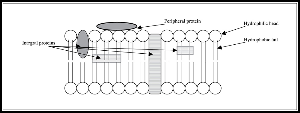
Figure 6.2 Cross-section of a cell membrane showing phospholipid bilayer.
The fluid mosaic model is the most accepted model for the arrangement of membranes. It states that the membrane consists of a phospholipid bilayer with proteins of various lengths and sizes interspersed with cholesterol among the phospholipids. These proteins perform various functions depending on their location within the membrane.
The fluid mosaic model consists of integral proteins, which are implanted within the bilayer and can extend partway or all the way across the membrane, and peripheral proteins, such as receptor proteins, which are not implanted in the bilayer and are often attached to integral proteins of the membrane. These proteins have various functions in cells. A protein that stretches across the membrane can function as a channel to assist the passage of desired molecules into the cell. Proteins on the exterior of a membrane with binding sites can act as receptors that allow the cell to respond to external signals such as hormones. Proteins embedded in the membrane can also function as enzymes, increasing the rate of cellular reactions.
The cell membrane is “selectively” permeable, meaning that it allows some molecules and other substances through, while others are not permitted to pass. The membrane is like a bouncer at a popular nightclub. What determines the selectivity of the membrane? One factor is the size of the substance, and the other is the charge. The bouncer lets small, uncharged polar substances and hydrophobic substances such as lipids through the membrane, but larger uncharged polar substances (such as glucose) and charged ions (such as sodium) cannot pass through. The other factor determining what is allowed to pass through the membrane is the particular arrangement of proteins in the lipid bilayer. Different proteins in different arrangements allow different molecules to pass through.
Cells are so small that you need a microscope to view them. Why is that? Cells must interact with their surrounding environment in order to survive and they must also bring in nutrients and remove waste across their membranes 24/7.
Types of Cell Transport
There are six basic types of cell transport:
1. Diffusion: the movement of molecules down their concentration gradient without the use of energy. It is a passive process during which substances move from a region of higher concentration to a region of lower concentration. The rate of diffusion of substances varies from membrane to membrane because of different selective permeabilities.
2. Osmosis: the passive diffusion of water down its concentration gradient across selectively permeable membranes. Water moves from a region of high water concentration to a region of low water concentration. Thinking about osmosis another way, water will flow from a region with a lower solute concentration (hypotonic) to a region with a higher solute concentration (hypertonic) (See Figure 6.4). Isotonic indicates there is no net movement of water across the membrane. This pro-cess does not require the input of energy. For example, visualize two regions—one with 10 particles of sodium per liter of water; the other with 15. Osmosis would drive water from the region with 10 particles of sodium toward the region with 15 particles of sodium (See Figure 6.3).
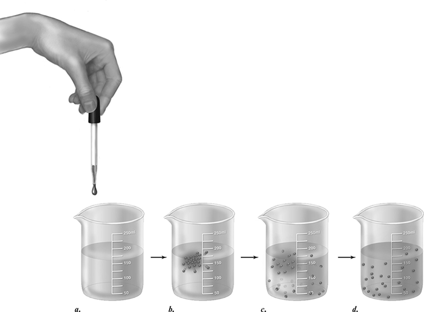
Figure 6.3 Diffusion. If a drop of colored ink is dropped into a beaker of water (a) its molecules dissolve (b) and diffuse (c). Eventually, diffusion results in an even distribution of ink molecules throughout the water (d). (Reproduced with permission from Raven P, Johnson G, Mason K, Losos J, Duncan T; Biology, 12th ed. New York: McGraw Hill; 2020)
ENE-2
Cells have membranes that allow them to establish and maintain internal environments that are different from their external environments.
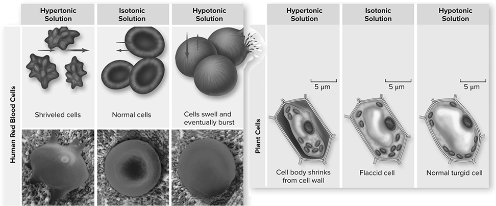
Figure 6.4 How solutes create osmotic pressure. In a hypertonic solution, water moves out of the cell, causing the cell to shrivel. In an isotonic solution, water diffuses into and out of the cell at the same rate, with no change in cell size. In a hypotonic solution, water moves into the cell. Direction and amount of water movement is shown with blue arrows (top). As water enters the cell from a hypotonic solution, pressure is applied to the plasma membrane until the cell ruptures. Water enters the cell due to osmotic pressure from the higher solute concentration in the cell. Osmotic pressure is measured as the force needed to stop osmosis. The strong cell wall of plant cells can withstand the hydrostatic pressure to keep the cell from rupturing. This is not the case with animal cells. (left, middle, right): ©David M. Phillips/Science Source (Reproduced with permission from Raven P, Johnson G, Mason K, Losos J, Duncan T; Biology, 12th ed. New York: McGraw Hill; 2020)
3. Facilitated diffusion: the diffusion of particles across a selectively permeable membrane with the assistance of the membrane’s transport proteins. These proteins will not bring any old molecule looking for a free pass into the cell; they are specific in what they will carry and have binding sites designed for molecules of interest. Like diffusion and osmosis, this process does not require the input of energy.
4. Active transport: the movement of a particle across a selectively permeable membrane against its concentration gradient (from low concentration to high). This movement requires the input of energy, which is why it is termed “active” transport. As is often the case in cells, adenosine triphosphate (ATP) is called on to provide the energy for this reactive process. These active-transport systems are vital to the ability of cells to maintain particular concentrations of substances despite environmental concentrations. For example, cells have a very high concentration of potassium and a very low concentration of sodium. Diffusion would like to move sodium in and potassium out to equalize the concentrations. The all-important sodium-potassium pump actively moves potassium into the cell and sodium out of the cell against their respective concentration gradients to maintain appropriate levels inside the cell. This is the major pump in animal cells.
5. Endocytosis: a process in which substances are brought into cells by the enclosure of the substance into a membrane-created vesicle that surrounds the substance and escorts it into the cell (Figure 6.5. This process is used by immune cells called phagocytes to engulf and eliminate foreign invaders.
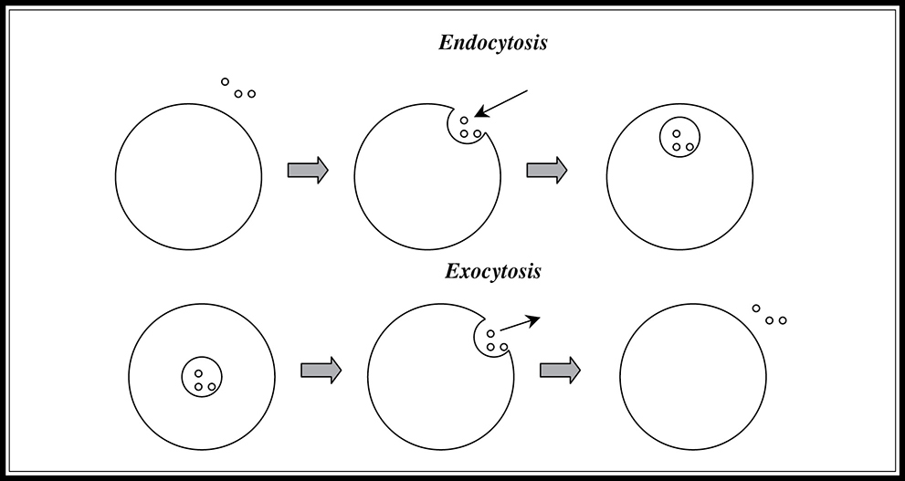
Figure 6.5 Endocytosis and exocytosis.
6. Exocytosis: a process in which substances are exported out of the cell (the reverse of endocytosis). A vesicle again escorts the substance to the plasma membrane, causes it to fuse with the membrane, and ejects the contents of the substance outside the cell (Figure 6.5. In exocytosis, the vesicle functions like the trash chute of the cell.
7. Pinocytosis: a process of bringing in droplets of extracellular fluid via tiny vesicles.
8. Receptor-mediated endocytosis: a specialized type of pinocytosis that moves specific molecules into a cell due to the budding of specific molecules with receptor sites on the cell membrane.
Water Potential
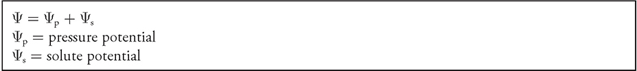
How does a plant defy gravity and stand up? How do fish survive in saltwater? Well, it has to do with water potential. Water potential (Ψ = Ψp + Ψs) indicates how freely water molecules can move in a particular environment or system. It is determined by the solute potential and pressure potential of each environment. Solute potential (Ψs), also called osmotic potential, depends on the amount of solute in a solution and decreases as the concentration of solute increases. It is negative in a plant cell and zero in distilled water. Pressure potential (Ψp), also called turgor potential, refers to the physical pressure exerted by objects or cell membranes on water molecules and increases with increasing pressure. Plant cells maintain a positive pressure to hold their shape, allowing them to stay rigid (See Figure 6.6).
ENE-2
Cells have membranes that allow them to establish and maintain internal environments that are different from their external environments.
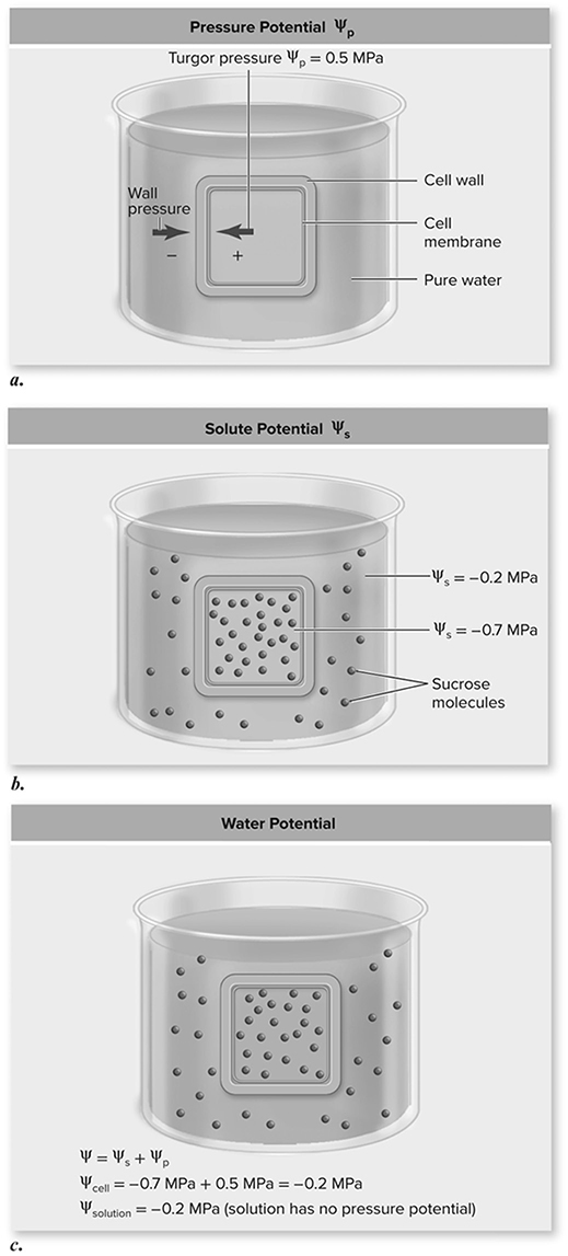
Figure 6.6 Determining water potential. a. Cell walls exert pressure in the opposite direction of cell turgor pressure. b. Using the given solute potentials, predict the direction of water movement based only on solute potential. c. Total water potential is the sum of ψs and ψp. Because the water potential inside the cell equals that of the solution, there is no net movement of water. (Reproduced with permission from Raven P, Johnson G, Mason K, Losos J, Duncan T; Biology, 12th ed. New York: McGraw Hill; 2020)
In an open container, water potential is equal to the solute potential due to the pressure potential being zero in an open container.
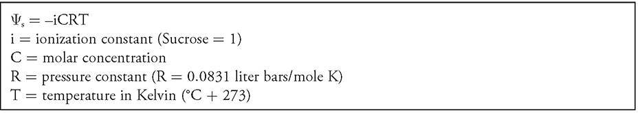
Osmoregulation is the ability to maintain water balance and allows organisms to control their internal environments by maintaining the right concentrations of solutes and the amount of water in their body fluids. The solute potential of a solution (Ψs = −iCRT) depends on the ionization constant (i), molar concentration (C), pressure constant (R), and temperature (T).
![]() Review Questions
Review Questions
For questions 1—4, please use the following answer choices:
A. Cell wall
B. Mitochondrion
C. Ribosome
D. Lysosome
1. This organelle is present in plant cells, but not animal cells.
2. Absence of enzymes from this organelle can lead to storage diseases such as Tay-Sachs disease.
3. This organelle is the host for the Krebs cycle and oxidative phosphorylation of respiration.
4. This organelle is synthesized in the nucleolus of the cell.
5. Which of the following best describes the fluid mosaic model of membranes?
A. The membrane consists of a phospholipid bilayer with proteins of various lengths and sizes located on the exterior portions of the membrane.
B. The membrane consists of a phospholipid bilayer with proteins of various lengths and sizes located in the interior of the membrane.
C. The membrane is composed of a phospholipid bilayer with proteins of uniform lengths and sizes located in the interior of the membrane.
D. The membrane contains a phospholipid bilayer with proteins of various lengths and sizes interspersed among the phospholipids.
6. Which of the following types of cell transport requires energy?
A. The movement of a particle across a selectively permeable membrane down its concentration gradient
B. The movement of a particle across a selectively permeable membrane against its concentration gradient
C. The movement of water down its concentration gradient across selectively permeable membranes
D. The movement of a sodium ion from an area of higher concentration to an area of lower concentration
7. Which of the following structures is present in prokaryotic cells?
A. Nucleus
B. Mitochondria
C. Cell wall
D. Golgi apparatus
8. Which of the following represents an incorrect description of an organelle’s function?
A. Chloroplast: the site of photosynthesis and energy production in plant cells
B. Peroxisome: organelle that produces hydrogen peroxide as a by-product of reactions involved in the breakdown of fatty acids, and detoxification of alcohol in the liver
C. Golgi apparatus: structure to which proteins, lipids, and other macromolecules are sent to be modified by the addition of sugars and other molecules to form glycoproteins
D. Rough endoplasmic reticulum: membrane-bound organelle lacking ribosomes on its cyto-plasmic surface, involved in lipid synthesis, detoxification, and carbohydrate metabolism
9. The destruction of which of the following would most cripple a cell’s ability to undergo cell division?
A. Microfilaments
B. Intermediate filaments
C. Microtubules
D. Actin fibers
10. Which of the following can easily diffuse across a selectively permeable membrane?
A. Na+
B. Glucose
C. Large uncharged polar molecules
D. Lipids
![]() Answers and Explanations
Answers and Explanations
1. A—Cell walls exist in plant cells and prokaryotic cells, but not animal cells. They function to shape and protect cells.
2. D—The lysosome acts like the stomach of the cell. It contains enzymes that break down proteins, lipids, nucleic acids, and carbohydrates. Absence of these enzymes can lead to storage disorders such as Tay-Sachs disease.
3. B—The mitochondrion is the power plant of the cell. This organelle specializes in the production of ATP and hosts the Krebs cycle and oxidative phosphorylation.
4. C—The ribosome is an organelle made in the nucleolus that serves as the host for protein synthesis in the cell. It is found in both prokaryotes and eukaryotes.
5. D—The fluid mosaic model says that proteins can extend all the way through the phospholipid bilayer of the membrane, and that these proteins are of various sizes and lengths.
6. B—Answer choice B is the definition of active transport, which requires the input of energy. Simple diffusion (answer choices A and D) and osmosis (answer choice C) are all passive processes that do not require energy input.
7. C—Prokaryotes do not contain many organelles, but they do contain cell walls.
8. D—This is the description of the smooth endoplasmic reticulum. We know that this is a tricky question, but we wanted you to review the distinction between the two types of endoplasmic reticulum.
9. C—Microtubules play an enormous role in cell division. They make up the spindle apparatus that works to pull apart the cells during mitosis (Chapter 9). A loss of microtubules would cripple the cell division process. Actin fibers (answer choice D) are the building blocks of microfilaments (answer choice A), which are involved in muscular contraction. Keratin fibers are the building blocks of intermediate filaments (answer choice B), which function as reinforcement for the shape and position of organelles in the cell.
10. D—Lipids are the only substances listed that are able to freely diffuse across selectively permeable membranes.
![]() Rapid Review
Rapid Review
Fluid mosaic model: plasma membrane is a selectively permeable phospholipid bilayer with proteins of various lengths and sizes interspersed with cholesterol among the phospholipids.
Integral proteins: proteins implanted within lipid bilayer of plasma membrane.
Peripheral proteins: proteins attached to exterior of membrane.
Diffusion: passive movement of substances down their concentration gradient (from high to low concentrations).
Osmosis: passive movement of water from the side of low solute concentration to the side of high solute concentration (hypotonic to hypertonic).
Facilitated diffusion: assisted transport of particles across membrane (no energy input needed).
Active transport: movement of substances against concentration gradient (low to high concentrations; requires energy input).
Endocytosis: phagocytosis of particles into a cell through the use of vesicles.
Exocytosis: process by which particles are ejected from the cell, similar to movement in a trash chute.
Water potential: (Ψ = Ψp + Ψs) indicates how freely water molecules can move in a particular environment or system. It is determined by the solute potential and pressure potential of each environment.
Hypertonic: a solution that contains a higher solute concentration when compared to inside the cell.
Hypotonic: a solution that contains a lower solute concentration when compared to inside the cell.
Isotonic: indicates there is no net movement of water across the membrane due to an equal concentration of solutes on both sides.
Pinocytosis: a process of bringing in droplets of extracellular fluid via tiny vesicles.
Phagocytosis: a process in which substances are brought into cells by the enclosure of the substance into a membrane-created vesicle that surrounds the substance and escorts it into the cell.