5 Steps to a 5: AP Biology 2017 (2016)
STEP 4
Review the Knowledge You Need to Score High
CHAPTER 15
Human Physiology
IN THIS CHAPTER
Summary: This chapter takes you on a tour of the human body and discusses how the various systems of the human body function on a daily basis.
Key Ideas
![]() Study this chapter well—human physiology comes up often on the AP exam.
Study this chapter well—human physiology comes up often on the AP exam.
![]() Passage of blood flow through the heart: vena cava → right atrium → right ventricle → lungs → left atrium → left ventricle → aorta → body and back.
Passage of blood flow through the heart: vena cava → right atrium → right ventricle → lungs → left atrium → left ventricle → aorta → body and back.
![]() The functional unit of the lung is the alveolus.
The functional unit of the lung is the alveolus.
![]() Four major thermoregulatory processes: conduction, convection, evaporation, and radiation.
Four major thermoregulatory processes: conduction, convection, evaporation, and radiation.
![]() The CNS consists of the brain and spinal cord. The PNS is broken down into the sensory and motor divisions.
The CNS consists of the brain and spinal cord. The PNS is broken down into the sensory and motor divisions.
![]() Three main types of muscle: skeletal, cardiac, and smooth.
Three main types of muscle: skeletal, cardiac, and smooth.
![]() Study the names, origins, and functions of the various hormones that appear in this chapter—this is a common subject for multiple-choice questions.
Study the names, origins, and functions of the various hormones that appear in this chapter—this is a common subject for multiple-choice questions.
![]() Learn about the difference between nonspecific and specific immunity.
Learn about the difference between nonspecific and specific immunity.
Introduction
Welcome to the tour of the human body. During this tour, we will discuss how our bodies work. We will be making eight stops, lunch will not be served, and we don’t want to hear any requests for bathroom breaks (although we will be learning about things of that nature). Buckle up—here we go.
BIG IDEA 4.B.2
Within multicellular organisms, specialization of organs contributes to overall function .
Circulatory System
Heart
Welcome to the heart. The human heart is a four-chambered organ whose function is to circulate blood by rhythmic contraction. The heart pumps oxygenated blood from the left ventricle out to the aorta (Figure 15.1 ). From there it travels through arteries to feed the organs, muscles, and other tissues of the body. The blood returns to the heart via the veins. The superior and inferior vena cavae return deoxygenated blood from the body to the heart. The blood reenters the heart through the right atrium, passes through to the right ventricle, and from there to the lungs to exchange carbon dioxide for more oxygen. At this point, the blood has made a complete cycle through the body. The blood is at its most oxygenated stage just after leaving the lungs as it enters the left side of the heart and travels into the aorta. The blood is in its least oxygenated stage as it reenters the right atrium of the heart. Structure–function relationships come up often on the AP Biology exam, and the circulatory system provides a good example you could add to an essay on the topic. The left ventricle of the heart is the thickest and most muscular part of the heart, and the most pressure is exerted on it. Why does this make sense functionally? Because the left ventricle is the portion of the heart that needs to pump the blood into the aorta and to the rest of the body. The left ventricle is structurally designed to fit its function. The right ventricle is smaller and less muscular because it only pumps blood a short distance to the lungs for gas exchange (the picking up of oxygen and the release of CO2 ).
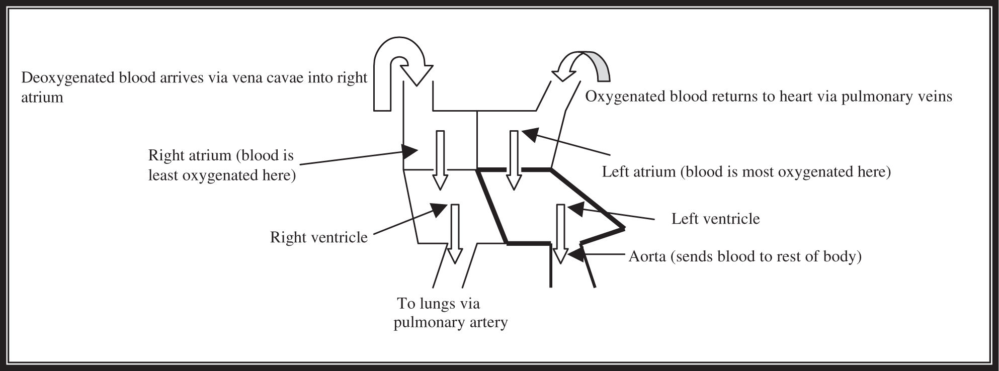
Figure 15.1 Oversimplified diagram of the heart and blood flow.
Blood
As we continue on our journey, if you look off to your right, you will see some of the blood and its components passing us now. If you look closely, you will see little red blood cells , which carry oxygen, traveling in the bloodstream. Thanks to a molecule termed hemoglobin, the red blood cells are able to carry and deliver oxygen throughout the body to hardworking organs and tissues. Iron is a major component of hemoglobin. If you do not have enough iron in your diet, your ability to deliver oxygen via the blood can be compromised, and you may develop anemia.
Blood is able to flow so efficiently because it contains primarily water. The liquid portion of the blood, the plasma, contains minerals, hormones, antibodies, and nutritional materials. Another common component seen in the bloodstream is the platelet, which is involved in the clotting of blood. You might ask, “What are the white cells flowing around?” The white blood cells are the protection system for our body. We will be seeing those up close when we talk about the immune system.
The lymphatic system is worth a brief mention here because it is an important part of the circulatory system. When blood flows through the capillaries of the body, proteins and fluid leak out during the exchange. The lymphatic system functions as the route by which these poor lost souls find their way back into the bloodstream. The lymphatic system also functions as a protector for the body because of the presence of structures known as lymph nodes,which are full of white blood cells that live to fight infection. If your neck sometimes swells when you are sick with the flu, for instance, it is probably the multiplication of white blood cells in the lymph nodes of your neck.
Diseases of the Cardiovascular System
Two diseases that you should be familiar with for the exam are hypertension and arteriosclerosis. Hypertension is high blood pressure and is a major cause of strokes and heart attacks. Arteriosclerosis is a big word that means hardening of the arteries. These hardened arteries become narrower and are a prime risk factor for death by embolism—the breaking off of a piece of tissue that lodges in an artery, blocking the flow of blood to vital tissues.
Respiratory System
We are going to head down to the lungs now. Please stay close because it will get a little loud in these windy tunnels. Air comes into the body through the mouth and the nose. We are currently in the nasal passages, and along with the air that came into the nose, we are being warmed and moistened in the nasal cavity before we head down toward the pharynx region, where the air and food passages cross. We will come back to this area again later on in the tour when we take the road that food uses to get from the mouth to the stomach. During inhalation, the air goes through a structure called the glottis into the larynx (human voicebox). From there, the air moves into the trachea, which contains rings of cartilage that help it maintain its shape. Each trachea is the tunnel that leads the air into the thoracic cavity . If you look outside your windows, you will notice some tiny arms waving at us as we go by. They are the cilia, which beat in rhythmical waves to carry foreign particles (like our tour bus) and mucus away from the respiratory tract.
BIG IDEA 2.A.3
The increased surface area of alveoli helps in gas exchange with the environment .
We are now at a fork in the road. Here the trachea divides into two separate tunnels: the two bronchi, which are also held open by cartilage rings, one going to the left lung, and one going to the right lung. Each bronchus divides into smaller branches, which divide into even smaller branches, which divide into tunnels called bronchioles. These bronchioles branch repeatedly until they conclude as tiny air pockets containing alveoli.
In Figure 15.2 , notice how thin the walls of the alveoli are. They are usually a single cell in thickness, are covered by a thin film of water, and are surrounded by a dense bed of capillaries. You might have questioned earlier exactly where the exchange of O2 and CO2 actually occurs—this is the place. The alveoli are considered to be the primary functional unit of the lung. Oxygen enters the alveolus the same way we just did—it dissolves in the water lining of the wall and diffuses across the cells into the bloodstream. At the same time, the CO2 , which is carried by the blood, primarily in the form of bicarbonate (HCO3 − ), passes out of the blood in a similar manner. The O2moves easily into the bloodstream because it is moving down its concentration gradient. Once there, it travels with the blood to the rest of the body.
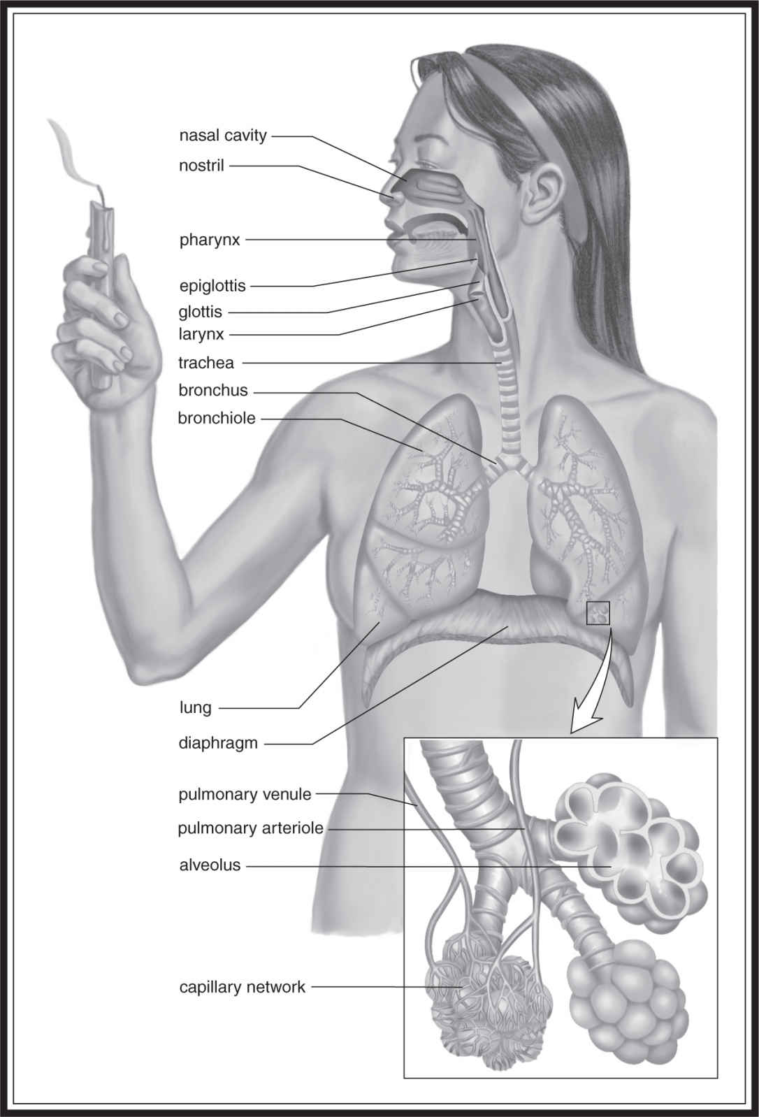
Figure 15.2 The human lungs, with close-up view of alveoli, bronchi, and bronchioles. (From Biology, 8th ed., by Sylvia S. Mader, © 1985, 1987, 1990, 1993, 1996, 1998, 2001, 2004 by the McGraw Hill Companies, Inc. Reproduced with permission of The McGraw-Hill Companies .)
Before we move on to the digestive system, we should discuss the mechanism by which breathing actually occurs. The rib cage and the diaphragm play important roles in the breathing process. Inhalation causes the volume of the thoracic cavity to increase. As a result, the air pressure in the chest falls below that of the atmosphere, and air flows into the body. This is accompanied by a contraction of the rib-cage muscles and the diaphragm, allowing for the increase in thoracic volume. After the air exchange occurs, the muscles relax, causing the diaphragm to move up against the lungs, reducing the thoracic volume. This causes the pressure in the lungs to exceed that of the atmosphere—driving the air containing CO2 out of the body.
Digestive System
Okay, folks, it is time to take the tour of the digestive system. Hang on tight—we are going to take a shortcut to the mouth as we are exhaled back through the system. Here we go: bronchioles, bronchi, trachea, larynx, pharynx, and mouth!
Here we sit in the oral cavity. This is where the digestion of food begins. Food is, of course, tasted in the oral cavity, and the teeth that help us chew (masticate) are performing a task called mechanical digestion. The liquid sloshing up against the windows of the bus is saliva, which contains enzymes such as amylase that help dissolve some of the food. Amylase breaks down the starches in our diet into simpler sugars like maltose, which are fully digested further down in the intestines. The saliva also acts as a lubricant to help the food move along the digestive pathway.
We need to carefully avoid the tongue, which functions to move food around while we chew and helps to arrange it into a ball that we swallow called a bolus . The tongue pushes the food toward the crossroad we visited during the tour of the respiratory system. You may notice that this time, as the swallowing occurs, we do not go through the glottis toward the lungs, but instead into the esophagus, which connects the throat to the stomach. The force created by the rhythmical contraction of the smooth muscle of the esophagus (currently pushing us toward the stomach) is called peristalsis.
After passing through the esophageal sphincter, which acts like a valve or trapdoor, food enters into the stomach, where more digestion will occur. The sphincter is usually closed in order to keep food from returning back up the esophagus to the mouth. In the stomach, the digestion occurs by a churning action that mixes the food and breaks it into smaller pieces. Folks, we would recommend that you do not step out of the bus here because the pH is way down in the 1.5–2.5 range, which provides quite an acidic environment. If you look closely along the edges of the stomach, you will see many glands. Some of these glands secrete gastric juice, composed of hydrochloric acid (HCl) and digestive enzymes, which helps in digestion and lowers the pH. The major enzyme of the stomach is pepsin, which breaks proteins down into smaller polypeptides that are handled by the intestines. The glands here secrete pepsinogen —the precursor to pepsin. Pepsinogen is activated into pepsin by HCl. Pepsin is picky and will function only in a particular range of pH values. This is a good thing because if it were active all the time, it would digest things it is not supposed to digest. Other glands secrete mucus to help line the stomach. It is this mucus that helps prevent the wall of the stomach from being digested along with the food.
Now we move on to the small intestine . To get to the small intestine, we need to pass through the Panama Canal of the body: the pyloric sphincter. For those of you interested in useful AP exam trivia, the small intestine is where most of the digestion and absorption occur. The terrain is a bit different in this organ. The walls are arranged into folds and ridges, which have more waving structures, this time called villi, similar to the cilia we saw in the respiratory tract. The walls in the small intestine contain something called a brush border, which is composed of a large amount of microvilli that increases the surface area of the small intestine to improve absorption efficiency. Digested nutrients absorbed in the small intestine are dumped into various veins that merge to form the hepatic portal vessel, which leads to the liver. The liver then gets first crack at the newly absorbed nutrients before they are sent to the rest of the body. As the food moves into the small intestine, it brings with it an acidity that promotes the secretion of numerous enzymes from the pancreas and the local glands. ( Important note to remember: hormones are vital to the turning on and off of the digestive glands.)
BIG IDEA 2.A.3
Villi create high surface area, helping in exchange of nutrients with the environment .
Those of you on the left side of the bus have a good view of the pancreatic duct as it expels lipase, amylase, trypsin, and chymotrypsin. Lipase is the major fat-digesting enzyme of the body. It receives some help in the handling of the fat from a product made in the liver called bile. Bile contains bile salts, phospholipids, cholesterol, and bile pigments such as bilirubin. The bile is stored in the gallbladder and is dumped into the small intestine upon the arrival of food. The bile salts help digest the fat by emulsifying it into small droplets contained in water. (Emulsification is a physical change—bile does not contain any enzymes.) Amylase continues the breakdown of carbohydrates into simpler sugars. Maltase, lactase, and sucrase break maltose, lactose, and sucrose, respectively, into monosaccharides. Trypsin and chymotrypsin work together to handle the digestion of the peptides in our diet. Trypsin cuts peptide bonds next to arginine and lysine; chymotrypsin cuts bonds by phenyalanine, tryptophan, and tyrosine. Like pepsin, these two proteolytic enzymes are secreted as inactive forms: trypsinogen and chymotrypsinogen. Trypsinogen is activated first to become trypsin, which, in turn, activates chymotrypsin. Some of you might ask “If the proteolytic enzymes only cut at certain sites, how do we finish digesting the proteins?” Trypsin and chymotrypsin are examples of enteropeptidases . It is the exopeptidases that complete the digestion of proteins by hydrolyzing all the amino acids of the remaining fragments.
After the small intestine comes the large intestine (which includes the cecum, colon, and rectum). The two meet up in the lower right corner of the abdomen. The colon has three main parts: the ascending, transverse, and descending colon . There are two major functions for this part of the system—the primary function is to reabsorb water and electrolytes. A second function is to serve as a passageway for the waste material as it moves toward the rectum. The food enters the large intestine, travels up the ascending colon, across the transverse colon, down the descending colon into the rectum, where it is stored until it gets eliminated . . . but we don’t need to go there. We’ve seen enough for now.
Control of the Internal Environment
BIG IDEA 2.D.2
Homeostatic mechanisms reflect common ancestry .
The next stop on our tour is the kidney (see Figure 15.3 for an overview of the human excretory system). The kidneys lie on the posterior wall of the abdomen. The renal artery and vein bring blood to and from the kidney, respectively. Kidneys are divided into two major regions: an outer region called the cortex, and an inner region called the medulla. These two regions are full of nephrons, the functional units of the kidney. The medulla is divided into structures called renal pyramids, which dump urine into the major and minor calyces . From here, the urine is sent toward the bladder via the ureter . When contracted to urinate, the bladder sends the urine through the urethra to the outside world.
We’ve pulled the bus right up to one of over a million nephrons in each kidney. The nephron is composed of a renal corpuscle, proximal convoluted tubule, loop of Henle, distal convoluted tubule, and collecting duct system . If you look closely, you will see that the renal corpuscle is made up of glomerular capillaries surrounded by Bowman’s capsule .
Osmoregulation and Excretion
The blood that enters via the renal artery is sent to the various nephrons by the branching of the renal artery into smaller and smaller vessels that culminate in the capillaries of the glomerulus. The blood pressure is the force that leads to the movement of solutes such as water, urea, and salts into the lumen of Bowman’s capsule from the glomerular capillaries. From here, the fluids pass down the proximal tubule, through the loop of Henle, and into the distal tubule, which dumps into the collecting duct. The various collecting ducts of the kidney collectively merge into the renal pelvis, which leads via the ureter to the bladder.
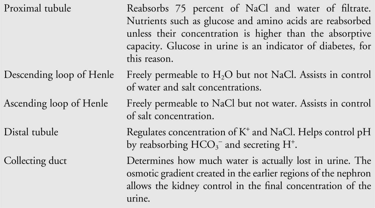
As we mentioned moments ago, fluid moves from the capillaries into the lumen of the nephron as a result of the force of blood pressure. During this process of filtration, the capillaries are able to let small particles through the pores of their endothelial linings, but large molecules such as proteins, platelets, and blood cells tend to remain in the vessel. As the filtrate progresses along the tubule, plasma solutes such as urea are added by the process of secretion, a selective process that helps to create a solute gradient. It is important to realize that much of what is dumped into the tubule originally is reabsorbed —nearly all the sugars, water, and organic nutrients. The combination of reabsorption and secretion help the nephron to control what gets released in the urine. The following chart outlines in detail what happens in the various parts of the nephron:
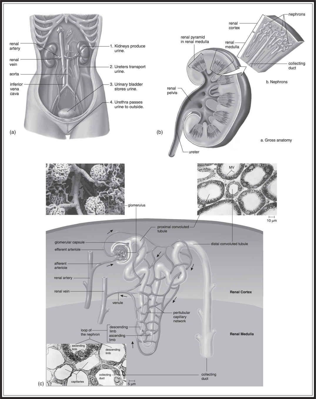
Figure 15.3 For legend see the following section.
Figure 15.3 The human excretory system on four size scales. (a) The kidneys produce urine and regulate the composition of the blood. Urine is conveyed to the urinary bladder via the ureter and to the outside via the urethra. Branches of the aorta, the renal arteries convey blood to the kidneys; renal veins drain blood from the kidneys into the posterior vena cava. (b) Urine is formed in two distinct regions of the kidney: the outer renal cortex and inner renal medulla. It then drains into a central chamber, the renal pelvis, and into the ureters. (c) Excretory tubules (nephrons and collecting ducts) and associated blood vessels pack the cortex and medulla. The human kidney has about a million nephrons, representing about 80 km of tubules. Cortical nephrons are restricted mainly to the renal cortex. Juxtamedullary nephrons have a long, hairpinlike portion that extends into the renal medulla. Several nephrons empty into each collecting duct, which drains into the renal pelvis.
(Adapted from Biology, 8th ed., by Sylvia S. Mader, © 1985, 1987, 1990, 1993, 1996, 1998, 2001, 2004 by the McGraw Hill Companies, Inc. Reproduced with permission of The McGraw-Hill Companies.)
BIG IDEA 2.C.1
Animals use feedback mechanisms (e.g., ADH) to maintain internal conditions .
The body controls the concentration of the urine according to the needs of the system. When dehydrated, the body can excrete a small volume of hypertonic concentrated urine (little water in the urine; it is dark yellow). But in times of excessive fluid, the body will excrete a large volume of hypotonic dilute urine to conserve the necessary salts (lots of water in the urine; it is clear). This is controlled by hormones and is discussed in more detail in a later section, but briefly: ADH (antidiuretic hormone) is released by the pituitary gland; it increases permeability of the collecting duct to water, leading to more concentrated urine. Aldosterone, released from the adrenal gland, acts on the distal tubules to cause the reabsorption of more Na+ and water to increase blood volume and pressure.
Thermoregulation
A fairly constant body temperature is important for many living organisms. The process by which this temperature is maintained is known as thermoregulation. A major organ involved in thermoregulation is the skin, which also plays a role in excretion through sweating. Four major thermoregulatory processes are conduction, convection, evaporation, and radiation. Conduction is the process by which heat moves from a place of higher temperature to a place of lower temperature. For example, let’s say that two people are sleeping in the same bed, and that person A is cold all the time. Person A would not make it through the night if it were not for this process. Since person B tends to be warmer than person A, person A takes advantage of conduction by pulling the heat from person B’s body to hers. Convection is heat transfer caused by airflow. Thinking about my baseline warmth (similar to that of person B), if it were not for our air conditioners in the summer, we would probably not be here today to write this book. But we curse convection in the winter as the cold wind removes heat from our bodies, making it feel that much colder outside. Evaporation is the process by which water leaves our bodies in the form of water vapor: sweat. Why do humid days feel so much warmer than nonhumid days? Because humidity increases the amount of water in the air, decreasing the driving force for water to leave our bodies. Radiation is the loss of heat through ejection of electromagnetic waves.
Before moving on to the nervous system, we must mention two more terms: endotherm and ectotherm. An endotherm is an organism whose body temperature is not dramatically affected by the surrounding temperature. We humans are endothermic creatures. Sure, a cold day can feel really cold, but at least it does not dramatically lower the human body temperature. Ectothermic animals are organisms whose body temperatures are affected by the surrounding temperature. Fish, reptiles, and amphibians are good examples of ectothermic organisms.
Nervous System
The nervous system is divided into two systems: the central nervous system (CNS) and the peripheral nervous system (PNS). The CNS contains the brain and the spinal cord. The PNS can be broken down into a sensory and a motor division. The sensory division carries information to the CNS while the motor division carries information away from the CNS. The motor division can be further broken down in the somatic nervous system (SNS), also known as the voluntary nervous system, and the autonomic nervous system (ANS). As indicated by its name, the SNS controls the voluntary contraction of muscles, while the ANS controls the involuntary activities of the body: smooth muscle, cardiac muscle, and glands. The ANS is divided into the sympathetic and parasympathetic divisions.
BIG IDEA 3.E.2
Animals have a nervous system that detects signals, transmits information, and produces responses .
Before delving into the various divisions of the nervous system, it is important to look at the mechanics of nerve cell transmissions (Figure 15.4 ). The functional unit of the nervous system is the neuron (nerve cell). Outside to the left of the bus is a nerve cell from the CNS. There are three main parts to a nerve cell: the cell body, the dendrite, and the axon. The cell body is the main body of the neuron. The dendrite is one of many short, branched processes of a neuron that help bring the nerve impulses toward the cell body. The axon is the longer extension that leaves from the neuron and carries the impulse away from the cell body toward the target cell. Some CNS nerve cells, as well as most PNS neurons, are myelinated neurons, which means that they have a layer of insulation around the axon, allowing for faster transmission. It is the cable Internet of the body.
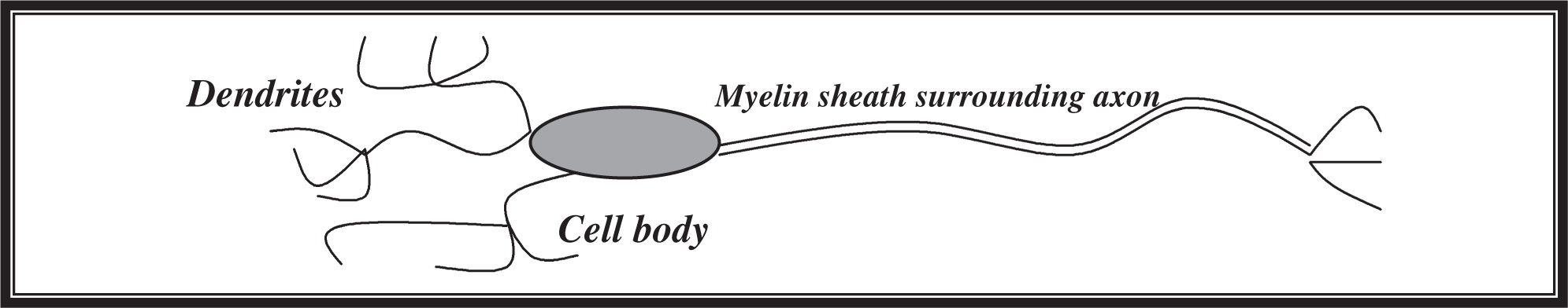
Figure 15.4 The components of a nerve cell (neuron).
The nerve cells can be divided into three main classes: sensory neurons, motor neurons, and interneurons. Sensory neurons receive and communicate information from the sensory environment. Interneurons function to make synaptic connections with other neurons. Located in the CNS, they tie together sensory input and motor output and are the intermediaries of the operation. Motor neurons take the commands of the CNS and put them into action as motor outputs. This relationship is the basis for the reflex arc, which is the basic unit of response in the CNS. A sensory neuron sends an impulse to the spinal cord, which is transmitted via a series of interneurons to a motor neuron whose impulse causes a muscular contraction.
BIG IDEA 4.A.4
Organisms exhibit complex properties due to interactions between constituent parts .
Whoa! Did you see that spark zip past just now? That was a perfect example of a nerve impulse. The membranes of these neurons all around us are full of pumps and special gated ion channels that allow the cell to change its membrane potential in response to certain stimuli. The opening of sodium channels causes the potential to become less negative, and the cell is depolarized. If the threshold potential is reached (electrical potential that, when reached, initiates an action potential), an action potential is triggered, which is the nerve impulse that we just saw zip by. Action potentials are quick changes in cell potential due to well-controlled opening and closing of ion channels. The cell also contains potassium channels that open slowly in response to depolarization. After a short period of time, the sodium channel closes, and potassium rushes out of the cell causing repolarization of the cell and lowering of the potential back down to its initial. Let’s move farther down this axon to see where this impulse is going.
BIG IDEA 3.D.2
Cells communicate with each other using chemical signals (e.g., neurotransmitters) .
Here we are at the end of the axon, sometimes called the synaptic knob. This is where calcium gates are opened in response to the changing potential, which causes vesicles to release substances called neurotransmitters into the synaptic gap between the axon and the target cell. These neurotransmitters diffuse across the gap, causing a new impulse in the target cell. Two of the most common neurotransmitters used in the body are acetylcholine and norepinephrine. Substances called cholinesterases function to clear the neurotransmitters from the synaptic gap after an action potential by binding to the neurotransmitters and recycling them back to the neuron.
The ANS regulates involuntary activities in the body. As mentioned earlier, it is subdivided into the parasympathetic and sympathetic divisions. For the most part, the parasympathetic response is one that promotes energy conservation: slower heart rate, decreased blood pressure, and bronchial muscle and urinary bladder constriction. The sympathetic response is one that prepares us for “fight or flight”—increased heart rate, dilated bronchial muscles, increased blood pressure, and digestive slowdown.
The CNS consists of the brain and spinal cord. The brain is divided into various sections that control the different regions of our bodies (Figure 15.5 ). The cerebellum is in charge of coordination and balance. The medulla oblongata is the control center for involuntary activities such as breathing. The hypothalamus is the thermostat and hunger-meter of the body, regulating temperature, hunger, and thirst. The amygdala is the portion of our brain that controls impulsive emotions and anger. The cerebrum is split into two “hemispheres” that connect to each other in the middle via the corpus callosum. Each half is divided into four different lobes, each specializing in various functions:
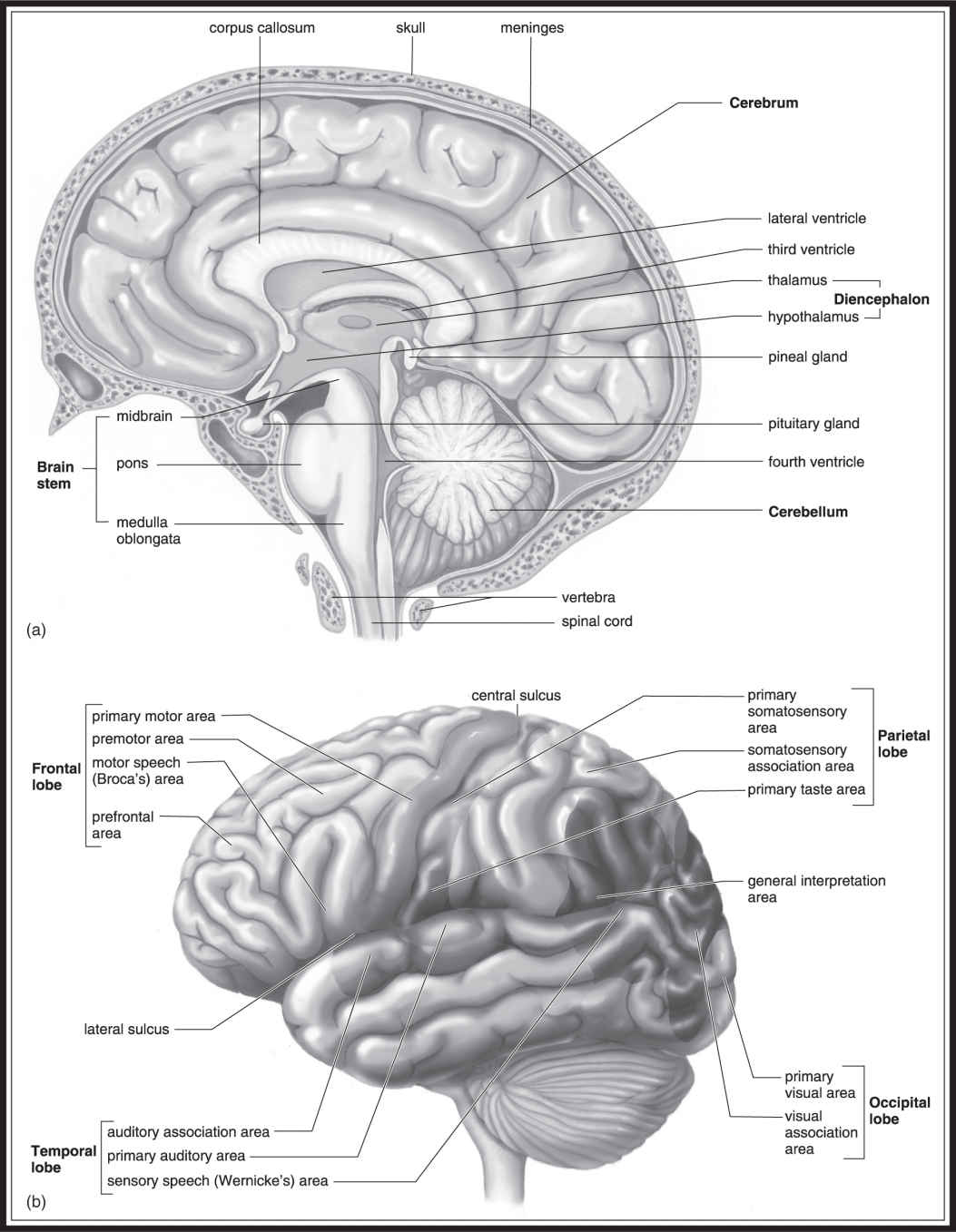
Figure 15.5 Major structures of the human brain. (a) Of the brain’s three ancestral regions, the forebrain is massively developed and contains the most sophisticated integrating centers. One of its subdivisions, the telencephalon, consists mainly of the cerebrum (cerebral hemispheres), which extends over and around most other brain centers. The diencephalon contains the thalamus and hypothalamus. The other two ancestral regions, the midbrain and hindbrain, make up the brian stem. (b) This rear view shows the bilateral nature of the brain components. The cerebral hemispheres, corpus callosum (large fiber tracts connecting the hemispheres), and basal ganglia are parts of the telencephalon. (From Biology, 8th ed., by Sylvia S. Mader, © 1985, 1987, 1990, 1993, 1996, 1998, 2001, 2004 by the McGraw Hill Companies, Inc. Reproduced with permission of The McGraw-Hill Companies.)

Muscular System
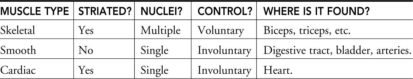
Our tour of the muscle types of the body will include a look at the types of muscles and a quick demonstration of muscle contraction. There are three main types of muscle: skeletal, smooth, and cardiac:
1. Skeletal muscle . Muscle type that works when you do pushups, lift a book, and do other voluntary activities. Skeletal muscle cells contain multiple nuclei. This muscle type has a striated appearance.
2. Smooth muscle . Involuntary muscle that contracts slowly and is controlled by the ANS. Smooth muscle cells contain a single nucleus. Found in the walls of arteries, digestive tract, bladder, and elsewhere. Smooth muscle is not striated in appearance.
3. Cardiac muscle . Involuntary muscle of the heart. Cardiac muscle cells contain a single nucleus. Cardiac muscle cells are striated in appearance.
Muscle cells are activated by the mechanism described earlier involving the action potentials and ion channels. When an action potential reaches a muscle cell, acetylcholine is released at the neuromuscular junction —the space between the motor neuron and the muscle cell. This neurotransmitter depolarizes the muscle cell and, through a series of intracellular reactions, causes the release of large amounts of stored calcium inside the cell, leading to muscle contraction. Muscle contraction stops when the calcium is taken back up by the sarcoplasmic reticulum of the cell.
CT teacher: “Know the functional units of the various systems discussed in this chapter. How the structure of these functional units relates to their function could be a nice essay.”
Folks, we are going to be treated to a demonstration of skeletal muscle contraction. Skeletal muscle consists of fiber bundles, which are composed of myofibrils. What are myofibrils? Good question. They are structures that are made up of a combination of myofilaments called thin filaments (actin ) and thick filaments (myosin ).
The Actin-Myosin “Tango”
It takes two to tango, and myosin and actin are up to the task. Myosin is the lead partner of this dynamic duo and powers muscle contraction. Myosin, the heart of the thick fibers, has a “head” and a “tail.” The tails of the numerous myosin molecules unite to form the “thick filament” seen in Figure 15.6 . The heads of the myosin molecules stick out from the thick filament and serve as the contact point with the actin. The head can exist in two forms: low and high energy. A relaxed muscle begins with the myosin heads in the low-energy form, attached to ATP. If the ATP is converted into ADP and phosphate, the myosin changes to the higher-energy form and is ready to dance. Myosin smoothly approaches its beloved partner, actin. When ready, the myosin and actin attach to each other, forming the “cross-bridge.” As they get ready to slide, myosin loses its ADP and phosphate, releasing its energy, and causing it to elegantly tilt its head to one side . . . sliding the beautiful actin toward the center of the sarcomere (Figure 15.6 b). The two part ways when myosin again binds to ATP, bringing us back to where we started (Figure 15.6 a).
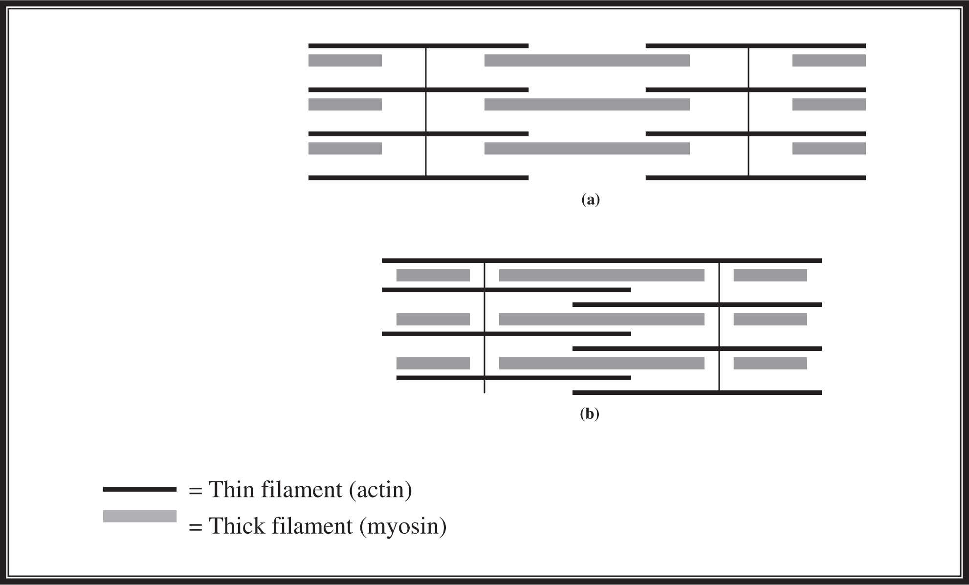
Figure 15.6 Actin-myosin interaction: (a) relaxed muscle; (b) contracted muscle.

Control mechanisms are often mentioned on the AP Biology exam and make good essay material. It would be really annoying and awkward if our muscles were contracting all the time. So, it makes sense that there must be some way to control the contraction. Myosin is only able to dance with actin if a regulatory protein, known as tropomyosin, is not blocking the attachment site on actin. The key to the removal of tropomyosin is the presence of calcium ions. Tropomyosin is also bound to another regulatory protein known as troponin. Calcium causes these two to do their own little dance and shuffle away from the actin-myosin binding site. This allows the actin-myosin dance to occur and muscle contraction to follow. When the calcium is gone, the dance is complete, and the filaments separate from each other.
What causes this calcium release seen in muscle contraction? This brings us back to the neuromuscular junction mentioned not too long ago. Nervous impulses from motor neurons cause the release of acetylcholine into the neuromuscular junction. Acetylcholine binds to the muscle cell and initiates a series of reactions that culminates in the dumping of calcium from its storage facility—the sarcoplasmic reticulum. This calcium finds troponin, binds to it, and lets the dance begin.
Endocrine System
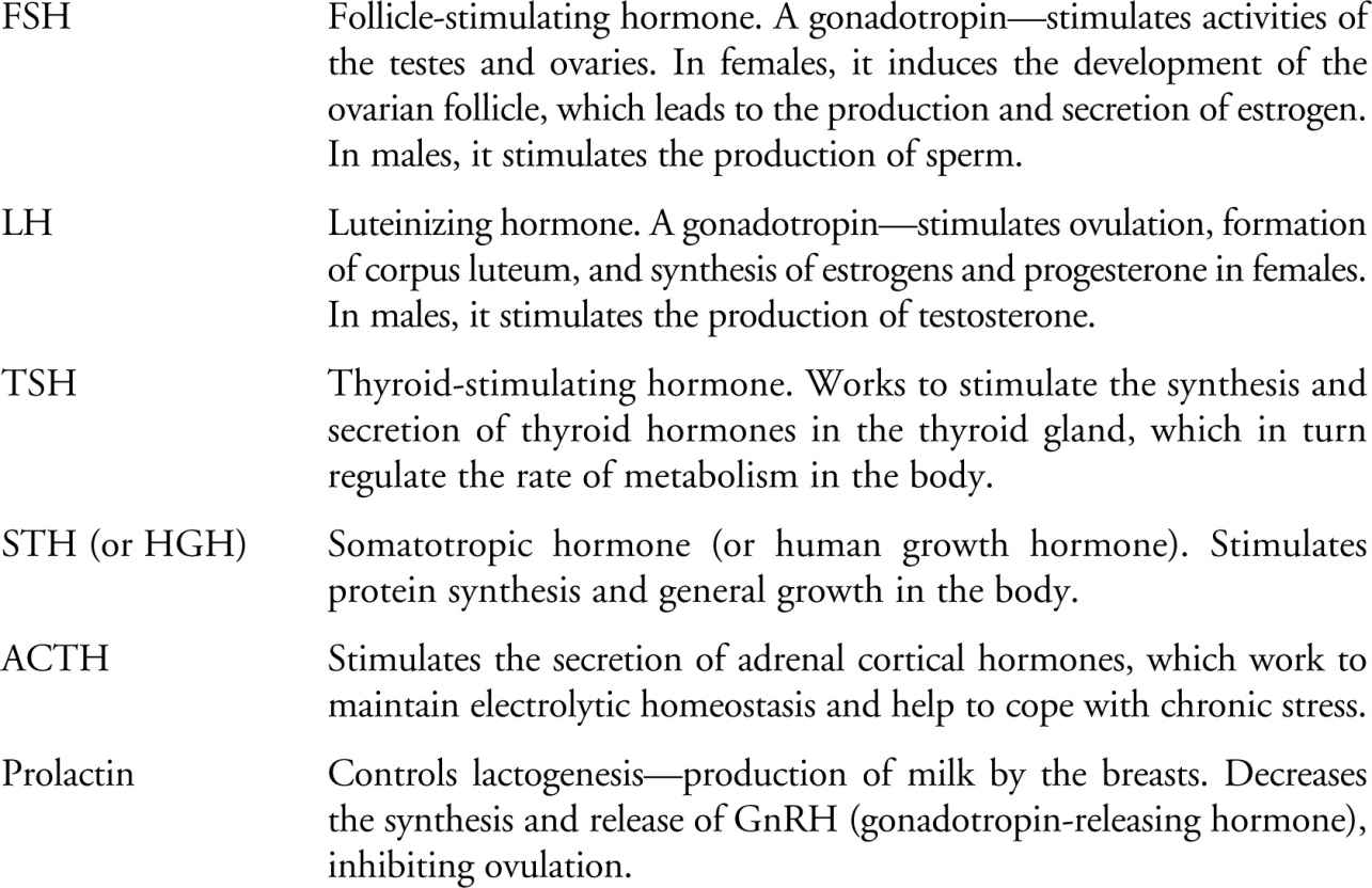
Endocrine signaling occurs when cells dump hormones into the bloodstream to affect cells in other parts of the body. Hormones are chemicals produced by glands such as the pituitary and distributed by the circulatory system to signal faraway target cells. Here we are at the pituitary gland. As you can see, it is not very big at all—it is the size of a pea and is divided into an anterior and a posterior division. The anterior pituitary gland is also called the adenohypophysis, and it produces six hormones: TSH, STH, ACTH, LH, FSH, and prolactin; the posterior pituitary gland, also known as the neurohypophysis, releases only two hormones: ADH and oxytocin (see definitions of these acronyms in the Glossary at the end of the book).
BIG IDEA 3.B.2
A variety of intercellular and intracellular signals mediate gene expression .
The two lobes of the pituitary gland differ in the way they deliver their hormones. If you look closely, you will see that there is a short stalk that connects the anterior portion of the pituitary gland to the brain. This stalk, called the hypothalamus, controls the output of hormones by the pituitary gland. The anterior pituitary is linked to the hypothalamus via the bloodstream. When the concentration of a particular anterior pituitary hormone is too low in the circulation, the hypothalamus will send releasing factors via the bloodstream that stimulate the production of the needed hormone. The posterior lobe of the pituitary gland is different—it is derived from neural tissue. Because of this, its connection to the hypothalamus is neural. ADH and oxytocin are produced by the nerve cell bodies that are located in the hypothalamus, where they are packaged into secretory granules and sent down the axons to be stored in the posterior pituitary. The posterior pituitary gland releases the hormones when appropriately stimulated by a nervous impulse from the hypothalamus. The following is a breakdown of the hormones you should be familiar with for the exam:
CT teacher: “Have a good general understanding of these hormones and their functions.”
Hormones of the anterior pituitary are
Hormones of the posterior pituitary are

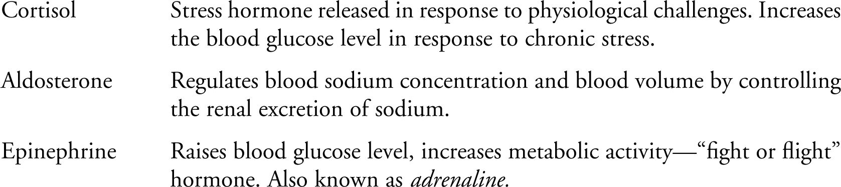
Hormones of the adrenal gland are
BIG IDEA 3.D.1
Activity within individual cells supports function for an entire organism (e.g., epinephrine stimulation of glycogen breakdown) .
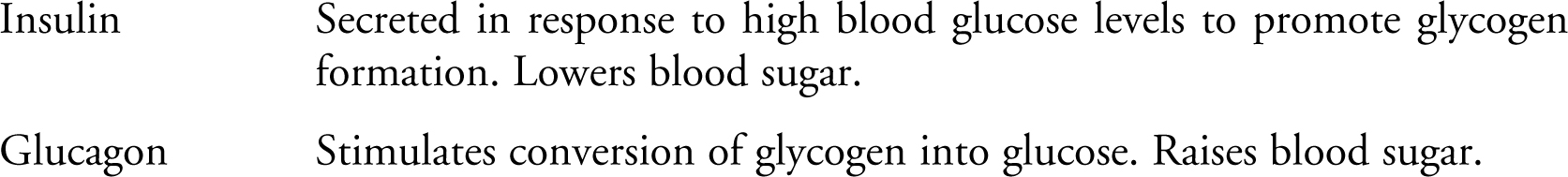
Pancreatic hormones are
The parathyroid hormone (PTH) increases serum concentration of Ca2 + , assisting in the process of bone maintenance.
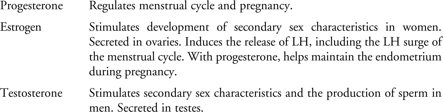
Sex hormones are
Thyroid hormones are

BIG IDEA 3.D.2
Cells communicate with each other from a distance via hormones .
The thymus hormone is thymosin, a hormone involved in the development of the T cells of the immune system.
The pineal gland hormone is melatonin, a hormone that is known to be involved in our biological rhythms (circadian). It is released at night.
How is the hormone secretion process of the body regulated? The two main types of regulation with which you should be familiar are negative feedback and positive feedback. Negative feedback occurs when a hormone acts to directly or indirectly inhibit further secretion of the hormone of interest. A good example of negative feedback involves insulin, which is secreted by the pancreas. When the blood glucose gets too high, the pancreas is stimulated to produce insulin, which causes cells to use more glucose. As a result of this activity, the blood glucose level declines, halting the production of insulin by the pancreas. Positive feedback occurs when a hormone acts to directly or indirectly cause increased secretion of the hormone. An example of this feedback mechanism is the LH surge that occurs prior to ovulation in females. Estrogen is released as a result of the action of FSH, and travels to the anterior pituitary to stimulate the release of LH, which acts on the ovaries to stimulate further secretion of estrogen.
Homeostasis
Homeostasis is the maintenance of balance. Hormones can work antagonistically to maintain homeostasis in the body. Two examples we will talk about are insulin/glucagon and calcitonin/PTH:
NYC teacher: “This could make a nice subquestion to an essay. Understand these relationships.”
1. Insulin/glucagon . Both are hormones of the pancreas and have opposing effects on blood glucose. Let’s say that you eat a nice sugary snack that pushes the blood glucose above its desired level. This results in the release of insulin from the pancreas to stimulate the uptake of glucose from the blood to the liver to be stored as glycogen. It also causes other cells of the body to take up glucose to be used for energy. Sometimes if you go a long time between meals, your blood glucose can dip below the desired level. This sets glucagon into action and causes its release from the pancreas. Glucagon acts on the liver to stimulate the removal of glycogen from storage to produce glucose to pump into the bloodstream. When the glucose level gets back to the appropriate level, glucagon release ceases. This back-and-forth dance works to keep the glucose concentration in our bodies relatively stable over time.
2. Calcitonin/PTH . Like glucose, the body has a desired blood calcium (Ca2 + ) level it tries to maintain. If it drops below this level, PTH is released by the parathyroid gland and works to increase the amount of Ca2 + in circulation in three major ways: it (a) releases of Ca2 + from bones, (b) increases absorption of Ca2 + by the intestines, and (c) increases reabsorption of Ca2 + by the kidneys. If the blood Ca2 + level gets too high, the thyroid gland releases calcitonin, which pretty much performs the three opposite responses to PTH’s work: it (a) puts Ca2 + into bone, (b) decreases absorption of Ca2 + by the intestines, and (c) decreases reabsorption of Ca2 + by the kidneys.
BIG IDEA 2.C.1
Animals use feedback to maintain homeostasis .
One last distinction we want to make before we move on is to touch on the difference between protein hormones and steroid hormones.
Protein hormones are too large to move into cells and thus bind to receptors on the surface of cells. In response to the binding of a protein hormone, a change occurs in the receptor that leads to the activation of molecules inside the cell, called second messengers, which serve as intermediaries, activating other proteins and enzymes that carry out the mission. The second messenger to know for this exam is cyclic adenosine monophasphate (cAMP), involved in numerous signal cascade pathways. Protein hormones activate cAMP through a multi-step process that begins with protein–hormone activation of relay proteins such as G proteins. These proteins are able to directly activate a compound known as adenyl cyclase, which in turn produces cAMP.
Since we discussed regulatory mechanisms earlier, it is important to point out that there are G proteins that function to inhibit cAMP and work antagonistically to hormones that activate cAMP.
Steroid hormones are lipid-soluble molecules that pass through the cell membrane and combine with cytoplasmic proteins. These complexes pass through to the nucleus to interact with chromosomal proteins and directly affect transcription in the nucleus of cells.
Immune System
What we are about to witness is an absolute treat. We just got word from the central office that the body we are touring has just received a vaccination. A vaccine is given to a patient in an effort to prime the immune system for a fight against a specific invader. This truly is a rare opportunity for us to see the immune system in action.
CT teacher: “Concentrate on the various cell types and the difference between specific and nonspecific defense.”
We have reentered the general bloodstream circulation of the body in an attempt to find some activity. While we are in transit, we will explain some basic immune system terms to you.
BIG IDEA 2.D.3
Organisms are affected by disruptions to their homeostasis (e.g., pathogens) .
The immune system is a two-tiered defense mechanism. It consists of nonspecific immunity and specific immunity. Nonspecific immunity is exactly how it sounds—it is the nonspecific prevention of the entrance of invaders into the body. Saliva contains an enzyme called lysozyme that can kill germs before they have a chance to take hold. Lysozyme is also present in our tears, providing a nonspecific defense mechanism for our eyes. The skin covering the entire body is a nonspecific defense mechanism—it acts as a physical barrier to infection. The mucous lining of our trachea and lungs prevent bacteria from entering cells and actually assists in the expulsion of bacteria by ushering the bacteria up and out with a cough. Finally, remember how we told you that you did not want to get out of the bus in the stomach? That is also the case for bacteria—it is a dangerous place for them as well. The acidity of the stomach can wipe out a lot of potential invaders.
BIG IDEA 2.C.2
Organisms respond to changes in their environment (e.g., infections) .
A nonspecific cellular defense mechanism is headed up by cells called phagocytes. These cells, macrophages and neutrophils, roam the body in search of bacteria and dead cells to engulf and clear away. Some assistance is offered to their cause by a protein molecule called complement. This protein makes sure that molecules to be cleared have some sort of identification displaying the need for phagocyte assistance. Complement coats these cells, stimulating phagocytes to ingest them. Cells involved in mechanisms that need cleanup assistance, such as platelets, have the ability to secrete chemicals that attract macrophages and neutrophils to places such as infection sites to help in the elimination of the foreign bacteria. They are nonspecific because they are not seeking out particular garbage . . . they are just looking for something to eat.
BIG IDEA 2.D.4
Animals have a variety of chemical defenses against infections that affect homeostasis .
A prime example of a nonspecific cellular response is inflammation. Let’s say that you pick up a tiny splinter as you grab a piece of wood. Within our tissues lie cells known as mast cells. These cells contain the signal histaminethat calls in the cavalry and initiates the inflammation response. Entrance of the splinter damages these mast cells, causing them to release histamine, which migrates through the tissue toward the bloodstream. Histamine causes increased permeability and blood flow to the injured tissue. The splinter also causes the release of signals that call in our nonspecific phagocytic cell friends, which come to the site of the injury to clear away any debris or pathogens within the tissue. The redness and warmth associated with inflammation occur because of the increase in blood flow to the area that occurs in this process.
The immune system also contains defense mechanisms, which are quite specific. One such defense mechanism involves a type of white blood cells called lymphocytes. There are two main flavors of lymphocytes: B cells and T cells. These cells are made in the bone marrow of the body and come from cells called stem cells. B cells mature in the bone marrow, and T cells mature in the thymus. B cells can differentiate into plasma cells and memory B cells, and the two main types of T cells are helper T cells and cytotoxic T cells. Cytotoxic T cells are the main players involved in cell-mediated immunity. Helper T cells, which assist in the activation of B cells, recognize foreign antigens on the surface of phagocytic cells and bind to these cells. After binding, they multiply to produce a bunch of T cells that pump out chemical signals, which bring in the B cells to respond.
BIG IDEA 3.D.2
Helper T cells communicate with other cells through direct contact .
We have arrived at the vaccination site in the left arm, and things are definitely heating up here. An antigen is a molecule that is foreign to our bodies and causes the immune system to respond. What is occurring right now is the process called the primary immune response. Every B cell has a specific (randomly generated) antigen recognition site on its surface. B cells patrol the body looking for a particular invader. When a B cell meets and attaches to the appropriate antigen, it becomes activated, and the B cell undergoes mitosis and differentiation into the two types of cells mentioned earlier: plasma cells and memory cells. The plasma cells are the factories that produce antibodies that function in the elimination of any cell containing on its surface the antigen that it has been summoned to kill. These antibodies, when released, bind to the antigens, immobilizing them and marking them for the macrophages to engulf and eliminate. This type of immune response falls under the category of humoral immunity —immunity involving antibodies.
BIG IDEA 4.C.1
Molecular variations in Fab enable cells to recognize a wider range of antigens .
Someone had a question? How do antibodies recognize the antigen they are designed for? Excellent question. Antibodies are protein molecules with two functional regions. One end is called the fragment antigen binding regionor Fab —this is what allows an antibody to recognize a specific antigen. It is designed by the plasma cell to have an Fab that binds to the antigen of interest. The other end, which binds to effector cells, is called the Fc region. There are five types of Fc regions, one for each of the five types of antibodies: IgA, IgD, IgE, IgM, and IgG. Each antibody type serves a slightly different function and is present in different areas of the body. When the antibodies bind to an antigen, complement gets involved, and this combination of antibodies and complement leads to the elimination of the invader.
We see a hand raised in the back. Yes, you are correct that we neglected to mention the memory cells. Very good. Memory cells contain the basis for the body’s secondary immune response to invaders. Memory cells are stored instructions on how to handle a particular invader. When an invader returns to our body, the memory cells recognize it, produce antibodies in rapid succession, and eliminate the invader very quickly. The secondary immune response is much more efficient than the primary response. This is why few people are infected by sicknesses such as chicken pox after they have had them once already—their memory cells protect them. One important fact that does come up on the exam is that the secondary immune response produces a much larger concentration of antibodies than does the primary response.
Well, this is too good to be true . . . we just got word that this body was just recently infected by a virus. This will allow us to look at the other side of the immune response: cell-mediated immunity. This type of immunity involves direct cellular response to invasion as opposed to antibody-based defense. The virus that infected this poor sap made it past the humoral immunity system because it entered into the host’s cells. This brings the cytotoxic T cells into play. The cells infected by the virus are forced to produce viral antigens, some of which show up on the surface of the cell. The cytotoxic T cells recognize these cells and wipe them out.
You might wonder how these T cells avoid killing all cells. All the cells of the body, except for red blood cells, have on their surface antigens called class I histocompatibility antigens (major histocompatibility complex [MHC]). The MHC I antigens for each person are slightly different, and the immune system accepts as friendly any cell that has the identical match for this antigen. Anything with a different MHC is foreign. This is the reason that organ donation often fails—the donor and the recipient have incompatible MHCs. There are also class II histocompatibility antigens, which are found on the surface of the immune cells of the body. These antigens play a role in the interaction between the cells of the immune system.
Well, we’d like to thank you for joining us on our tour of the body. We’ve seen a lot of things today and—whoa! We’ve been hit by something—and hit hard! Oh, dear. . . . Folks, we don’t want you to be alarmed, but it appears that our rival tour company has played a bit of a practical joke on us. Apparently as we were observing the B cell’s interaction with the vaccine, they attached a series of antigens and complement proteins to the surface of our bus. That loud noise you just heard was the sound of a macrophage taking us in . . . oh, dear, this is bad. Oh, no . . . folks, brace yourselves! The macrophage is about to ——— (transmission ended).
![]() Review Questions
Review Questions
1 . In what form is most of the carbon dioxide of the body transported in the bloodstream?
A. Complexed with hemoglobin
B. As CO2
C. As HCO3 −
D. As CO
E. As ferredoxin
For questions 2–5, use the following answer choices:
A. LH
B. FSH
C. Estrogen
D. Aldosterone
E. TSH
2 . This hormone is involved in the regulation of the body’s metabolic rate.
3 . This gonadotropin induces ovulation in females.
4 . This hormone is involved in the regulation of the body’s sodium concentration.
5 . This hormone is vital to the maintenance of the endometrium during pregnancy.
6 . The major emulsifier of fats in the digestive system is
A. lipase.
B. amylase.
C. trypsin.
D. chymotrypsin.
E. bile salts.
7 . Which cell type keeps humans from being infected by the same organism twice?
A. Plasma cell
B. Memory cell
C. Macrophage
D. Neutrophil
E. Phagocyte
8 . Which of the following muscle sites does not contain smooth muscle?
A. Aorta
B. Bladder
C. Esophagus
D. Quadriceps
E. Renal artery
9 . Which of the following is the functional unit of the respiratory system?
A. Bronchus
B. Bronchioles
C. Alveolus
D. Larynx
E. Trachea
10 . Which of the following regions of the brain controls breathing?
A. Cerebellum
B. Medulla
C. Cerebrum
D. Hypothalamus
E. Amygdala
11 . Which of the following is the major digestive enzyme of the stomach?
A. Trypsin
B. Chymotrypsin
C. Pepsin
D. Amylase
E. Sucrase
12 . Which of the following is not an example of nonspecific immunity?
A. Lysosyme of saliva
B. Skin
C. Mucous lining of the lungs and trachea
D. Lower pH of the stomach
E. Plasma cells
13 . Which of the following is true about the filtrate of the glomerulus?
A. It contains little or no glucose.
B. It contains little or no protein.
C. It contains little or no sodium.
D. It contains little or no urea.
E. It contains little or no potassium.
14 . Which of the following is not a hormone secreted by the anterior pituitary?
A. TSH
B. FSH
C. ADH
D. LH
E. STH
15 . Which of the following scenarios would be least likely to initiate a response from the sympathetic nervous system of the body?
A. Getting called on in class by the teacher when you do not know the answer
B. Seeing a cop while you are driving too fast on the highway
C. Walking through the woods and seeing a bear in the near distance
D. Waking from a midafternoon nap as sunlight strikes your face
E. Hearing a dish break on the kitchen floor right behind you
![]() Answers and Explanations
Answers and Explanations
1 . C —Most of the carbon dioxide traveling through the bloodstream of the body is in the form of the bicarbonate ion—HCO3 – . Oxygen is the one that likes to complex with hemoglobin. You should try to avoid answer choice D when possible; carbon monoxide is poisonous. Ferredoxin has nothing to do with the transport of CO2 —it is involved in photosynthesis.
2 . E —The thyroid gland is important to the maintenance of the body’s metabolic rate. An increase in TSH leads to an increase in thyroxin (thyroid hormone), which leads to an increase in the metabolic rate of the body.
3 . A —The LH surge brought about by the combined effect of FSH and LH during the menstrual cycle leads to ovulation in females. This is essentially the release of an egg from its holding pattern in the ovary that allows it to move toward the uterus.
4 . D —Aldosterone is released by the adrenal gland in an effort to maintain appropriate levels of sodium in the body. Its main site of action is the kidney.
5 . C —Estrogen and progesterone work together to maintain the endometrium, which is the site of attachment for the growing fetus during pregnancy. Without these two hormones, the endo-metrium sloughs off and is lost.
6 . E —Lipase is the major fat-digesting enzyme of our body. Bile salts help digest the fat by emulsifying it into small droplets contained in water. Amylase digests carbohydrates. Trypsin and chymotrypsin break down polypeptides.
7 . B —Memory cells are produced after the body first reacts to a foreign invader. The next time the body is exposed to that invader, it can respond much more quickly and efficiently. Plasma cells produce the antibodies designed to wipe out the antigens. Macrophages and neutrophils are both types of phagocytes, which generally roam around looking for nonspecific garbage to pick up and destroy.
8 . D —The quadriceps is the only muscle type on this list that is not a smooth muscle. It is a skeletal muscle involved in voluntary movements.
9 . C —The alveolus is the functional unit of the lung. It is the true site of gas exchange during the respiratory process. The trachea, larynx, bronchus, and bronchioles are all tubes that the air passes through on its way to this exchange center.
10 . B —The medulla oblongata controls the involuntary actions of the body, including respiration. The cerebellum controls balance, and the cerebrum is in charge of higher thinking. The hypothalamus monitors the concentration of many substances throughout the body, and determines when certain hormones of the pituitary should be released or cut down on. The amygdala controls our emotions and is associated with rage and anger.
11 . C
12 . E —Plasma cells are designed to produce antibodies that combat a particular antigen. They are a great example of specific immunity. All the other answer choices are examples of nonspecific immunity.
13 . B —The filtrate in the glomerulus contains almost everything that is in the blood plasma except for large proteins, which are unable to fit through the pores. Glucose does pass into the filtrate but is usually reabsorbed if present in normal concentrations in the blood. Sodium and potassium are always present in the filtrate. Urea is one of the major waste products that the excretory system is attempting to eliminate, so it is definitely present in the filtrate.
14 . C —ADH is secreted by the posterior pituitary.
15 . D —A sympathetic response is one that comes in a time of fight or flight. It is designed to get you ready for action. All the other choices are things that rev you up, whereas waking from a nap as sunlight strikes your face is a rather passive and tranquil experience that doesn’t usually make you want to flee the scene or fight a great battle.
![]() Rapid Review
Rapid Review
The following terms are important in this chapter:
Circulatory system: blood flow—left side of heart → aorta → via arteries to organs, muscles → into the venous system of the body (vena cava) → right side of heart → lungs (pick up O2 and release CO2) → left side of heart.
Respiratory pathway: nose/mouth → pharynx → larynx → trachea → bronchi → bronchioles → alveoli (functional unit of the lungs; this is where gas exchange occurs).
Digestive system: digestion begins in mouth, continues in the stomach, and completes in the intestines.
• Amylase: enzyme that breaks down starches in the diet (mouth and small intestine).
• Pepsin: main digestive enzyme of the stomach that breaks down proteins.
• Lipase: major fat digesting enzyme of the body (small intestine).
• Trypsin and chymotrypsin: major protein digesting endopeptidases of the small intestine.
• Bile: contains phospholipids, cholesterol, and bile salts (major emulsifier of fat).
• Maltase, lactase, and sucrase: carbohydrate digesting enzymes of the small intestines.
• Most of the digestion and absorption of food occurs in the small intestine.
• Function of the large intestine is to reabsorb water and to pack the indigestible food into feces.
Excretory system: kidneys lie on the posterior wall of the abdomen. Kidney is divided into the cortex and the medulla. The functional unit of the kidney is the nephron. The medulla is divided into renal pyramids, which dump the urine produced into the minor and major calyces → renal pelvis → bladder via the ureter → out of the body via the urethra.
• Most of what is filtered out of the glomerulus is reabsorbed—nearly all the sugar, vitamins, water, and nutrients. If sugar appears in urine, it is because there is too much in the blood (diabetes).
• Two important hormones of the excretory system are ADH (controls water absorption) and aldosterone (controls sodium reabsorption).
Muscular System
Endocrine System
Anterior pituitary hormones
• FSH: stimulates production of eggs or sperm.
• LH: stimulates ovulation, increases estrogen/progesterone release.
• TSH: increases release of thyroid hormone.
• STH: increases growth.
• ACTH: increases secretion of adrenal cortical hormones.
• Prolactin: controls lactogenesis, decreases secretion of GnRH.
Pancreatic hormones
• Insulin: increases glycogen formation.
• Glucagon: increases glycogen breakdown.
Parathyroid hormone (PTH) increases blood Ca2+ involved in bone maintenance.
Posterior pituitary hormones
• ADH: stimulates H2 O reabsorption in kidneys.
• Oxytocin: stimulates uterine contraction and milk ejection.
Adrenal gland hormones
• Aldosterone: regulates blood sodium concentration.
• Cortisol: chronic stress hormone.
Sex hormones
• Progesterone: involved in menstrual cycle and pregnancy.
• Estrogen: made in ovaries; increases release of LH (LH surge); develops female secondary sex characteristics.
• Testosterone: (testes): stimulates sperm production; develops male secondary sex characteristics.
Negative feedback: hormone acts to directly, or indirectly, inhibit further release of the hormone of interest.
Positive feedback: hormone acts to directly, or indirectly, cause increased secretion of the hormone.
Nervous system: divided into two parts: central nervous system (CNS) and peripheral nervous system (PNS).
• SNS: controls skeletal muscles and voluntary actions.
• ANS: controls involuntary activities of body.
• ANS: sympathetic (prepare for fight): increased heart rate, increased blood pressure, digestive slowdown, dilate bronchial muscles; parasympathetic (conserve energy): decreased heart rate, decreased blood pressure, bladder constriction.
• Brain: cerebellum (coordination/balance); medulla (involuntary actions such as breathing); hypothalamus (regulates hunger, thirst, temperature); amygdala (emotion control center).
Immune system:
• Nonspecific immunity: nonspecific prevention of entrance of invaders into the body (skin, mucus).
• Specific immunity: multilayered defense mechanism: (1) first line of defense—phagocytes, macrophage, neutrophils, complement; (2) second line of defense: B cells (plasma/memory), T cells (helper/cytotoxic).
• Primary immune response: antigen invader → B cell meets antigen → B cell differentiates into plasma cells and memory cells → plasma cells produce antibodies → antibodies eliminate antigen (humoral immunity ).
• Secondary immune response: antigen invader → memory cells recognize antigen and pump out antibodies much quicker than primary response → antibodies eliminate antigen.
• Cell-mediated immunity: involves T cells and direct cellular response to invasion. Defense against viruses.
CHAPTER 15
Human Physiology
1 . Filtration in the kidneys occurs at the level of the
(A) albumin.
(B) glomerulus.
(C) epithelial cells.
(D) ureter.
2 . Which type of muscle is found in the walls of arteries, the digestive tract, and the bladder?
(A) Skeletal muscle
(B) Striated muscle
(C) Smooth muscle
(D) Cardiac muscle
3 . Which of the following lobes of the brain controls vision?
(A) Frontal lobe
(B) Parietal lobe
(C) Occipital lobe
(D) Temporal lobe
4 . Which of the following hormones is known to stimulate uterine contraction and milk ejection?
(A) FSH
(B) Prolactin
(C) Cortisol
(D) Oxytocin
![]() Answers and Explanations
Answers and Explanations
1 . B —The glomerulus has small holes that allow small molecules and water to pass through, but it keeps back cells and large proteins.
2 . C —Smooth muscle is involuntary muscle that contracts slowly and is controlled by the autonomic nervous system. It is NOT striated in appearance.
3 . C —The frontal lobe controls speech and the motor cortex. The parietal lobe controls speech, taste, reading, and somatosensory functions. The temporal lobe controls hearing and smell. The occipital lobe controls vision.
4 . D —FSH stimulates the production of eggs or sperm. Prolactin controls lactogenesis and decreases the secretion of GnRH. Cortisol is the chronic stress hormone. Oxytocin is the winner here.