CCEA GCSE Biology - Denmour Boyd, James Napier 2017
Unit 1
Cells
Specification points
This chapter covers specification points 1.1.1 to 1.1.11. It covers the structures of animal, plant and bacterial cells; using a light microscope; magnification and size; resolution and the electron microscope; movement of substances into and out of cells by diffusion; stem cells and specialisation of cells in multi-celled organisms.
In Double Award Science, it covers specification points 1.1.1 to 1.1.5 and covers the structures of animal, plant and bacterial cells, and the specialisation of cells in multi-celled organisms.
Cells
Living organisms are made up of microscopic units called cells.
Animal cells
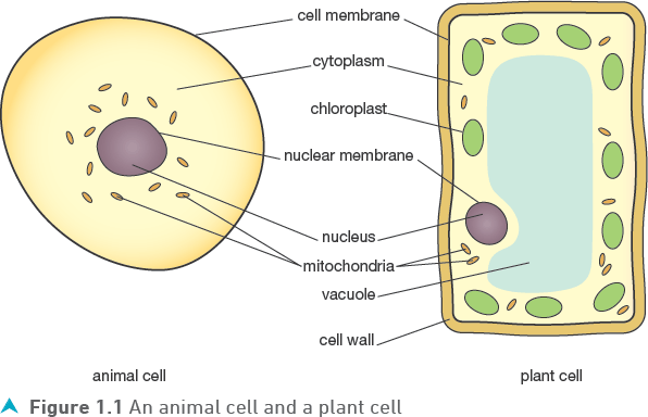
Animal cells are surrounded by a selectively permeable cell membrane that forms a boundary to the cell and controls what enters or leaves. The main part of the cell is the cytoplasm and this is where chemical reactions take place.
The nucleus, surrounded by a nuclear membrane, contains several threadlike structures: the chromosomes. Each chromosome is made of a molecule of DeoxyriboNucleic Acid (DNA) which has part of the organism’s genetic information coded in its structure. For this reason, the nucleus is sometimes referred to as the control centre of the cell.
![]()
Mitochondria are structures in the cytoplasm within which the chemical reactions of cell respiration take place.
Tip
Cells like muscle cells need a lot of energy and so have many more mitochondria in their cytoplasm.
Plant cells
Like animal cells, plant cells have a cell membrane, cytoplasm with mitochondria and a nucleus containing chromosomes. However, in addition they have:
• a cellulose cell wall, which is a stiff structure immediately outside the cell membrane that provides support
• a large permanent vacuole in the cytoplasm containing cell sap, that when full pushes the cell membrane against the cell wall, making the cell rigid and providing more support
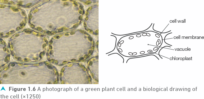
• chloroplasts in the cytoplasm that contain chlorophyll, which traps light and helps the plant make food during photosynthesis. Chloroplasts are not found in all plant cells — they are only present in green parts of the plant, particularly the leaves.
Bacterial cells
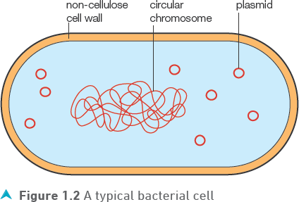
Bacteria are microscopic single-celled organisms (microorganisms). They are neither plant nor animal, largely because their cell structure is very different. They have a cell membrane surrounding cytoplasm but there is no nucleus. The genetic material (DNA) is in the form of a circular chromosome within the cytoplasm. Small rings of DNA, called plasmids, are also present, as shown in Figure 1.2. A cell wall is present but it is not made of cellulose.
Test yourself
1 Draw a table to compare the structures found in animal, plant and bacterial cells.
2 What is the function of:
a) a cell membrane
b) a chloroplast
![]()
c) a mitochondrion?
3 Give two structures in bacterial cells that contain DNA.
Observing cells using a microscope
Although a few cells, like birds’ eggs, are large and can be seen with the naked eye, the vast majority can only be seen using a microscope. In school, you will use a light microscope in which light shines through a thin layer of cells on a slide.
Making slides
Most slides containing animal cells will already be prepared for you but you should get a chance to prepare temporary slides containing animal and plant cells (pages 6 and 7). Figure 1.3 outlines the procedure for making a temporary slide containing onion skin cells.
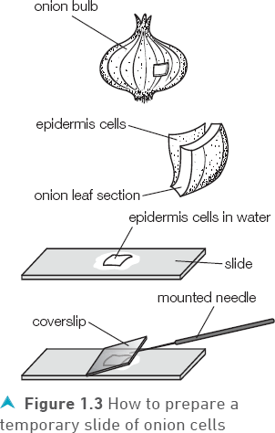
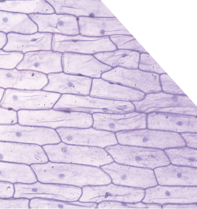
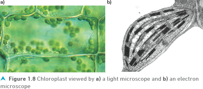
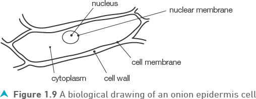
Forceps are used to peel the thin, transparent layer of epidermis cells from the inside of a small section of onion leaf. The epidermis cells are then placed on a microscope slide with a few drops of water. Iodine solution or methylene blue can be used instead of water — these chemical stains colour the cells, making certain parts such as the nucleus more obvious.
A coverslip is then lowered onto the onion epidermis using a mounted needle or forceps. It is better to lower the coverslip one end first as this helps to prevent trapping air bubbles. The coverslip will protect the lens of the microscope should it have contact with the slide and will prevent the cells drying out.
Tip
Sometimes you will see black rings in your slide preparation — these are probably air bubbles trapped under the coverslip.
Tip
When moving the slide on the stage to focus on a different part of the field of view, moving the slide away from you moves what you see towards you and vice versa.
Using a microscope
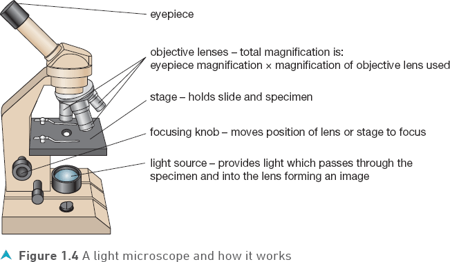
When using a light microscope (Figure 1.4), it is important to start by using the low power objective lens. With the low power lens (×4 or ×10) the circle of light you see in the eyepiece (the field of view) is wider and allows you to see more of the cells on the slide. This makes it easier to find what you are looking for and to focus a sharp image.
After you observe the cells at low power you may want to see some of them in more detail. To do this, first move the slide slightly so that the cell or area of cells you want to look at in greater detail is in the centre of the field of view. One of the larger, high power, objective lenses (×20 or ×40) can then be carefully rotated into place and focused.
Great care is needed while rotating a high-power lens into position above the slide as the end of the high-power lenses are very close to the slide and can be damaged while focusing.
Test yourself
4 Why are chemical stains used in temporary slides?
5 What is the field of view?
6 If the microscope you are using has an eyepiece with ×10 magnification, what is the total magnification of the image you see when using an objective lens with a magnification:
a) ×10?
b) ×40?
Show you can
Describe how to use a microscope to observe a specimen on a slide.
![]()
Example
a) Incorrectly drawn scratchy lines with gaps
b) Correctly drawn continuous lines with no gaps

Recording what you see in the microscope
Drawing
Although it is possible to take digital photographs with some microscopes, drawing remains the usual way of recording the cell(s) you see in a microscope. Good biological drawings use biological knowledge to select the amount of detail to include and are:
• made in pencil with lines that are firm, continuous and not ’scratchy’ with gaps
• large, have the same proportions and are a faithful representation of the cell(s) being observed
• labelled using separate ruled lines, spread out around the drawing with each line starting as a bullet point on the structure and ending with a clearly written label
• given a title including some idea of its magnification or size.
Magnification of a drawing or photograph
When a photograph is taken or a drawing is made of the cell you see in the microscope, the magnification is bigger again. Magnification in this situation is the number of times the length of the image (in the photograph or drawing) is larger than the actual length of the cell and can be summarised by the relationship:
![]()
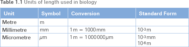
The relationship depends on the measurement of length which should use the same units for both the Length of Image and the Actual length. The units should also be appropriate for what is being measured. The units of measurement most often used in Biology are the metre (m), millimetre (mm) and micrometre (µm).
The length of a cell is most appropriately measured in micrometres, but the measurements you can make from photographs or drawings will be in millimetres, so you will need to be able to convert millimetres into micrometres. To do this multiply the number of millimetres by 1000.
![]()
Examination questions usually ask you to calculate either the Actual length (size) or the Magnification of a cell or organism. If you remove what you are asked to calculate from the relationship, then the relationship tells you what to do in the calculation, for example:

Test yourself
7 What is the relationship between the length of an image, its magnification and its actual length?
8 If the image of a cell measures 15 mm, how many micrometres does that convert to?
9 If the image of a cell in a photograph measures 5 mm in diameter and the magnification is given as ×400, what is the actual diameter of the cell?
![]()
Calculating magnification using a scale bar
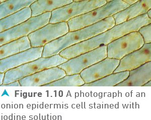
In some images, the magnification is represented by a line, called a scale bar, drawn on or near the photograph or drawing, as in Figure 1.7. A scale bar has a label showing the Actual length of the bar before it was magnified in the image. The length of the scale bar Image can therefore be measured in millimetres, converted to micrometres and divided by the Actual length to calculate the Magnification.
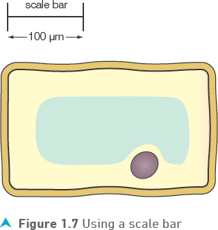
Example
Calculate the magnification of the cell in Figure 1.7
Electron microscopes
Although magnification is an important property of microscopes, continually increasing the magnification of an image beyond a certain point does not increase the detail that you can see. If you look at a digital photograph with greater and greater magnification, it eventually appears as a matrix of coloured squares and the detail is lost. The ability of a microscope to let us see detail is its resolution. The best light microscopes can resolve details which are 0.2 µm apart and require a magnification of about ×1500 so that our eyes can see this detail. This is enough to see larger cell structures.
Test yourself
10 What is the resolution of a microscope?
Biologists use electron microscopes, which pass a beam of electrons through a specimen, to investigate the detail of the structures inside cells. Electron microscopes have much greater resolution, being able to show details that are about ![]() µm apart.
µm apart.
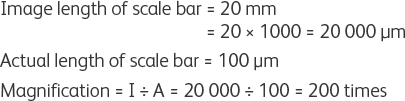
The highest-powered electron microscopes can even show the detail of large molecules such as proteins. To see this level of detail with our eyes we need to use magnifications up to ×500 000. Figure 1.8 shows that when using a light microscope to observe a chloroplast, too much magnification makes the image ’fuzzy’ but when an electron microscope is used, the membranes around and inside each chloroplast can clearly be distinguished because of the higher resolution.
Show you can
Explain why simply increasing the magnification of a light microscope image does not necessarily help our understanding of cell structures.
![]()
Tip
Electron microscopes are large, very expensive and require special rooms in a laboratory. Another disadvantage is that the techniques used to prepare tissues for observation are complex and cause the death of the tissue.
![]()
Tip
Practical 1.1 is not prescribed for Double Award Science students, although, they are expected to make a temporary slide and use a light microscope to examine and identify the structures of a typical plant and animal cell.
Prescribed practical
Biology Practical 1.1: Using a light microscope to examine and identify the structures of a typical plant and animal cell and record them in a drawing
Observe and record the structures of an onion skin cell
Procedure
1 Prepare a temporary slide of onion epidermis cells, stained with iodine solution. Remember to wear eye protection.
2 Observe the cells on the slide using the low power objective lens of the microscope and select an area which has a single layer of cells.
3 Change to a high power objective and select a cell which shows clearly most of the following structures:
• cell wall
• nucleus
• cytoplasm
• vacuole.
4 Make a large biological drawing of the cell and label the structures you observe.
Sample results and questions
1 Make a large biological drawing of the cell.
2 Explain the benefit of staining the cell.
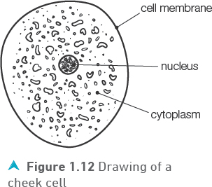
Observe and record the structures of a cheek cell
Procedure
1 Rub a clean cotton bud gently over the inside of your cheek. Remember to wear eye protection.
2 Smear the end of your cotton wool bud on the surface of a clean microscope slide. Dispose of the cotton bud safely.
3 Add one drop of methylene blue stain to the smear and slowly lower a cover slip on top of the methylene blue.
4 Observe the cells on the slide using the low power objective lens of the microscope and select an area which has cells well spread out.
5 Change to a high power lens and observe one or two cells in detail.
6 Make a large biological drawing of the cheek cell and label the structures you observe.
Sample results
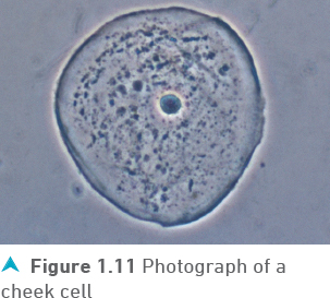
![]()
Movement of substances into and out of cells
Cells need substances such as oxygen, water, dissolved food molecules and mineral ions to be able to enter through the cell membrane, while carbon dioxide and nitrogen wastes need to be able to pass out. At the same time, the cell membrane prevents the passage of other molecules. For this reason, the cell membrane is described as selectively permeable.
Many of these substances move by diffusion, the random movement of a substance (usually a simple, soluble molecule) from where it is in a high concentration to where the concentration is lower. Three factors affect the rate of diffusion:
• The difference in the concentration between the high and low concentration areas is the concentration gradient. The larger the difference, the faster the diffusion.
• Temperature is another factor which affects diffusion. Higher temperatures increase the kinetic energy of the molecules, making them move faster and increasing the rate of diffusion.
• The larger the surface area through which the diffusion takes place, the faster diffusion occurs.
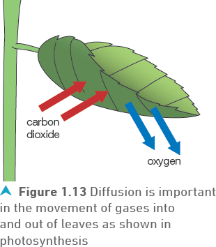
Diffusion, (Figure 1.13), is particularly important in gas exchange, the movement of oxygen and carbon dioxide between the air and living organisms, for example in leaves (page 17) and in the lungs (page 38).
![]()
The effect of surface area and volume on diffusion
The rate at which substances diffuse into and out of cells depends on several factors, most importantly the surface area of the cell and its volume. Figure 1.14 shows how surface area and volume change as a cell becomes larger.
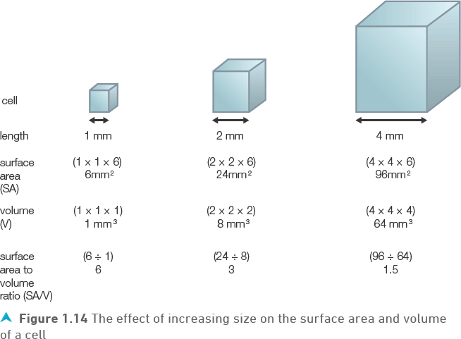
The importance of these changes is that the larger the cell volume, the greater the amount of substances (such as oxygen) the cell needs for its reactions. At the same time, the larger the surface area the greater the amount of oxygen that can diffuse into the cell. Although the calculations in Figure 1.14 show that both the volume and surface area increase with size, the volume increases faster. This means that the surface area available to allow oxygen in for each mm3 of cell volume (SA/V) halves each time the cell doubles in size. Eventually, a cell will be so large that there is not enough surface area to allow in the oxygen that the cell needs and it will die. For this reason, there is a limit on the size of single-celled organisms.
Multi-celled organisms
Most organisms have become multi-celled, making them much larger than single cells. However, as we have learned, organisms need a large enough surface area (large SA/V ratio) to be able to efficiently exchange substances with their environment. Being multi-celled does not solve this problem by itself as the body cells are closely packed together, increasing the volume but not the surface area.
Tip
A further problem in multi-celled organisms is communication between cells in different parts of the organism, which has led to the development of nerves and hormones.
Large active multi-celled organisms have developed special gas exchange organs inside their body (lungs) which have a greatly increased surface area. This creates another problem — the gas exchange surface is often far away from many of the other body cells. A transport system such as the circulatory system has therefore developed in many organisms.
Tissues, organs, organ systems and organisms
In single-celled organisms all the life processes are carried out by the one cell but in multi-celled organisms the cells differentiate and become specialised. They develop a structure which best adapts them to the function they carry out, for example red blood cells (page 88) and plant palisade cells (page 18).
Specialised cells are not just randomly spread through an organism but are organised into groups in an effective manner. There are different levels of organisation:
1 Cells with the same specialised structure and function are grouped together and are called a tissue. Examples of animal tissues include blood and skin.
2 An organ is a structure made of several types of tissue that carries out a particular function:
✵ the heart contains muscle, nerve and blood tissues and its function is to pump blood around the body
✵ the leaf is an example of an organ in a plant which contains several different tissues and has the function of photosynthesis.
3 Organs which operate together to carry out a particular function are linked together into organ systems. Table 1.2 summarises three of the organ systems in humans.
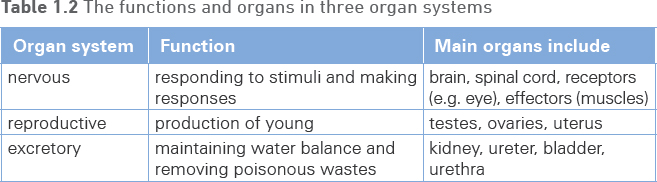
![]()
Stem cells
During the very early stages in the growth of living organisms, the embryo contains only a few unspecialised cells. They are called stem cells. These simple cells have, unlike other cells, two important abilities:
• to continue dividing by cell division to produce more stem cells
• to differentiate into a wide variety of specialised cell types.
Embryonic stem cells of this type can be collected from embryos not used in fertility treatments or from the umbilical cord and placenta. In adult animals, most stem cells have changed permanently into specialised cells and lost their ability to divide and specialise further. However, it is possible to collect stem cells from some adult tissues like bone marrow, but they can only differentiate into different types of blood cells.
In plants the situation is slightly different. Stem cells are found in each of the apical growing points or meristems at the end of shoots and roots, shown in Figure 1.15. The cells they produce retain the ability to divide until they are in their final position in the root, stem or leaf and even then, under certain conditions the specialisation can be reversed. This property has allowed scientists to develop techniques of cloning capable of producing large numbers of genetically identical plants in a very short time.
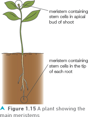
![]()
The use of stem cells in medicine
The use of stem cells is becoming increasingly important in medicine as it offers the potential benefits of treatments for a range of diseases. One example is the treatment of the cancer of white blood cells, leukaemia. In this cancer, the white blood cells produced in the bone marrow function abnormally and lose the ability to fight disease. The first stage of the treatment uses chemotherapy and radiotherapy to destroy the cancerous white blood cells in the patient’s body. This also stops the patient’s bone marrow producing new blood cells. The patient is then given a transplant of bone marrow containing stem cells from a matching healthy donor. If successful, these donor stem cells will multiply and produce healthy blood cells in the patient.
However, there are risks with this type of treatment.
• The use of chemotherapy and radiotherapy to destroy the patient’s white blood cells before the stem cell transplant leaves the patient with no immune system and in danger of infections from the environment or from the donor.
• The stem cells may divide in an uncontrolled way and produce tumours or unwanted cell types.
Some people also have ethical issues with using embryos as a source of stem cells as it necessitates the destruction of the embryos. For that reason the government carefully controls the limits of research in this area.
New research, including stem cell research, is validated (or rejected) by other scientists, expert in the subject area. This process of peer review involves a rigorous examination of new scientific advances.
Test yourself
11 Where are stem cells found in humans and in plants?
12 Why are stem cells different from other types of cells?
13 Give one advantage of cloning plants.
Practice questions
1 An understanding of stem cells is increasingly important.
a) Describe two ways stem cells are different from normal cells.
(2 marks)
b) Give two places from which stem cells can be collected.
(2 marks)
c) Explain one reason why some people have ethical issues with the use of stem cells.
(1 mark)
d) Stem cells can be used in the treatment of the blood cancer leukaemia, where the white blood cells lose the ability to fight disease.
i) Explain how a stem cell transplant is used in this treatment.
(1 mark)
ii) Chemotherapy and radiotherapy are used before the stem cell transplant. Explain why this increases the risk to the patient.
(1 mark)
iii) Give one other risk caused by a stem cell transplant.
(1 mark)
2 Figure 1.16 shows an image of a bacterium and a scale bar to indicate its magnification.
a) Give two features of a bacterial cell which are not found in a plant cell.
(2 marks)
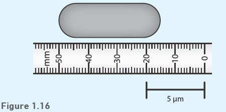
b) Use the scale bar and the millimetre scale to calculate the magnification of the bacterium.
(3 marks)
3 a) What is diffusion?
(2 marks)
b) Explain how temperature affects diffusion.
(2 marks)
4 a) What happens to the surface area, the volume, and the surface area to volume ratio, of a cell as it grows larger?
(3 marks)
b) Explain why these changes are important for the survival of a cell.
(2 marks)
c) i) What are the benefits of differentiation to multi-celled organisms?
(3 marks)
ii) Give one example of a tissue, an organ and an organ system.
(3 marks)
d) Explain why multi-celled organisms have developed special gas exchange surfaces.