CONCEPTS IN BIOLOGY
PART V. THE ORIGIN AND CLASSIFICATION OF LIFE
21. The Nature of Microorganisms
21.3. The Kingdom Protista
The kingdom Protista is a taxonomic category of convenience. Scientists do not actually think that the organisms in this group are closely related to one another. The only characteristic they share is that they are the simplest eukaryotic organisms. Many of them are single-celled, but others are multicellular and show a degree of cellular specialization. The ancestors of this group are thought to have formed about 2 billion years ago.
There is great diversity among the more than 250,000 species. Because of this diversity, it is a constant challenge to separate the kingdom Protista into meaningful subgroups. Furthermore, research continually reveals new evidence about the members of this group and their evolutionary relationships, requiring changes in taxonomy. Usually, the species are divided into three general groups, based on their mode of life: algae, autotrophic unicellular organisms; protozoa, heterotrophic unicellular organisms; and funguslike protists. These categories are helpful in discussing the major roles of these organisms but do not reflect how the organisms have evolved. For example, many kinds of flagellated protists are photosynthetic, but other, closely related organisms are not. Thus, two closely related organisms would be placed in the categories protozoa or algae, depending on their ability to perform photosynthesis. Figure 21.7 shows some current ideas about how some of these groups are related.
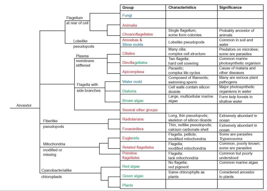
FIGURE 21.7. Relationships Among Members of the Protista
In this figure, organisms that are photosynthetic are shown in green and are generally known as algae. Those that are shown in red are typically hetertrophs and are generally known as protozoa. Those shown in blue are funguslike. Some groups, such as the Euglenids and Dinoflagellates, have some members that are photosynthetic and others that are not.
Algae
Algae are protists that contain chlorophyll in chloroplasts and therefore carry on photosynthesis. Many of these organisms are single-celled, but some groups are multicellular. Although most live in the ocean and bodies of freshwater, they can also be found in other moist places, such as soil and the surface of other organisms in rainforests and other moist habitats. Plankton is a collection of small, floating or weakly swimming organisms. Algae are the major components of the phytoplankton that consists of the photosynthetic plankton that is the basis of most aquatic food chains. Cyanobacteria are the other major component of phytoplankton. Zooplankton consists of nonphotosynthetic plankton, including aquatic protozoa and tiny animals. Benthic organisms live attached to the bottom or to objects in the water. Many benthic algae form denses “forests” of large seaweeds in shallow sea water. The large number of benthic and planktonic algae makes them an important source of atmospheric oxygen (O2). It is estimated that over 50% of the oxygen in the atmosphere is produced by marine algae. Furthermore, because algae are important producers in marine food chains, disruptions to the marine algal community can have serious implications for the production of fish and shellfish.
Because algae require light, they are found only near the surface of the water. Even in the clearest water, photosynthesis does not usually occur any deeper than 100 meters. Benthic forms are found in shallow water and are common along the ocean shoreline. Some phytoplankton have flagella or other methods of locomotion, which assist them in remaining near the surface. Others maintain their position by storing food as oil, which is less dense than water and enables the cells to float near the surface.
The various kinds of algae reproduce both sexually and asexually. However, their primary method of reproduction is asexual cell division. Like the cyanobacteria, in warm, nutrient-rich waters, various kinds of algae can reproduce rapidly by asexual reproduction and cause an algal bloom. The population can become so large that clumps of algae float on the surface or in the case of single-celled algae, the water may become colored or murky.
Single-Celled Algae
There are several common kinds of single-celled algae. Euglenids are single-celled algae that move by flagella. They have an outer covering, called a pellicle, which gives them shape but is flexible. Euglenids vary in terms of their metabolism. Some lack chloroplasts and are heterotrophs. Others have chloroplasts and are autotrophs. However, even among those that have chloroplasts, many are able to consume food and behave as heterotrophs, particularly when light levels are low. Most of them, like the common Euglena, are found in freshwater. They are widely studied because they are easy to culture.
Diatoms are extremely common single-celled algae found in freshwater, marine, and soil environments. They are a major component of the phytoplankton of the oceans and serve as a food source for zooplankton and many kinds of filter-feeding organisms, such as whales, clams, and barnacles. A few species are heterotrophs or parasites. They are typically brownish in color. Although they do not have cilia or flagella, they are able to move with a sort of gliding motion. They are unique because their cell walls contain silicon dioxide (silica). The walls fit together like the lid and bottom of a shoe box; the lid overlaps the bottom. The silica-containing walls have many pores, which form interesting patterns. Because their cell walls contain silicon dioxide, they readily form fossils. The fossil cell walls have many tiny holes and can be used in a number of commercial processes. They are used as filters for liquids and as abrasives in specialty soaps, toothpastes, and scouring powders.
Dinoflagellates, along with diatoms, are important food producers in the ocean’s ecosystem. They are also very common in freshwater and brackish water. All members of this group of algae have two flagella, which is the reason for their name (di = two), and an outer cellulose covering made up of plates. Although many dinoflagellates are photosynthetic, some are heterotrophs and others are parasites. Some change from autotroph to heterotroph, depending on environmental conditions.
Some species of dinoflagellates have symbiotic relationships with marine animals, such as the reef corals; the dinoflagellates provide a source of nutrients for the reef-building coral. Corals that live in the light and contain dinoflagellates grow 10 times faster than corals without this symbiont. Thus, in coral reef ecosystems, dinoflagellates form the foundation of the food chain.
Some forms of dinoflagellates produce toxins. Because many of the toxin-producing dinoflagellates are reddish in color, a bloom of these organisms is called a red tide. Often, fish and other vertebrates such as birds and mammals are killed by exposure to the toxins. Although the toxins do not seem to harm shellfish, such as oysters, consuming the toxin along with the shellfish can cause sickness or death. Red tides usually occur in the warm months and are more common in tropical and semitropical waters. During red tide episodes, people are warned not to swim in areas that have a red tide or harvest fish or shellfish for food. Commercially available shellfish are tested for toxin content; if they are toxic, they are not marketed.
The dinoflagellate Pfiesteria piscidia has been responsible for the death of millions of fish in estuaries of the eastern United States. These dinoflagellates release toxins that paralyze fish. The dinoflagellates then feed on the fish. They have also been responsible for human and wildlife poisoning. It appears that blooms of these organisms may be triggered by high amounts of nutrients in the water as a result of runoff from feedlots and agricultural land.
Many marine forms of dinoflagellates are bioluminescent; they are responsible for the glow seen at night in ocean waves or in a boat’s wake. Figure 21.8 shows examples of euglenids, diatoms, and dinoflagellates.
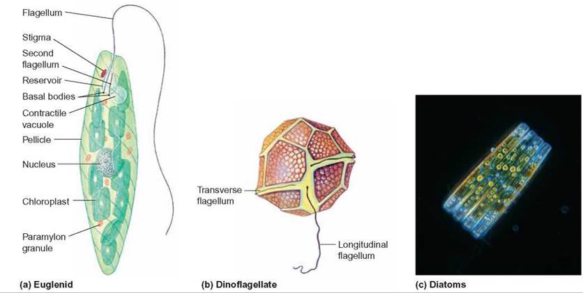
FIGURE 21.8. Single-Celled Algae
Three very common kinds of single-celled algae are the euglenids, dinoflagellates, and diatoms.
Multicellular Algae
Many kinds of algae are multicellular and can be quite large, with some specialization of cells and body parts. These algae are commonly known as seaweed. They are found in shallow water attached to objects. Two types, red algae and brown algae, are mainly marine forms. The green algae are primarily freshwater species.
Red algae live in warm oceans and attach to the ocean floor by means of a holdfast structure. They are found from the splash zone, the area where waves are breaking, to depths of 100 meters. Some red algae become encrusted with calcium carbonate and are important in reef building. Other species are commercially important, because they produce agar and carrageenin. Agar is widely used as a jelling agent for growth media in microbiology. Carrageenin is a gelatinous material used in paints, cosmetics, and baking. It is also used to make gelatin desserts harden faster and ice cream smoother. In Asia and Europe, some red algae are harvested and used as food.
Brown algae are found in cooler marine environments. Most species of brown algae have a holdfast organ. Colonies of these algae can reach 100 meters in length. Brown algae produce alginates, which are widely used as stabilizers in frozen desserts, as emulsifiers in salad dressings, and as thickeners to give body to foods such as chocolate milk and cream cheeses; they are also used to form gels in such products as fruit jellies.
The Sargasso Sea is a large mat of free-floating brown algae between the Bahamas and the Azores. It is thought that this huge mass (as large as the European continent) is the result of brown algae that have become detached from the ocean bottom, have been carried by ocean currents, and have accumulated in this calm region of the Atlantic Ocean. This large mass of floating algae provides a habitat for a large number of marine animals, such as marine turtles, eels, jellyfish, and innumerable crustaceans. Figure 21.9 shows examples of red and brown algae.
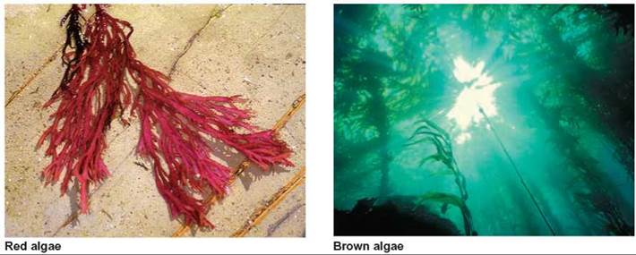
FIGURE 21.9. Red and Brown Algae
Red and brown algae are primarily marine organisms. Most of them grow attached to the ocean bottom or other organisms in their environment.
Green algae are found primarily in freshwater ecosystems, although a few kinds live in oceans. Some are single-celled and have flagella; some lack flagella and form strings, which either float in the water or grow on surfaces. The members of this group can also be found growing on trees, in the soil, and even on snowfields in the mountains. Like land plants, green algae have cellulose cell walls and store food as starch. Green algae also have the same types of chlorophyll as do plants. Biologists believe that land plants evolved from the green algae. Figure 21.10 shows a variety of green algae.
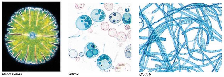
FIGURE 21.10. Green Algae
Some green algae are single-celled; others form colonies.
Protozoa
Protozoa are members of the kingdom Protista; they are eukaryotic, heterotrophic, single-celled organisms that lack cell walls. Generally, protozoa lack all types of chlorophyll, but some organisms may contain chloroplasts at some times in their lives and lack them at others. One common way to classify the protozoa into subgroups is by their method of locomotion. Although this is a convenient way to subdivide the organisms for the purposes of discussion, it is clearly not a valid phylogenic grouping.
Flagellates
Flagellates are an extremely diverse group of organisms that have flagella and lack cell walls and chloroplasts. They live in any moist environment, including marine waters and freshwater, moist soil, and as parasites or symbionts. Some flagellates have an extremely simple structure, suggesting that they may be the most primitive of all eukaryotic organisms. Some feed by absorbing simple organic molecules through their cell membranes; others engulf food particles or other organisms.
Many kinds of flagellates are mutualistic or parasitic. Termites are insects that eat wood but cannot digest it. Their guts contain mutualistic flagellated protozoa capable of digesting cellulose. Thus, the termite benefits from a food source and the flagellate benefits from a good place to live and a continuous supply of food.
There are many examples of parasitic flagellates (figure 21.11). One is Trichomonas vaginalis, that can live in the reproductive tract of both men and women and is the cause of a common sexually transmitted disease. Often, it doesn’t cause any symptoms but sometimes causes itching and a discharge. The symptoms are more common in women than men. Trypanosomes, which cause sleeping sickness in humans and domestic cattle, primarily in Africa are another example of a parasitic flagellate. The parasite develops in the circulatory system and moves to the cerebrospinal fluid surrounding the brain. When this occurs, the infected person develops the “sleeping” condition, which, if untreated, is eventually fatal.
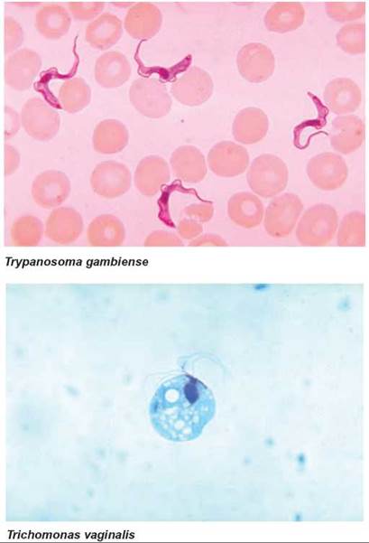
FIGURE 21.11. Flagellates
Several flagellated protozoa are parasites. Trypanosoma gambiense causes sleeping sickness. It is shown here among red blood cells. Trichomonas vaginalis is the cause of a common sexually transmitted disease.
Giardia lamblia is a flagellated protozoan that contaminates freshwater throughout the world. Because Giardia is a common intestinal parasite of deer, beaver, and many other animals, even “pure” mountain streams in wilderness areas are likely to be contaminated. Infection usually causes diarrhea, intestinal gas, and nausea, although it does not usually cause life-threatening illness. The most effective way to eliminate the spores formed by this protozoan is to filter out particles as small as 1 micrometer from the water or boil it for at least 5 minutes before drinking.
Choanoflagellates are colonial flagellates that many biologists believe are ancestral to all multicellular animals, because the simplest animals, sponges, contain cells that are extremely similar in structure to free-living choanoflagellates.
Amoeboid Protozoans
Amoeboid protozoans have extensions of their cell surface called pseudopods in which the cytoplasm flows. They range from the well-known Amoeba, with its constantly changing, lobelike pseudopods to species with thin, fiberlike pseudopods (figure 21.12). Most amoeboid protozoans are free-living and feed on bacteria, algae, or even small, multicellular organisms. Amoeba uses pseudopods to move about and to engulf food.
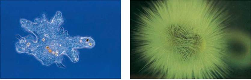
FIGURE 21.12. Amoeboid Protozoa
Amoeboid protozoa have extensions of their cell surface called pseudopods. Pseudopods contain moving cytoplasm. Some, such as Amoeba, have large, lobelike pseudopods, which change shape as the cell moves and feeds. Others have long, filamentous pseudopods that trap organisms and transport food molecules to the central cell from the objects they feed on.
Some forms are parasitic. Entamoeba histolytica is responsible for the disease known as amoebic dysentery. People become infected with this protozoan when they travel to parts of the world that have poor sewage and water treatment facilities and often have contaminated water.
Radiolarians and foraminiferans are two specialized groups of amoeboid protozoans that are extremely common in the oceans. Both kinds have long, thin pseudopods and float in the ocean, feeding on organic material and other living organisms. However, the radiolarians have a kind of skeleton composed of silicon dioxide, and the foraminiferans have a skeleton of calcium carbonate. When these organisms die, their cells disintegrate but their skeletons remain and sink to the bottom of the sea. Extensive limestone deposits were formed from the accumulated skeletons of ancient foraminiferans. The white cliffs of Dover, England, were formed from such shells.
Apicomplexa
All members of the Apicomplexa are nonmotile parasites with a sporelike stage in their life cycles. The disease malaria, one of the leading causes of disability and death in the world, is caused by members of the Apicomplexa. About 3.3 billion people live in malaria-prone regions of the world. There are about 250 million new cases of malaria each year, and the disease kills about one million people annually.
The organisms that cause malaria have a complex life cycle involving transmission by a mosquito vector (figure 21.13). While in the mosquito vector, the parasite goes through the sexual stages of its life cycle. One of the best ways to control this disease is to eliminate the vector, which usually involves using a pesticide. Many of us are concerned about the harmful effects of pesticides in the environment. However, in the parts of the world where malaria is common, the harmful effects of pesticides are of less concern than the harm generated by the disease. Many diseases of insects, birds, and mammals are also caused by the members of this group.
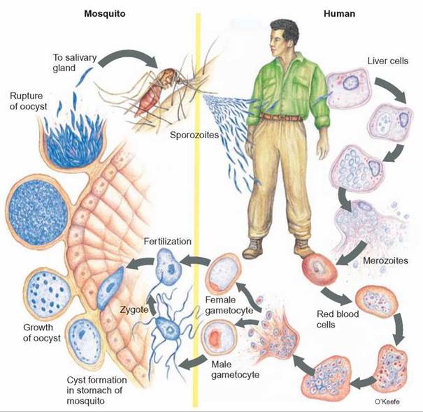
FIGURE 21.13. The Life Cycle of Plasmodium vivax
Plasmodium vivax is one of the members of the Apicomplexa that causes malaria. The life cycle requires two hosts, the Anopheles mosquito and the human. Humans get malaria when they are bitten by a mosquito carrying the larval stage of Plasmodium. The larva undergoes asexual reproduction and releases thousands of individuals, which invade the red blood cell. Their release from massive numbers of infected red blood cells causes the chills, fever, and headache associated with malaria. Inside the red blood cell, more reproduction occurs to form male gametocytes and female gametocytes. When the mosquito bites a person with malaria, it ingests some gametocytes. Fertilization occurs and zygotes develop in the stomach of the mosquito. The resulting larvae are housed in the mosquito’s salivary gland. Then, when the mosquito bites someone, some saliva containing the larvae is released into the person’s blood and the cycle begins again.
Ciliates
Ciliates are a group of protozoans with a complex cellular structure and numerous short, flexible extensions from the cell called cilia (figure 21.14). The cilia move in an organized, rhythmic manner and propel the cell through the water. Some types of ciliates, such as Paramecium, have nearly 15,000 cilia per cell and move at a rapid speed of 1 millimeter per second. Most ciliates are free-living cells found in freshwater and salt water or damp soil, where they feed on bacteria and other small organisms. Ruminant animals have large numbers of ciliates in their digestive systems, where they are part of the complex ecology of the ruminant gut (see Outlooks 21.2).
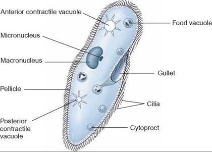
FIGURE 21.14. Ciliates
Ciliates, such as Paramecium, have a complex cell structure and a large number of cilia on their surface, which propel them through the water. They feed on a variety of organisms.
Ciliates have a complex cellular structure with two kinds of nuclei. Most have a macronucleus and one or more micronuclei. The macronucleus is involved in the day-to-day running of the cell, whereas the micronuclei are involved in sexual reproduction. Sexual reproduction involves a process called conjugation, in which two cells go through a series of nuclear divisions equivalent to meiosis and exchange some of their nuclear material. Although the exchange does not result in additional cells, it does result in cells that have a changed genetic mixture.
Funguslike Protists
Funguslike protists have a motile reproductive stage but they do not have chitin in their cell walls, which differentiates them from true fungi. There are two kinds of funguslike protists: slime molds and water molds.
Slime Molds
Slime molds are amoeba-like organisms that crawl about and digest dead organic matter. Some slime molds look like giant amoebae. They are essentially a large mass several centimeters across, in which the nucleus and other organelles have divided repeatedly within a single large cell (figure 21.15). No cell membranes partition this mass into separate segments. They vary in color from white to bright red or yellow, and they can reach relatively large sizes (45 centimeters in length) when in an optimum environment.
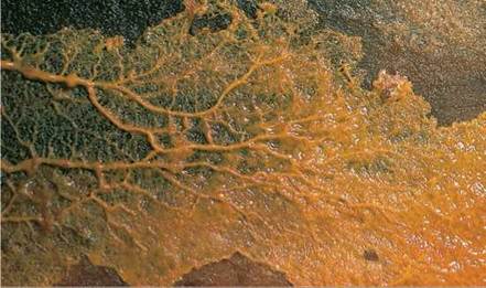
FIGURE 21.15. Slime Mold
Slime molds grow in moist conditions and are important decomposers. As slime molds grow, additional nuclei are produced by mitosis, but there is no cytoplasmic division. Thus, at this stage, a slime mold is a single mass of cytoplasm with many nuclei.
Other kinds of slime mold exist as large numbers of individual, amoeba-like cells. These haploid cells get food by engulfing microorganisms. They reproduce by mitosis. When their environment becomes dry or otherwise unfavorable, the cells come together into an irregular mass. This mass glides along rather like an ordinary garden slug and is labeled the sluglike stage. This sluglike form may flow about for hours before it forms spores. When the mass gets ready to produce spores, it creates a stalk with cells that have cell walls. At the top of this specialized structure, cells are modified to become haploid spores. When released, these spores may be carried by the wind and, if they land in a favorable place, may develop into new amoeba-like cells.
Water Molds
Water molds were once thought to be fungi. However, they differ from fungi in two fundamental ways. Their cell walls are made of cellulose, not chitin, and water molds have a flagellated reproductive stage. Thus, they are considered to be more closely related to the diatoms and brown algae than to fungi. Although called water molds, they live in many moist environments, not just in bodies of water (figure 21.16).
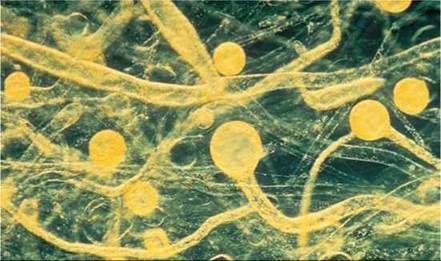
FIGURE 21.16. Water Mold
Rapidly reproducing water molds quickly produce a large mass of filaments. These filaments cause the fuzzy growth often seen on dead fish and other dead material in the water.
Water molds are important saprophytes and parasites in aquatic ecosystems. They are often seen as fluffy growths on dead fish or other organic matter floating in water. A parasitic form of water mold is well known to people who rear tropical fish; it causes a cottonlike growth on the fish. Although these organisms are usually found in aquatic habitats, they are not limited to this environment. Some species cause downy mildew on plants such as grapes. In the 1880s, this mildew almost ruined the French wine industry when it spread throughout the vineyards. A copper-based fungicide called Bordeaux mixture—the first chemical used against plant diseases—was used to save the vineyards. A water mold was also responsible for the Irish potato blight. In the nineteenth century, potatoes were the staple of the Irish diet. Cool, wet weather in 1845 and 1847 damaged much of the potato crop, and more than a million people died of starvation. Nearly one- third of the survivors left Ireland and moved to Canada or the United States.
21.3. CONCEPT REVIEW
14. Why is the kingdom Protista not considered a valid phylogenetic group?
15. What is phytoplankton?
16. List three different categories of organisms that are considered algae.
17. List two major kinds of marine phytoplankton.
18. List the two major kinds of multicellular marine algae.
19. Describe a characteristic for each of the following:
a. apicomplexa
b. ciliates
c. flagellates
d. foraminifera
20. Why are water molds and slime molds not considered to be Fungi?