CONCEPTS IN BIOLOGY
PART VI. PHYSIOLOGICAL PROCESSES
26. The Body's Control Mechanisms and Immunity
26.2. Nervous System Function
The nervous, endocrine, and immune systems are the three major systems of the body that play key roles by integrating stimuli and generating the appropriate responses necessary to maintain homeostasis.
The nervous system is well suited to managing the rapid adjustments that must take place within the body. As we discuss the structure and function of the nervous system it is useful to have a basic understanding of how it is organized. The nervous system is organized in a fashion similar to a computer. Information from various input devices (sense organs) is delivered to the central processing unit (brain) by way of wires (sensory nerves). The information is interpreted in the central processing unit. Eventually, messages can be sent by way of cables (motor nerves) to drive external machinery (muscles and glands). The following sections describe the major structural and functional characteristics of the nervous system.
The Structure of the Nervous System
The nervous system consists of a network of cells, with fibrous extensions, that carry information along specific pathways from one part of the body to another. A neuron, or nerve cell, is the basic unit of the nervous system. A neuron consists of a central body, called the soma or nerve cell body, which contains the nucleus. It has several long extensions called nerve fibers. There are two kinds of nerve fibers: axons, which carry information away from the cell body, and dendrites, which carry information toward the cell body (figure 26.2). Most neurons have one axon and several dendrites.
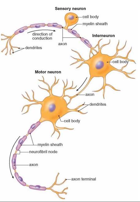
FIGURE 26.2. Structure of Neurons
Neurons consist of a nerve cell body, which contains the nucleus, and several fibrous extensions. The fibers that carry impulses to the nerve cell body are dendrites. The fiber that carries the impulse away from the cell body is the axon. Sensory neurons have a long dendrite that carries information from the sense organ to the cell body. Motor neurons have a long axon that carries information from the cell body to a muscle or gland. Most neurons other than sensory neurons have many dendrites but only one axon.
Neurons are arranged into two major systems. The central nervous system, which consists of the brain and spinal cord, is surrounded by the skull and the vertebrae of the spinal column. The spinal cord is a collection of nerve cells and nerve fibers surrounded by the vertebrae that conveys information to and from the brain. The central nervous system receives input from sense organs, interprets information, and generates responses. The peripheral nervous system is located outside the skull and spinal column; it consists of bundles of long axons and dendrites called nerves. In the peripheral nervous system, cranial nerves connect to the brain, whereas spinal nerves connect to the spinal cord. There are two sets of neurons in the peripheral nervous system: the sensory neurons and the motor neurons. Sensory neurons have long dendrites that carry input from sense organs to the central nervous system. Motor neurons carry messages from the central nervous system to muscles and glands. Motor neurons have one long axon that runs from the spinal cord to a muscle or gland. Some motor nerves constituting the somatic nervous system, control the skeletal (voluntary) muscles. Other motor nerves, collectively called the autonomic nervous system, control the smooth (involuntary) muscles, the heart, and glands. Figure 26.3 summarizes the different structural and functional portions of the nervous system.
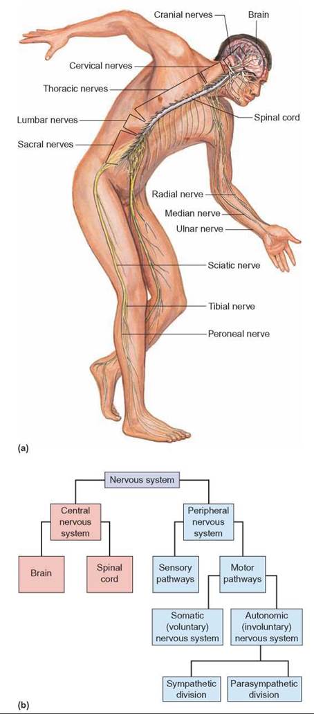
FIGURE 26.3. Organization of the Nervous System
(a) A generalized view of the central nervous system and some of the nerves of the peripheral nervous system. (b) The chart shows how the various functional portions of the nervous system are related to one another.
The Nature of the Nerve Impulse
Because most neurons have long, fibrous extensions, information can be passed along the nerve cell from one end to the other. The nerve impulse is the message that travels along a neuron. A nerve impulse is not like an electric current; instead, it involves a specific sequence of chemical events at the cell membrane.
Because all cell membranes are differentially permeable, it is difficult for some ions to pass through the membrane, and the combination of ions inside the membrane is different from that outside it. Cell membranes also contain proteins that actively transport specific ions from one side of the membrane to the other. Active transport involves the cell’s use of adenosine triphosphate (ATP) to move materials from one side of the cell membrane to the other. Because ATP is required, cells lose this ability when they die. One of the ions that is actively transported from cells is the sodium ion (Na+). At the same time sodium ions are being transported out of cells, potassium ions (K+) are being transported into the normal resting (not stimulated) cells. However, there are more sodium ions transported out than potassium ions transported in.
Because a normal resting cell has more positively charged Na+ ions on the outside of the cell than on the inside, a small but measurable voltage exists across the membrane of the cell. (Voltage is a measure of the electrical charge difference that exists between two points or objects.) The voltage difference between the inside and the outside of a cell membrane is about 70 millivolts (0.07 volt). The two sides of the cell membrane are, therefore, polarized in the same sense that a battery is polarized, with a positive (+) and a negative (—) pole. A resting neuron has its positive pole on the outside of the cell membrane and its negative pole on the inside of the membrane (figure 26.4).
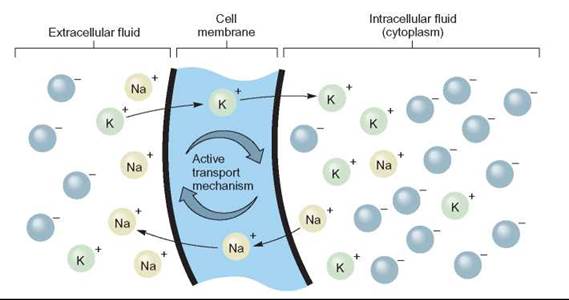
FIGURE 26.4. The Polarization of Cell Membranes
All cells, including neurons have an active transport mechanism that pumps Na+ out of cells and simultaneously pumps K+ into them. The end result is that there are more Na+ ions outside the cell and more K+ ions inside the cell. In addition, negative ions, such as Cl—, are more numerous inside the cell. Consequently, the outside of the cell is positive (+) compared with the inside, which is negative (—).
When a cell is stimulated at a specific point on the cell membrane, the cell membrane changes its permeability and lets sodium ions (Na+) pass through it from the outside to the inside. The membrane is thus depolarized; it loses its difference in charge as sodium ions diffuse into the cell from the outside. Sodium ions diffuse into the cell because, initially, they are in greater concentration outside the cell than inside. When the membrane becomes more permeable, they are able to diffuse into the cell, toward the area of lower concentration. The depolarization of one point on the cell membrane causes the adjacent portion of the cell membrane to change its permeability as well, and it also depolarizes. Thus, a wave of depolarization passes along the length of the neuron from one end to the other (figure 26.5). The depolarization and passage of an impulse along any portion of the neuron is a momentary event. As soon as a section of the membrane has been depolarized, potassium ions diffuse out of the cell. This reestablishes the original polarized state, and the membrane is said to be repolarized. Subsequently, the continuous active transport of sodium ions out of the cell and potassium ions into the cell restores the original concentration of ions on both sides of the cell membrane. This is similar to a “wave” passing around a stadium. The “wave” is initiated by specific individuals as they stand up and “wave” (depolarization). People adjacent participate as they are affected by the initiators while at the same time the originators are sitting down (repolarization). As long as the next people in line participate, the “wave” continues. There is an “impulse” that moves around the stadium but the individuals remain in place.
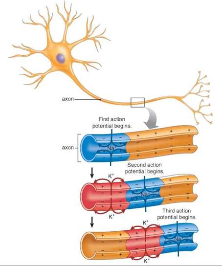
FIGURE 26.5. A Nerve Impulse
When a neuron is stimulated, a small portion of the cell membrane depolarizes as Na+ flows into the cell through the membrane. This encourages the depolarization of an adjacent portion of the membrane, and it depolarizes a short time later. In this way, a wave of depolarization passes down the length of the neuron. Shortly after a portion of the membrane is depolarized, the ionic balance is reestablished. It is repolarized and ready to be stimulated again.
When the nerve impulse reaches the end of the axon, it stimulates the release of a molecule that stimulates depolarization of the next neuron in the chain.
Activities at the Synapse
The synapse is the space between the fibers of adjacent neurons in a chain. Many chemical events occur in the synapse that are important in the function of the nervous system. When a neuron is stimulated, an impulse passes along its length from one end to the other. When the impulse reaches a synapse, a molecule called a neurotransmitter is released into the synapse from the axon. It diffuses across the synapse and binds to specific receptor sites on the dendrite of the next neuron. When enough neurotransmitter molecules have been bound to the second neuron, an impulse is initiated in it as well. Several kinds of neurotransmitters are produced by specific neurons. These include dopamine, epinephrine, acetylcholine, and several other molecules. The first neurotransmitter to be identified was acetylcholine. Acetylcholine molecules are neurotransmitters manufactured in the soma; they migrate down the axon, where they are stored until needed.
As long as a neurotransmitter is attached to its receptor, it continues to stimulate the neuron. Thus, if acetylcholine continues to occupy receptors, the neuron continues to be stimulated again and again. Acetylcholinesterase, an enzyme, destroys acetylcholine and prevents this from happening. (The breakdown products of the acetylcholine can be used to remanufacture new acetylcholine molecules.) The destruction of acetylcholine allows the second neuron in the chain to return to normal (figure 26.6). Thus, it will be ready to accept another burst of acetylcholine from the first neuron a short time later. Neurons must also constantly manufacture new acetylcholine molecules, or they will exhaust their supply and be unable to conduct an impulse across a synapse.
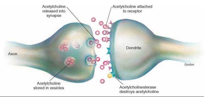
FIGURE 26.6. Activities at the Synapse
When a nerve impulse reaches the end of an axon, it releases a neurotransmitter into the synapse. In this illustration, the neurotransmitter is acetylcholine. When acetylcholine is released into the synapse, acetylcholine molecules diffuse across the synapse and bind to receptors on the dendrite, initiating an impulse in the next neuron. Acetylcholinesterase is an enzyme that destroys acetylcholine, preventing continuous stimulation of the dendrite.
Certain drugs, such as curare and strychnine, interfere with the activities at the synapse. Curare blocks the synapse and causes paralysis, whereas strychnine causes neurons to be stimulated continually. Many insecticides are nerve poisons and, therefore, are quite hazardous.
Because of the way the synapse works, impulses can go in only one direction: Only axons secrete acetylcholine, and only dendrites have receptors. This explains why there are sensory and motor neurons to carry messages to and from the central nervous system.
The Organization of the Central Nervous System
The brain consists of several regions, each with a specific function. Certain parts of the brain are involved in controlling fundamental functions, such as breathing and heart rate. Others are involved in generating emotions, others decode sensory input, and others coordinate motor activity. The human brain also has considerable capacity to store information and create new responses to stimuli.
The functions of the brain can be roughly divided into three major levels: automatic activities, basic decision making and emotions, and thinking and reasoning. If we begin with the spinal cord and work our way forward, we will proceed from the more fundamental, automatic activities of the brain to its more complex, thinking portions. The medulla oblongata is at the base of the brain, where the spinal cord enters the skull. It controls fundamental activities, such as blood pressure, breathing, and heart rate. Most of the fibers of the spinal cord cross from one side of the body to the other in the medulla oblongata. This is why the left side of the brain affects the right side of the body.
The cerebellum is the large bulge at the base of the brain that is connected to the medulla oblongata. The primary function of the cerebellum is the coordination of muscle activity. It receives information from sense organs, such as the portions of the ear involved in balance, the eyes, and pressure sensors in muscles and tendons. The cerebellum uses this information to make adjustments in the strength and sequence of muscle contractions, so that the body moves in a coordinated fashion.
The pons is the region of the brain that is connected to the anterior end of the medulla oblongata. It also connects to the cerebellum and to higher levels of the brain. It is involved in controlling many sensory and motor functions of the sense organs of the head and face. The pons is also connected to a portion of the brain known as the midbrain that is connected to regions of the brain that control many automatic activities, but also are involved in some level of decision making. The primary regions are the thalamus and the hypothalamus.
The thalamus is the region of the brain that relays information between the cerebrum and lower portions of the brain. It also provides a level of awareness in that it determines pleasant and unpleasant stimuli and is involved in sleep and arousal. The hypothalamus is the region of the brain involved in regulating sleep cycles; it is important in emotions such as anger, fear, pleasure, and the sensations that accompany hunger, sexual response, and pain. Several other, more automatic functions are regulated in this region, such as body temperature, blood pressure, and water balance. The hypothalamus also is connected to the pituitary gland and influences the manufacture and release of its hormones. Figure 26.7 shows the relationship of the various, more primitive parts of the brain.
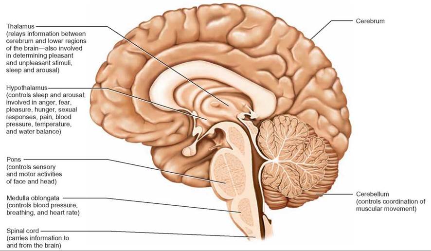
FIGURE 26.7. Functions of the More Primitive Brain Regions
The brain is organized into several levels of function. The more primitive regions of the brain connected to the spinal cord monitor and manage many essential functions automatically.
The cerebrum is the thinking part of the brain. It receives, interprets, and integrates information from sense organs and generates responses that involve the actions of muscles and glands. It is also the largest portion of the brain in humans. The two hemispheres of the cerebrum cover all other portions of the brain except the cerebellum. The surface of the cerebrum has been extensively mapped as to the locations of many functions. Abilities such as memory, language, the control of movement, the interpretation of sensory input, and thought are associated with specific areas of the cerebrum. Figure 26.8 illustrates the cerebrum and the locations of specific functions.
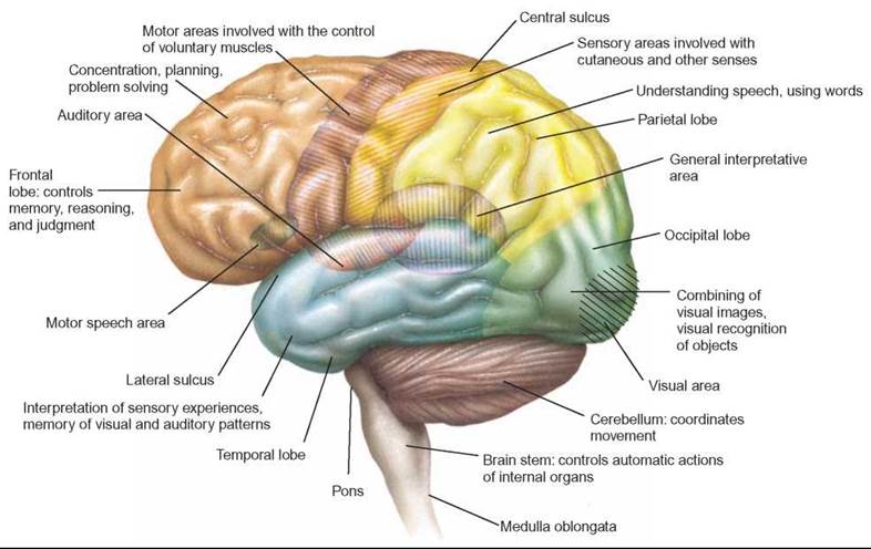
FIGURE 26.8. Specialized Areas of the Cerebrum
Each portion of the cerebrum has particular functions.
The function of the brain is not determined by structure alone. Many parts of the brain have specialized neurons, which produce specific neurotransmitter molecules used only to stimulate particular neurons that have the proper receptor sites. As scientists learn more about the functioning of the brain, they are finding more kinds of specialized neurotransmitter molecules, allowing for the treatment of many types of mental and emotional diseases. Manipulating these neurotransmitter molecules can help correct inappropriate functioning of the brain. However, the brain is still not completely understood. Scientists are at an early stage in their search to comprehend this organ, which sets us apart from other animals (How Science Works 26.1).
HOW SCIENCE WORKS 26.1
How Do We Know What the Brain Does?
Scientists know a great deal about the function of the brain, although there is still much more to learn. Certain functions have been identified as residing in specific portions of the brain, as a result of many kinds of studies over the past century. For example, persons who have had specific portions of their brains altered by damage from accidents or strokes have been studied. Their changes in behavior or the way they perceive things can be directly correlated with the portion of the brain that was damaged. During surgeries that require the brain to be exposed, a local anesthetic can be given and the patient can be conscious while the surgery is taking place. (The brain perceives pain from pain receptors throughout the body; however, because the brain does not have many pain receptors within it, touching or manipulating the brain does not cause pain to be perceived.) Specific portions of the brain can be stimulated and the patient can be asked to describe his or her sensations and the patient's motor functions can be observed.
Many kinds of experiments have also been done with animals, in which specific portions of the brain have been destroyed and the animals' changes in behavior noted. Electrodes have been inserted into the brains of animals to stimulate certain portions of the brain.
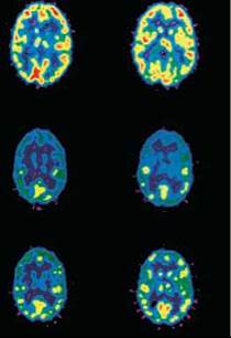
Modern brain-imaging techniques are used to observe changes in the electrical activity of specific portions of the brain without electrodes or other invasive procedures. This allows researchers to present stimuli to human subjects and determine which parts of the brain alter their activity. In addition to localizing the part of the brain that responds, it is also possible to determine what parts of a complex stimulus are most important in changing brain activity. For example, these techniques have revealed that languages learned by adults are processed in different places in the brain than the languages they learned as children and that the brain has a built-in mechanism for recognizing unexpected words or musical notes.
Although much is known about the brain, there is still much to learn. Current experiments are seeking ways to regenerate neurons that have been damaged. A better understanding of the chemical events that take place in the brain would enable us to cure many kinds of debilitating mental illnesses.
26.2. CONCEPT REVIEW
3. Describe how the changing permeability of the cell membrane and the movement of sodium ions cause a nerve impulse.
4. What is the role of acetylcholine in a synapse? What is the role of acetylcholinesterase?
5. List the differences between the central and peripheral nervous systems and between the motor and sensory nervous systems.