Biology For Dummies
Part IV Systems Galore! Animal Structure and Function
Chapter 16
Checking Out the Plumbing: Animal Digestive and Excretory Systems
In This Chapter
Looking at how different types of animals consume and remove food
Taking an in-depth look at the human digestive system
Playing with the building blocks of healthy food choices
Discovering how waste products leave the human body
After an animal ingests or absorbs food, its digestive system immediately starts breaking down the food to release the nutrients within it. After the useful nutrients are absorbed into the bloodstream, the animal eliminates solid wastes through its large intestine and nitrogenous wastes through its urinary system.
Want to know more about the ins and outs of digestive and excretory systems? Then you’re in the right place. In this chapter, we give you the lowdown on the various ways animals obtain food and eliminate wastes. We then present the workings of the human digestive system and the fate of food and wastes in your body. After all, everyone should know the scoop on their poop.
Obtaining Food and Breaking It Down
All animals need food as a source of energy and materials for growth (as explained in Chapter 5), but the various types of animals have different strategies for obtaining the food they need.
Bulk feeders take big bites out of food. They use teeth, claws, tentacles, or pinchers to tear off pieces of food and ingest them. Eagles, people, frogs, and snakes are all examples of bulk feeders.
Filter feeders strain liquids to capture tiny particles suspended in the fluid. They have structures such as gills or very fine tentacles that trap small particles out of the water. Clams, for example, pull seawater through their bodies and pass it over their mucus-covered gills, trapping the small organisms and organic matter that were suspended in the water. Other examples of filter feeders include sponges, whale sharks, and gray whales.
Fluid feeders suck nutrient-rich fluids from other organisms. They’re often parasites that live inside a host and use the host’s fluids as a source of food. Aphids and mosquitoes, for example, puncture other organisms and draw fluid from them — aphids draw the sugary sap out of plants, and mosquitoes draw blood out of animals. Ticks, leeches, and lampreys are some additional fluid feeders you may be familiar with.
Substrate feeders live right in or on their food and eat as they move through it. Despite its complex name, this type of animal obtains its food pretty simply. Just think of earthworms and maggots. As earthworms burrow through the soil, they ingest the soil, digest the small organisms and organic matter within it as it passes through them, and then release the undigested matter as worm casings out their back ends. Similarly, when flies lay their eggs in bodies, the maggots that hatch out of the eggs eat their way through the food (the decomposing organism) until they reach the outside.
 Four main events occur from the moment food enters an animal’s body until the time its wastes are released:
Ingestion occurs when an animal takes food into its digestive tract.
Digestion occurs when the animal’s body gets busy breaking down the food. Two types of digestion exist:
• Mechanical digestion physically breaks down food into smaller and smaller pieces. It begins when an animal consumes the food and continues until the food enters its stomach.
• Chemical digestion uses enzymes and acids to break down chewed or ground-up food into even smaller pieces. It also begins as soon as food is consumed and the enzymes in the mouth go to work. Chemical digestion continues as the food moves through the stomach and small intestine and encounters enzymes and acids in the stomach and enzymes in the small intestine.
Absorption occurs when cells within the animal move small food molecules from the digestive system to the insides of the cells.
Elimination occurs when material that can’t be digested passes out of an animal’s digestive tract.
The Ins and Outs of Digestive Systems
The basic way an animal’s digestive system works has a great deal to do with whether it can spend a few hours between meals or whether it has to keep consuming food constantly just to stay alive. The following sections take a look at the two types of digestive systems and help you distinguish between animals that need to take in food all the time and those that don’t.
Incomplete versus complete digestive tracts
Of the animals that you can see without a microscope, the ones with the most primitive digestive system are animals with incomplete digestive tracts, meaning they have a gut with just one opening that serves as both mouth and anus. (Gross, we know.) Jellyfish are a classic example of an animal with an incomplete digestive tract.
Increasing in complexity are animals that have gut tubes (where foods are digested and nutrients are absorbed) with a mouth at one end and an anus at the other. Although simple, this type of system is considered a complete digestive tract. You’re probably quite familiar with one particular animal that possesses this digestive system — you.
The benefit of a complete digestive tract is that it allows thorough digestion before excretion occurs. Organisms with incomplete digestive tracts release undigested food along with their wastes. An organism with a complete digestive tract doesn’t have to take in food constantly to replace food that’s excreted before the nutrients could be acquired from it.
Continuous versus discontinuous feeders
Animals that must “eat” constantly because they take food in and then push it out soon afterward are called continuous feeders. Most of these animals are either permanently attached to something (think clams or mussels) or incredibly slow movers. Animals that are discontinuous feedersconsume larger meals and store the ingested food for later digestion. These animals are generally more active and somewhat nomadic.
 The ability to “eat and run” serves a preying carnivore well. If an animal such as a lion were a continuous feeder that had to hunt and eat constantly, it’d be exhausted and spend much more time out in the open savanna, increasing the chance that it could become prey for another predator.
Although you may find yourself snacking and grazing constantly throughout a day, you’re really a discontinuous feeder. You can consume food rapidly, but you digest it gradually so you don’t have to eat again for several hours.
You and all the other animals that are discontinuous feeders must have a place in the body to store food as it slowly digests. In humans, this organ is the stomach.
Traveling through the Human Digestive System
You know that the mouth is where you put your food, but did you realize it’s also part of your digestive system (see Figure 16-1)? Well, it is. The act of chewing (the technical term for it is mastication) is the first step in food digestion for humans. Chewed-up food travels from your mouth, down your esophagus, and into your stomach. From your stomach, it passes into the small intestine. Materials that can’t be digested and absorbed move into the large intestine and then pass out of your body through your rectum and anus. In the sections that follow, we fill you in on the role that each of these parts of the digestive system plays in digestion.
Figure 16-1: The organs of the digestive system.
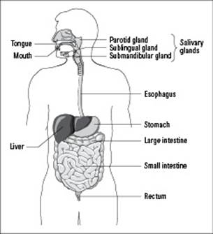
From LifeART®, Super Anatomy 1, © 2002, Lippincott Williams & Wilkins
The busiest stop of all — your mouth
Your mouth does more than let you shout at the television during sporting events or talk to that cute guy or gal sitting next to you in biology class. It also kicks off the entire process of digestion. And don’t think your teeth are the only participants in this process just because they mash your food into smaller and smaller bits. Your whole mouth gets a piece of the pie, so to speak. In addition to what your teeth do, your
Taste buds detect the nutrients that make up the food you’re eating — such as carbohydrates, proteins, and fats — so the cells of your digestive system can release the right enzymes to chemically digest your food.
Saliva starts chemically digesting your food thanks to salivary amylase, an enzyme found within saliva that splits apart the bonds between glucose molecules in a long chain of starch.
You know how you salivate just before you’re about to eat something? That’s the effect of your eyes or nose sensing something delicious and sending a message to your brain that you’re about to open your mouth and take a bite. It’s also your mouth’s way of preparing for digestion by producing saliva containing salivary amylase.
After your teeth have chewed, your taste buds have sent along the information about what you’re eating, and the enzymes in your saliva have started breaking apart starches, you’re ready to swallow. This action involves your tongue pushing the chewed food to the back of your throat and you squeezing food down your esophagus and into your stomach. (The esophagus is the tube that connects your mouth to your stomach.)
The squeezing of food through a hollow muscular structure is called peristalsis, and it occurs throughout the digestive system to move food down the esophagus and into the stomach and then again to move food through the intestines.
When the swallowed food drops into your stomach, it’s referred to as a bolus. At this point, salivary amylase stops breaking apart the starch molecules, and the gastric juice in your stomach, which contains enzymes as well as hydrochloric acid (HCl), takes over chemical digestion.
If you eat too much, your stomach produces more acid, and the contents of your overly full stomach can be forced back up into your esophagus, which runs in front of the heart. The unpleasant result? Heartburn.
The inner workings of your stomach
When food particles reach your stomach, the organ churns them up, and the enzyme pepsin starts breaking down the food’s proteins into smaller chains of amino acids (see Chapter 4 for more on enzymes). Then the whole goopy substance gets squirted into the top of your intestines via the pyloric valve, otherwise known as the gate between your stomach and small intestine. Your pyloric sphincter muscle occasionally opens the valve, allowing your stomach’s contents into your small intestine a little bit at a time.
Perhaps you’re wondering why pepsin doesn’t destroy the proteins that make up the tissues in your digestive tract. Well, when pepsin is secreted, it’s in its inactive form, called pepsinogen. Because pepsinogen is inactive, it doesn’t damage the cells that make it. When it’s in the cavity of the stomach, pepsinogen gets converted to its active form, pepsin, by losing a few dozen of its amino acids. The lining of the stomach isn’t affected by pepsin because pepsin acts only on proteins, and the stomach is covered with mucus (a substance made from fats) that protects the protein-containing tissues — that is, unless you have a stomach ulcer (see the related sidebar nearby for more on stomach ulcers).
The long and winding road of your small intestine
When food molecules hit your small intestine, they get broken down into even smaller units (so your cells can absorb them) with a little help from your liver and pancreas.
The true cause of stomach ulcers
When the lining of the stomach is eroded by digestive enzymes and stomach acid, the resulting condition is referred to as a stomach ulcer. For years the medical community thought stomach ulcers were largely caused by stress, worry, frustration, and other negative emotions. That belief changed in 1982 when two Australian doctors, Barry Marshall and Robin Warren, detected a bacterium called Helicobacter pylori (or H. pylori) in the stomachs of people with ulcers. They proposed that H. pylori in fact caused the ulcers, but no one believed them. To prove their point, Dr. Marshall, who didn’t have any stomach ulcers, did the unthinkable — he drank a culture of live H. pylori! After drinking the culture, Dr. Marshall developed ulcers, proving that infection with the bacterium could indeed cause stomach ulcers.
So just how does a stomach ulcer form? Well, H. pylori inflames the lining of the stomach, causing the organ’s protective mucus to disappear and allowing the enzymes and stomach acids secreted during digestion to eat away at the proteins in the tissue of the stomach wall. Stomach ulcers can be painful, and if they bleed or cause a hole (or perforation) through the wall of the stomach, they can become a medical emergency. Fortunately, antibiotic therapy can often successfully treat the condition by removing the H. pylori bacteria from the stomach.
The liver secretes bile (a yellow-brown or greenish fluid) into the small intestine. Bile salts emulsify (suspend in water) fats, helping them mix into the liquid in the intestine so they can be digested more easily. Meanwhile, the pancreas releases pancreatic juice into the mixture that contains the following enzymes to help chemically digest fats and carbohydrates:
Lipase breaks apart fat molecules into fatty acids and glycerol.
Pancreatic amylase breaks long carbohydrates into disaccharides, which are short chains of two sugars. The disaccharidases then break apart into monosaccharides that can be absorbed by the cells lining your small intestine.
Trypsin and chymotrypsin are enzymes that break apart peptide fragments. After they break the peptides down into small chains, aminopeptidases finish them off by breaking apart the peptides into individual, absorbable amino acids.
Just like pepsin in the stomach, trypsin and chymotrypsin are originally secreted in their inactive forms, trypsinogen and chymotrypsinogen. To protect the cells of your digestive system, the enzymes aren’t activated until they reach the cavity of your small intestine.
Don’t let the word small fool you. The small intestine is much longer than the large intestine (10 feet long versus 5 feet long). The term small intestine refers to the fact that this part of the intestine is narrower in diameter than the large intestine; the large intestine is wider in diameter but shorter in length.
After several hours in your digestive system, the carbohydrates, fats, and proteins from your food are all in their smallest components: monosaccharides (such as glucose), fatty acids and glycerol, and amino acids (see Chapter 3 for details on these molecules). Now they can leave your digestive system in order to be used by all the cells in your body (as explained in the next section).
Absorbing the Stuff Your Body Needs
During the digestion process, the nutrients your body can use are absorbed into the cells lining your small intestine. The rest of the material that can’t be further digested or used passes on to the large intestine. The next sections outline how nutrients move throughout your system and how your liver constantly works to make sure you have the right amount of blood glucose (blood sugar) necessary to keep you going.
How nutrients travel through your body
If digestion is all about obtaining the nutrients your cells need to function, then how exactly do those nutrients get out of your digestive system and into the rest of your body?
First, they pass directly into the cells of your small intestine via active transport. In other words, energy obtained from adenosine triphosphate (ATP) molecules is expended to move sugars (which came from consumed carbohydrates) and amino acids (which came from consumed proteins) into the intestinal cells (see Chapter 4 for the full scoop on active transport). From there, the nutrients are able to participate in capillary exchange, a type of trading system that allows cells to exchange nutrients and waste.
Capillary exchange relies on two things: capillaries, teeny-tiny blood vessels with extremely thin walls, and interstitial fluid, the fluid that fills every space between every cell in your body, cushioning and hydrating the cells.
Did you know that 60 percent of a human’s body weight is from fluid? Of that 60 percent, 20 percent is extracellular fluid, fluid that exists outside the cells. This extracellular fluid is made up mostly of interstitial fluid (16 percent) and plasma (4 percent; we fill you in on plasma in Chapter 15). The remaining 40 percent of fluid in your body is intracellular fluid, fluid that exists inside the cells of your body; it’s called cytoplasm.
The nutrients gained from digested food diffuse through the walls of your small intestine, through the capillaries’ walls, across the interstitial fluid, and into your cells. At the same time, waste produced by your cells’ metabolic processes diffuses out of the cells, across the interstitial fluid, and into the capillaries, where it can be carried to your kidneys for excretion. (To discover how your kidneys remove wastes from your body, head to the later related section in this chapter.)
Although the sugars and amino acids inside the capillaries get shuttled through the bloodstream to the liver, the products of fat digestion have a different fate. They get coated with proteins and acquire a new name: chylomicrons. Instead of being carried through the bloodstream, the chylomicrons get transported through the lymph system, which deposits lymph fluid into veins near the heart.
Glucose regulation
Perhaps the most important stop digested sugars make as they travel through your bloodstream is your liver. The liver can detect abnormalities in the levels of various substances in your blood, such as glucose, and correct them.
If the level of glucose is too high (a condition called hyperglycemia), the liver removes some of the glucose from the blood and turns it into the storage polysaccharide glycogen. If excess glucose is still in the blood after the liver has made enough glycogen, the liver switches its metabolic process to storing the extra glucose as fat. The fat molecules are then carried away by your bloodstream and deposited around your body in your adipose tissue (fat).
If the level of glucose in the blood is too low (a condition called hypoglycemia), the liver converts some of its stored glycogen back into glucose and puts the glucose into the blood. If all the glycogen stores are used up, the liver starts to break down stored fats to obtain glucose for your cells.
Having just the right amount of glucose in your bloodstream is essential because glucose is your brain’s main source of fuel. Glucose is so important, in fact, that your body will literally digest itself in order to get glucose to your brain. In extreme cases, such as starvation, when the breakdown of glycogen and fat isn’t enough to restore blood glucose to normal levels, the body starts breaking down proteins to get the energy molecules it so desperately needs. Proteins in the muscles are broken down into amino acids, which can be converted into glucose. This may not sound so bad until you realize that the heart is a muscle. When your body starts breaking down proteins in your heart, death is a serious possibility.
What’s for Dinner? Making Wise, Nutritious Food Choices
It’s a shame humans have such sensitive taste buds. If you didn’t have such an evolved sense of taste, maybe you’d be like other animals and just eat what’s part of your natural diet only when you’re truly hungry. But, alas, food tastes good, and humans are often tempted to put really cheap fuel in their systems. Would you do that to your car on a regular basis? Or would you rather use the premium stuff to make sure your car’s engine doesn’t knock and ping?
If you want to keep your bodily systems from knocking and pinging, we strongly encourage you to follow the nutrition recommendations made by the United States Department of Agriculture (USDA). Its MyPyramid, an updated version of the classic Food Guide Pyramid, serves as a way of visualizing the proportion of items from different food groups that should make up your diet. More detailed information, such as the difference between whole grains and refined carbohydrates in the bread, cereal, and pasta group, are available on the USDA’s MyPyramid Web site at www.mypyramid.gov.
For now, why not check out the following sections? They provide the basic facts about the essential nutrients your body needs: carbohydrates, proteins, fats, and minerals and vitamins.
Carbohydrates: The culprits of your food cravings
Carbohydrates are compounds of carbon, hydrogen, and oxygen that supply your body with short-term energy (we cover the chemical structure of carbs in greater detail in Chapter 3). Carbohydrate molecules, such as sugars, break down quickly. You’ve witnessed this yourself if you’ve ever held a marshmallow over a campfire too long.
Glucose is the most important carbohydrate molecule. You can acquire it directly from foods containing carbohydrates (such as breads, pastas, sweets, and fruits). However, your body also creates glucose when it breaks down proteins and fats.
When it comes to your diet, all carbohydrates are not created equal.
Foods made with whole grains are high in fiber and vitamins. They help prevent heart disease and constipation. Eating breads and cereals made with whole grains, as well as the grains themselves (think brown rice, quinoa, and bulgur wheat), is a way to get plenty of good carbs in your diet.
 Lots of products advertise that they’re “made with whole grains,” but that assertion doesn’t tell you what the proportion of whole grains in the food actually is. When in doubt, check the fiber content of the food. Foods that are high in fiber are made with lots of whole grains.
Foods made with refined grains are low in fiber and vitamins. They break down very quickly and cause a rapid rise in blood glucose. White bread, flour, and pasta — as well as cookies, cakes, and scones — are made with refined grains. It’s best to avoid these types of carbs as much as you can.
Proteins: You break down their chains; they build yours
Every muscle, cell membrane, and enzyme in your body is made from proteins. So, to create more muscle fibers, new cells, and other elements that help your body run, you need to take in protein (to get an idea of a protein’s chemical structure, see Chapter 3).
Proteins are made from amino acids. Your body requires nine amino acids in particular to construct proteins, which is why those nine amino acids are referred to as essential amino acids (we list them for you in Table 16-1). Humans can synthesize 11 amino acids from a variety of starting compounds that aren’t necessarily derived from amino acids themselves. Because these amino acids are made in the body, they’re considered nonessential amino acids; it’s not essential that you consume foods containing them because you can get them another way. We list nonessential amino acids in Table 16-1 too.
|
Table 16-1 The Amino Acids Humans Can Consume |
|
|
Essential Amino Acids |
Nonessential Amino Acids |
|
Histidine |
Alanine |
|
Isoleucine |
Arginine |
|
Leucine |
Asparagine |
|
Lysine |
Aspartate |
|
Methionine |
Cysteine |
|
Phenylalanine |
Glutamate |
|
Threonine |
Glutamine |
|
Tryptophan |
Glycine |
|
Valine |
Proline |
|
Serine |
|
|
Tyrosine |
|
When you think of protein sources, you probably think of meat. There’s a good reason for that. Both your muscles and the muscles of other animals are made from protein. When you eat meat — whether it’s beef, chicken, turkey, pork, or fish — you’re consuming the protein-containing muscle tissue of another animal.
Beans, nuts, and soy are additional sources of protein, but the protein they contain is plant protein, not animal protein. Plant proteins are considered incomplete because they don’t contain enough of some of the amino acids humans need. Because animals acquire essential amino acids plus make their own nonessential amino acids, animal protein is considered complete — it has all the amino acids humans need.
Vegetarians must combine certain foods to make sure they’re getting all the necessary amino acids.
Fats: You need some, but don’t overdo it
Your body needs fats to make tissues and hormones and insulate your nerves (just like a rubber coating often insulates wires). Fat is also a source of stored energy. It gives your body shape, reduces heat loss by insulating your organs and muscles, and cushions your body and organs (much like shock absorbers).
Fat supplies long-term energy, which is why you don’t start to “burn fat” until 20 minutes or more into an aerobic workout. First, your body quickly burns off the glucose that’s readily available in your cells. Then it starts breaking down fat molecules and converting them into glucose for fuel.
Lipoproteins and your risk for heart disease
Lipoproteins are compounds made from a fat and a protein. Their job is to carry cholesterol around your body through the bloodstream. You’re capable of producing four types of lipoproteins:
High-density lipoproteins (HDLs)
Low-density lipoproteins (LDLs)
Very low-density lipoproteins (VLDLs)
Chylomicrons
Sometimes in the news you read or hear about HDL being “good” cholesterol and LDL being “bad” cholesterol. However, HDL and LDL are lipoproteins, not cholesterol molecules. They just attach to and transport cholesterol. Here’s what’s “good” and “bad” about the lipoproteins.
Chylomicrons are very small, newly created lipoproteins that fall into the VLDL category. VLDLs have very little protein and a lot of fat. As VLDLs travel through your bloodstream, they lose some lipids, pick up cholesterol, and become LDLs. The LDLs deliver the cholesterol to cells in your body that need it, but along the way, VLDLs and LDLs can squeeze through blood vessel walls. While doing that, the cholesterol can get stuck to the wall of the blood vessel, causing deposits called plaque to form. If enough cholesterol gets stuck, an artery may get clogged, which means blood can’t flow through. If that happens, a heart attack or stroke may occur. So, although LDLs help the body by transporting cholesterol, if you have too many of them, the cholesterol may start to block your blood vessels, increasing your risk of heart disease, heart attack, and stroke.
HDLs, on the other hand, are the lipoproteins that contain more protein than lipid, which makes them denser and gives them their name. Because they’re so dense, HDLs can’t squeeze through the blood vessel walls. Instead, they shuttle cholesterol right out of the body. They aren’t able to deposit cholesterol in blood vessels because they can’t get into them, so they don’t increase your risk of heart disease, heart attack, or stroke. Remember: You always want to have more of these dense little guys floating in your blood than LDLs or VLDLs.
Although fats are important nutrients for your body, good and bad kinds exist.
Unsaturated fats are good for you. Plant oils, such as olive and flaxseed, are excellent sources of unsaturated fats, as are fish oils.
Saturated fats are unhealthy. Animal fat, like the fat on meat, and the fat found in butter are saturated fats.
Contrary to popular belief, fat doesn’t make people fat. Consuming more fuel than you burn leads to the production and deposit of fatty tissue, whether that fuel comes from fats, proteins, or carbohydrates.
Minerals and vitamins: The fuel for your enzymes
In addition to carbohydrates, proteins, and fats, your body needs certain minerals and vitamins to help your enzymes function (see Chapter 4 for more on enzymes).
Minerals are inorganic molecules that are part of the Earth (think iron, zinc, and calcium). Your body doesn’t need a ton of minerals to stay healthy, but some are essential for its proper functioning. The essential minerals are called major minerals, and the minerals that you need only in very small amounts are called trace elements. Table 16-2 lists all the major minerals and trace elements your body needs.
|
Table 16-2 The Minerals Humans Need |
|
|
Major Minerals |
Trace Elements |
|
Calcium |
Chromium |
|
Chloride |
Copper |
|
Magnesium |
Fluoride |
|
Phosphorus |
Iodine |
|
Potassium |
Iron |
|
Sodium |
Manganese |
|
Sulfur |
Molybdenum |
|
Selenium |
|
|
Zinc |
|
Vitamins are organic molecules that exist naturally in all living things. They’re made up of the same carbon, hydrogen, oxygen, and nitrogen atoms as carbohydrates, proteins, and fats are. Any given vitamin falls into one of two categories:
Fat-soluble: These vitamins (which include vitamins A, D, E, and K) need to be “dissolved” in fat molecules (or phospholipids) so that cells can use them. The phospholipids carry the “dissolved” vitamins through the bloodstream and into your cells.
Water-soluble: These vitamins (which include vitamin C and all the B vitamins) often act with enzymes to speed up reactions.
Exploring the Human Excretory System
When the human body breaks down food, it uses as many of the nutrients as possible to fuel cellular processes. Of course, you can only squeeze so much out of any given nutrient. What’s left is waste that your body removes via your excretory system, which consists of your large intestine and kidneys. The next sections introduce you to these organs and how they remove waste from your body to keep you from getting sick.
Getting to know your large intestine and how it eliminates solid wastes
After the usable nutrients from food are absorbed into the bloodstream from the small intestine, the leftover material continues on to the large intestine (also called the colon). This is where fecal matter (or feces) is created. Feces pass out of your large intestine and into the rectum, which acts like a holding tank. When the rectum is full, you feel the need to defecate (remove fecal material). This feat, signaling the end of the digestive process, is performed through the anus.
As the large intestine converts the leftover material into feces, it absorbs water and some electrolytes from the material and returns that water to the body to prevent dehydration. If too much water is absorbed, constipation occurs; if too little water is absorbed, diarrhea occurs.
Why you must wash your hands
Although the bacteria that produce vitamin K in your intestines are helping to keep you healthy, they can be extremely harmful if they get anywhere else in your body. One of the bacteria that lives in your colon is Escherichia coli (or E. coli). Any strain of E. coli can cause diarrhea and vomiting when ingested; serious E. coli contamination can also contribute to sepsis (bacteria in the bloodstream, traveling around your body causing infections elsewhere), which can lead to coma and death.
The number one way that E. coli gets into food is from dirty hands. When you wipe yourself after defecating, some of the bacteria that were excreted with your feces can easily get onto your hands. If you don’t wash your hands to remove any potential bacteria lingering there and then you pick up food with your hands and eat it, you may ingest the bacteria and make yourself sick. All the more reason to never forget to wash your hands after going to the bathroom!
The large intestine absorbs ions (such as sodium) into its cells from the material passing through it. Sodium ions are necessary for many cellular processes, such as the active transport of materials across cell membranes (a process described in Chapter 4). The large intestine also collects (from the bloodstream) ions to be excreted, helping to regulate the amount of ions in your body. If the amount of ions in your body isn’t in the normal range, serious effects occur. For example, if your level of sodium and potassium ions (also called electrolytes) is abnormal, the ability for muscles to contract properly or for nerves to send impulses correctly is affected, which can interfere with your heartbeat and potentially cause a heart attack.
Flowing through how your kidneys remove nitrogenous wastes
Nitrogenous wastes — unnecessary, excess materials containing nitrogen and resulting from the breakdown of proteins and nucleic acids — are released from the body in urine. In humans, the kidneys are the organs responsible for the production of urine.
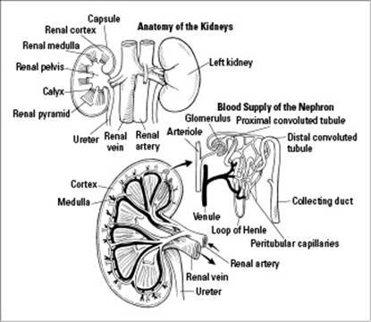
You have two kidneys, one on each side of your back, just below your ribs. Like most organs in the human body, the function of a kidney is closely tied to its structure (which we depict in Figure 16-2). As you can see, each kidney has three distinct areas:
The renal cortex, which is the outer layer
The renal medulla, which is the middle layer
The renal pelvis, which becomes a ureter
Each kidney contains more than 1 million nephrons, microscopic tubules that make urine. Each nephron contributes to a collecting duct that carries the urine into the renal pelvis. From there, the urine flows down the ureter, which is the tube that connects the kidney to thebladder.
Urine is spurted from the ureter into the top of the bladder continuously. The bladder holds a maximum of about one pint of urine, but you begin to feel the need to urinate when your bladder is only one-third full. When your bladder is two-thirds full, you start to feel really uncomfortable.
Figure 16-2:Structure of the kidneys and the nephrons inside the kidneys.
Urinary problems
Incontinence, the inability to hold urine in the bladder, often becomes an embarrassing problem for men and women later in life. Basically, the need to urinate becomes more urgent and frequent, and sometimes urine leaks out of the bladder uncontrolled. Incontinence is thought to be more prevalent in women than men due to the stress put on the sphincter muscles associated with the bladder during pregnancy and childbirth. Men, however, have an additional urinary issue: enlargement of the prostate gland.
The prostate sits right below the bladder and surrounds the urethra. Its function is to add fluid to semen as the semen passes through the urethra in the penis. As the prostate enlarges (which starts approximately when men reach age 50), it presses on the urethra, squeezing urine back up into the bladder and making urinating painful. If the condition progresses far enough, urine can back up into the kidneys, which can lead to kidney disease. As people age, the number of nephrons (the microscopic tubules that make urine) in a kidney declines, and the overall size of the kidney decreases, which can contribute to reduced renal function and lead to serious problems in older people.
Urine leaves the body through the urethra (see Figure 16-3), a tube at the bottom of the bladder that opens to the outside of the body. It’s held closed by a sphincter muscle. When you want to start urinating, the sphincter muscle relaxes, opening the urethra and letting the urine out.
Figure 16-3: The urinary system.
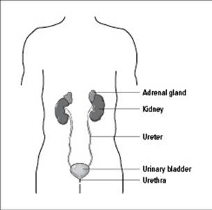
From LifeART®, Super Anatomy 1, © 2002, Lippincott Williams & Wilkins