Must Know High School Biology - Kellie Ploeger Cox 2019
PART FIVE Forms of Life
Animal Circulation and Respiration
MUST ![]() KNOW
KNOW
![]() The circulatory and respiratory systems work together to move oxygen and carbon dioxide between the tissues and the outside environment.
The circulatory and respiratory systems work together to move oxygen and carbon dioxide between the tissues and the outside environment.
![]() A high surface-area-to-volume ratio helps facilitate diffusion.
A high surface-area-to-volume ratio helps facilitate diffusion.
![]() Gases diffuse from high partial pressure to low partial pressure.
Gases diffuse from high partial pressure to low partial pressure.
![]() Oxygenated and deoxygenated blood does not mix in the most advanced four-chambered heart.
Oxygenated and deoxygenated blood does not mix in the most advanced four-chambered heart.
Circulatory System
All the cells of your body need to obtain oxygen and get rid of carbon dioxide. When these gases (CO2 and O2) are dissolved in water, they are more than happy to diffuse into (and out of) body cells. However movement by diffusion is fast only over very short distances. It is necessary for each and every cell in your body to have a source of oxygen nearby. The circulatory system takes care of oxygen delivery. I want you to keep the must know concept in mind as you continue reading. When oxygen (or any dissolved gas) moves from one area to another, it must do so down its concentration gradient. This means the gas will move from an area of high concentration (where there is a lot of the gas dissolved in the fluid) to an area of low concentration (where there is less of the gas dissolved in the fluid).
Some critters (such as flatworms and sea jellies) lack circulatory systems and instead rely only on diffusion to move oxygen into their body tissues. As pointed out by our must know concept, oxygen will move down its concentration gradient from the surrounding fluid into the oxygen-starved tissues. In order to make this an effective means of transfer, the critters’ body plans have evolved to maximize tissue exposure to the environment. For example, a flatworm is, well, flat, and a sea jelly has a hollow cavity called a gastrovascular cavity:
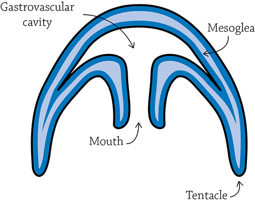
Cross section of a sea jelly
What these guys have in common (besides both being invertebrates) is that their body plans have maximal surface-area-to-volume ratios (SA:VOL), meaning there is a lot of surface area for the given volume of the body.
![]()
The higher the SA:VOL ratio, the better the diffusion rates. This means, for example, a tiny cell can deal with substances moving in and out much better than a large cell:

This is significant from an evolutionary perspective! The first cells to evolve were the small prokaryotic cells. Diffusion was sufficient for these early life-forms because prokaryotic cells have a high SA:VOL ratio. As cells evolved into the more complex eukaryotic cells, they became bigger. Their SA:VOL ratios decreased to the point that they could no longer rely on simple diffusion. In order to compensate, internal compartments helped by subdividing the contents and increasing the membrane’s surface area. These “compartments” are the eukaryotic organelles!
By far, most animals must rely on a circulatory system in order to reach all those oxygen-hungry cells. There are two options: an open circulatory system or a closed circulatory system.
Open Circulatory System
An open circulatory system is “open” because the circulating fluid (called “hemolymph”) freely washes through open cavities in the body, bathing organs with oxygen-rich fluids. Eventually, through the pumping of the simple heart and contractions of the muscles, the fluid is drawn through pores and funneled back into the heart. This works for arthropods (such as insects and crustaceans) and some mollusks (such as clams and snails). It is not very efficient, however, because the oxygen-rich and oxygen-poor blood can mix in the body. This just isn’t going to cut it for more active and physiologically complex critters … which is why natural selection has resulted in the second option: the closed circulatory system.
Closed Circulatory System
Consider for a moment the glorious octopus. Like the clams and the snails mentioned above, it is a member of Phylum Mollusca. But unlike all the other mollusks, members of Class Cephalopoda (octopi, squids, and cuttlefish) are graced with a closed circulatory system. Octopi and others of its class are avid hunters who need to quickly and nimbly chase down their prey. Such rapid movement requires a lot of energy, more than the inefficient open circulatory system of their peers can provide. This evolutionary event is a perfect illustration of the benefits of a closed circulatory system—its increased efficiency leads to much greater energy production.
A closed circulatory system is composed of a heart, blood, and vessels (arteries, veins, and capillaries) through which the blood flows. Because the blood is confined to vessels, it results in higher blood pressure, necessary for effective oxygen delivery in large and active species. The heart pumps blood into vessels that decrease in size, increase in branching, and supply blood to all the tissues of the body. Along with most cephalopods, annelids and all vertebrates benefit from a closed circulatory system.
Vertebrate Circulatory System
Our circulatory system is ridiculous … the total length of all vessels would circle Earth. Twice. Along with the muscle behind the movement (the heart), we have arteries to move blood away from the heart, and as they branch off into smaller arterioles, they provide pathways to all the organs in our body. Eventually the branching reaches maximum branchy-ness as tiny capillaries, the site of gas transfer between the blood and the cells of the body tissues. As suggested by our must know concept, the movement of the gases between the cells and the capillaries is based on concentration gradients. The blood then makes its way back to the heart as capillaries fuse into venules and finally the larger veins.
There are different circulation pathways, depending where on the chain of evolutionary events you stand. Fish, for example, are the first vertebrates to evolve, and they possess the simplest option of a two-chambered heart powering a single circulation pathway.
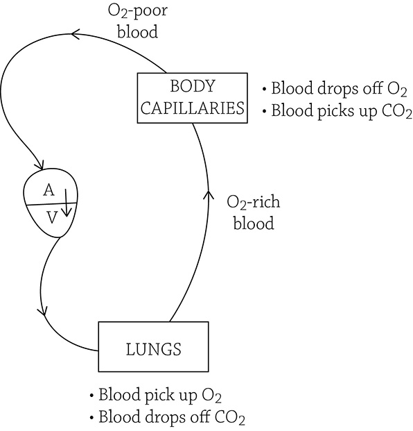
Single-circuit circulation pathway. A = atrium; V = ventricle
Blood passes through the heart only once per circuit, as it is powered out to the gills to pick up oxygen and release carbon dioxide. Then it travels on to the body tissues to drop off that oxygen (and pick up waste carbon dioxide). Blood pressure is pretty low at this point, since it traveled the entire circuit with a single pump of the heart. When the blood returns, it collects in the atrium and is pumped out through the ventricle for another loop. Once evolution took that great step from water onto land, there needed to be an option with a bit more power. Therefore, the amphibians of today (the closest vertebrate cousin to the fish) and reptiles (one more evolutionary step onto land) have a new-and-improved double-circulation pathway with a three-chambered heart. This provides better blood flow because the heart repressurizes the blood before it goes to the body tissues and again when it travels to the lungs. Yet there is still some mixing of oxygenated and deoxygenated blood in that single ventricle:
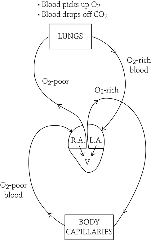
Double circulation with three-chambered heart. R.A. = right atrium; L.A. = left atrium; V = ventricle
Once we get to endotherms such as birds and mammals, we see the most efficient of systems: double circulation with a four-chambered heart. As it was for amphibians, there is a circuit that pumps blood to the lungs, and another circuit that pumps blood to the rest of the body. This keeps blood pressure strong, even when traveling to the farthest reaches of your toes. Now, however, mammals and birds have a four-chambered heart, with the right and left sides separated by a muscular wall called a septum. This prevents the oxygen-rich blood from the pulmonary circuit mixing with the oxygen-poor blood of the systemic circuit.
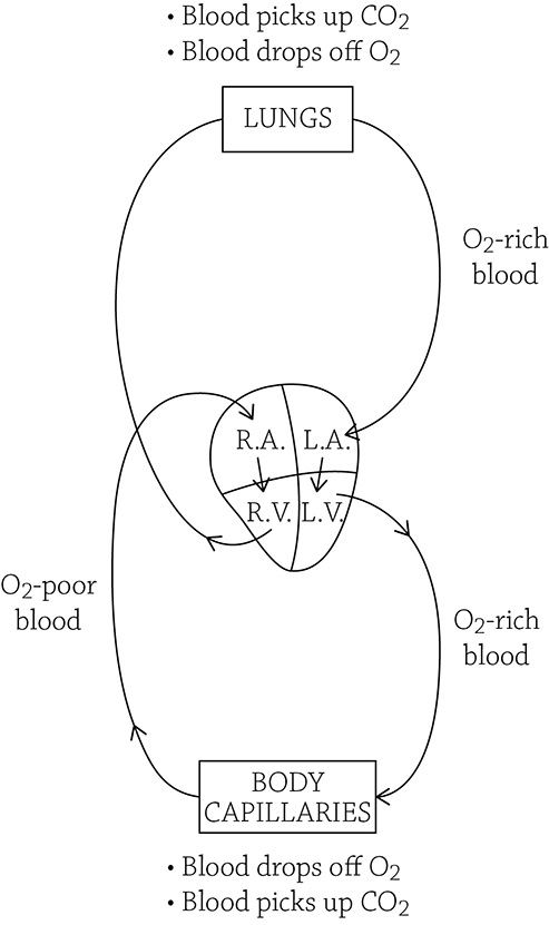
Double circulation with four-chambered heart
Since mammals and birds use about 10 times as much energy as their ectothermic cousins, it is necessary to deliver much more fuel and oxygen to the tissues.
Mammalian Details
The heart provides all the force needed to propel blood through the entire body. The atria receive the blood from the body: the left atrium gets the freshly oxygenated blood from the lungs, and the right atrium gets the spent, oxygen-poor blood from the body tissues. Once in the small atria, the blood passes through one-way valves into the larger ventricles, which then pump it away from the heart out to either the lungs (right ventricle’s pulmonary circuit) or the rest of the body (left ventricle’s systemic circuit). Since the blood has to travel much farther through the systemic circuit, it makes sense that the walls of the left ventricle are thicker and stronger.
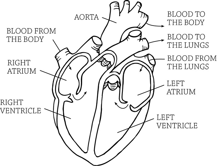
Mammalian heart anatomy
Author: Firkin. https://openclipart.org/detail/274066/heart-diagram-annotated
Blood pressure is a measurement of the force the blood exerts on the walls of the blood vessels; systolic pressure is the higher number (the force when the heart contracts) and diastolic pressure is the lower number (the pressure when the heart is at rest).
Arteries take blood away from the heart. Considering the heart is the source of all blood pressure, the arteries must be strong to withstand the force of the beating heart. The walls of arteries tend to be thick and muscular. The strongest artery of the body is the one closest to the heart: the aorta. Veins, on the other hand, bring blood back to the heart after gas exchange occurs. Their walls are floppy and thin, since the pumping force of the heart is so far away. Blood pressure in some veins can be so low that there are even one-way valves to prevent backflow!
Capillaries are the location of gas exchange (both oxygen and carbon dioxide) between the blood and the surrounding environment. Gas diffusion works best over close distances when the blood is traveling at slow speed, and capillary structure is perfect for creating both of these conditions.
The walls of capillaries are thin and porous (only one endothelial cell thick!). This greatly helps diffusion by easing the passage of gases through the capillary wall. The width of a capillary is also surprisingly narrow. In some locations, red blood cells must squeeze through in single file. The width of a single red blood cell is about 7 µm; capillary diameters range from 5 to 10 µm.
A capillary bed is a network of capillaries and the site for gas exchange in tissues. As blood enters through arterioles, the vessel branches again and again into the tiny capillaries. This leads to a huge increase in total cross-sectional area through which blood flows. The increase in area results in a drop in blood pressure; the blood flow slows down significantly, allowing time for diffusion to occur. This is a perfect example of form helping function!
Oxygen is ferried around the circulatory system by red blood cells. These cells are chock full of a chemical pigment whose sole job is to hang onto the oxygen until it is needed in the body. The pigment molecule responsible for grabbing onto oxygen is hemoglobin. It is composed of four subunits, each able to snag an oxygen molecule from the alveolar air. The cool thing about hemoglobin, it relates back to a concept we learned way back in Chapter 1. Remember allosteric enzymes? They had multiple subunits, and they could change their shapes into an active form or an inactive form. Well, hemoglobin is an example of a specific kind of allosteric regulation called cooperativity. Here, the binding of an oxygen molecule at one subunit will cause a shape change in the other subunits, making them even better at grabbing oxygen. That means if there’s oxygen waiting to be picked up, it’s going to happen quickly. And the opposite is true, too. When the blood reaches an area of the body that is low in oxygen, one molecule will break free and diffuse out of the blood into the oxygen-starved tissues, and all the other subunits will revert back to the shape that isn’t as good at holding onto oxygen. That way, when it is time to unload oxygen, it happens all at once.
![]()
Keep in mind the big picture. Why, exactly, do all the cells in your body need oxygen? Because cells need oxygen to produce ATP (the process of cellular respiration):
C6H12O6 + 6CO2 → 6CO2 + 6H2O + energy (ATP)
Respiratory System
At its most basic, the respiratory system supplies oxygen to the cells of the body. The specifics of how the respiratory system managed to funnel oxygen throughout the body is different, depending on the critter. Those flatworms and sea jellies, with their high surface areas, can manage without a specific respiratory system and rely on diffusion directly from the environment. Fish have gills, highly vascular tissues that absorb oxygen from the surrounding water. Insects have these funky internal tubes that run throughout their bodies and manage to supply every cell with air; the tubes themselves are connected to openings spaced along the surface of the bug’s body. Spiders have book lungs, fascinating structures that are so highly folded, it looks like the pages from a book. Yet as we did in the circulatory system, we will focus on the mammalian model (though all of these examples are fascinating!).
Mammalian Respiratory System
Gas exchange occurs across respiratory surfaces, where the circulatory system crosses paths with the respiratory system. Our must know concept is the same for the respiratory system as it was for the circulatory system: movement of respiratory gases is based on concentration gradients. In order to help diffusion of gases (oxygen and carbon dioxide) occur, the respiratory surfaces must be moist and have a high surface area. In mammals, this occurs at the dead-end passages of the lungs, tiny air sacs called alveoli:
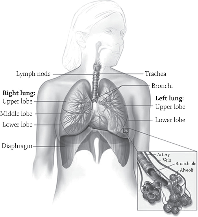
Alveoli of the respiratory tract
Author: National Cancer Institute (NCI). https://visualsonline.cancer.gov/details.cfm?imageid=7235
Human lungs contain literally millions of alveoli, and the total surface area is about 50 times that of our skin (or, more disturbingly, the size of a tennis court). Oxygen from the air entering the alveoli quickly diffuses across the epithelia into the capillaries surrounding each alveolus, where it’s whisked away to body tissues in need of oxygen. Meanwhile, the waste carbon dioxide moves from the capillaries and into the alveoli airspace, where it will be exhaled and removed from the body. The way to determine the direction gases will diffuse is through something called partial pressure of gases. The more gas in a mixture of gases, the higher its partial pressure. A gas will diffuse from a region of high partial pressure to low partial pressure (once again, our must know concept).
![]()
This sounds a lot like other diffusion scenarios. Diffusion, in general, is movement of something from a region of high concentration to low concentration. Osmosis (diffusion of water) occurs from a region of high water potential to low water potential. Diffusion of gases occurs from a region of high partial pressure to low partial pressure.
The carbon dioxide and oxygen partial pressures in the body vary, depending on the location. For example, blood that just dropped off oxygen to body cells and picked up the cells’ waste carbon dioxide will have a higher CO2 partial pressure and a lower O2 partial pressure. The opposite partial pressures would occur in the blood that just entered the tissues of the body, carrying with it a fresh supply of oxygen; here it would have a high O2 partial pressure and a low CO2 partial pressure. The pressures of each gas determine the directions they will move. Here are two examples:
![]() Scenario 1 A capillary bed supplying body tissues with oxygen
Scenario 1 A capillary bed supplying body tissues with oxygen
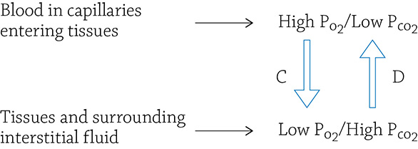
This is a model of partial pressures of gases in the capillaries supplying body tissues with oxygen-rich blood. The tissues, meanwhile, have been producing carbon dioxide as a waste product of cellular respiration:
A. The partial pressure of oxygen (PO2) is higher in the freshly oxygenated blood compared to the PO2 in the tissues; oxygen will diffuse from the bloodstream into the surrounding tissues.
B. The blood PCO2 is low because blood just left the alveoli where it dumped of excess carbon dioxide to be exhaled; the carbon dioxide will now diffuse from the tissues into the bloodstream, so it can travel back to the lungs.
![]() Scenario 2 Blood supply arriving at the lungs to pick up oxygen and dump off carbon dioxide:
Scenario 2 Blood supply arriving at the lungs to pick up oxygen and dump off carbon dioxide:
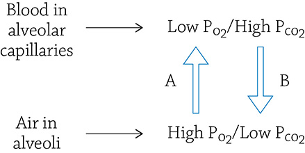
This shows what happens when a fresh inhalation of air settles into the alveoli.
A. The partial pressure of oxygen (PO2) is lower in the blood compared to the PO2 in the air; oxygen will diffuse from the air into the bloodstream.
B. The blood PCO2 is high because it just returned from picking up waste CO2 from body tissues; the carbon dioxide will diffuse from the bloodstream into the alveoli.
REVIEW QUESTIONS
1. In order for an organism to rely on direct diffusion for oxygen transport (without the aid of a circulatory system), what quality must its body plan have?
2. Why is a four-chambered heart more efficient than a three-chambered heart?
3. The place of gas exchange between the respiratory system and the circulatory system occurs in the ______________ of the respiratory system and the surrounding ______________ of the circulatory system.
4. Where would the partial pressure of oxygen be higher than the partial pressure of carbon dioxide?
5. How did surface area play a role in eukaryotic cell evolution?
6. The mammalian ______________ ventricle has a thicker wall than the ______________ ventricle. The ______________ ventricle needs to be stronger because it powers the blood through the entire body (the ______________ circuit). The ______________ ventricle only needs to power the blood out to the ______________ (the ______________ circuit).
7. How are the circulatory system and the respiratory system related in their functions?
8. Choose the correct term from each of the following pairs: Blood coming from the lungs and entering the tissues has a high oxygen/carbon dioxide partial pressure. The tissues have a high oxygen/carbon dioxide partial pressure because the cells have been producing a lot of oxygen/carbon dioxide through the process of cellular respiration. These partial pressures facilitate the movement of oxygen from the tissues/blood into the tissues/blood.