Biology of Humans
7. Neurons: The Matter of the Mind
In the previous chapter, we learned how muscles contract when they receive messages from nerve cells. In this chapter, we will discover how nerve cells communicate with muscle cells and with one another. In Chapter 8, “The Nervous System," we will explore the parts and functions of the brain and spinal cord.
Cells of the Nervous System
The nervous system integrates and coordinates all the body's varied activities. Its two primary divisions, which are the subject of Chapter 8, are (1) the central nervous system, consisting of the brain and spinal cord; and (2) the peripheral system, consisting of all the nervous tissue in the body outside the brain and spinal cord. Both these major divisions of the nervous system are composed of two types of specialized cells. Neurons (nerve cells) are excitable cells that generate and transmit messages. Outnumbering the neurons by about 10 to 1, neuroglial cells (also called glial cells) support and protect neurons.
Neuroglial Cells
The nervous system has several types of glial cells, each with a different job to do. Some glial cells provide structural support for the neurons of the brain and spinal cord. Glial cells also provide a steady supply of chemicals, called nerve growth factors, that stimulate nerve growth. Without nerve growth factors, neurons die. Other glial cells form insulating sheaths around axons that, as described shortly, are the long projections extending from certain neurons. This sheath, called the myelin sheath, has several important functions that are also described shortly. Scientists now know that glial cells can communicate with one another and with neurons.
· The brain cells we use to plot and plan our way to victory in a chess match have the same design and mode of functioning as the cells that tell our muscles to contract or that carry information from our sensory organs to our brain. They are neurons, the basic functional units of the nervous system.
Neurons
The basic unit of the nervous system is the neuron, or nerve cell. Neurons, which are responsible for an amazing variety of functions, can be grouped into the three general categories depicted in Figure 7.1.
• Sensory (or afferent) neurons conduct information toward the brain and spinal cord. These neurons generally extend from sensory receptors, which are structures specialized to gather information about the conditions within and around our bodies.
• Motor (or efferent) neurons carry information away from the brain and spinal cord to an effector—either a muscle, which will contract, or a gland, which will secrete its product, as a response to information from a sensory or interneuron.
• Association neurons, commonly called interneurons, are located between sensory and motor neurons. They are found only within the brain and spinal cord, where they integrate and interpret the sensory signals, thereby "deciding" on the appropriate response. Interneurons are by far the most numerous nerve cells in the body; they account for more than 99% of the body's neurons.
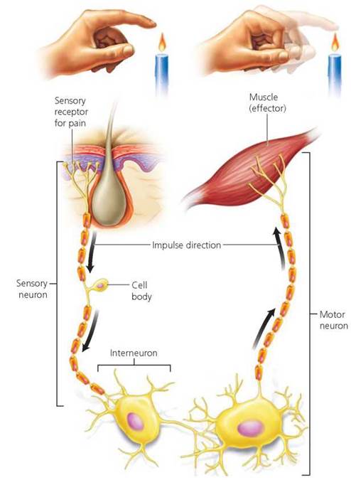
FIGURE 7.1. Neurons may be sensory neurons, interneurons, or motor neurons. This diagram traces the pathway of an impulse from a sensory receptor to an interneuron and from there to a motor neuron and its effector. Sensory receptors detect changes in the external or internal environment. An interneuron usually receives input from many sensory neurons, integrates that information, and if the input is appropriate, stimulates a motor neuron. The motor neuron then causes a muscle or a gland (an effector) to respond.
We can appreciate the specific roles of each type of neuron by considering the symptoms of a progressive disease called amyotrophic lateral sclerosis (ALS), also known as Lou Gehrig's disease. In ALS, motor neurons throughout the brain and spinal cord die and stop sending messages to skeletal muscles. Without stimulation from motor neurons, the muscles gradually weaken, and the person loses control over arms, legs, and body. The cause of death is respiratory failure, because the muscles that control breathing (the diaphragm and rib muscles) eventually die. Sensory neurons and interneurons are not affected by ALS, so awareness and reasoning do not deteriorate.
Structure of Neurons
The shape of a typical neuron is specialized for communicating with other cells (Figure 7.2).
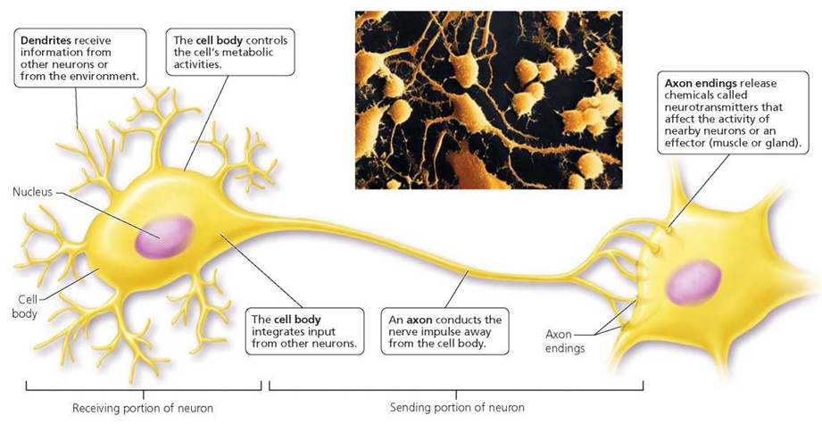
FIGURE 7.2. The structure of a neuron
Axons and Dendrites
A neuron has many short, branching projections called dendrites, which provide a huge surface for receiving signals from other cells. Such signals travel toward an enlarged central region called the cell body, which has all the normal organelles, including a nucleus, for maintaining the cell. When a neuron responds to an incoming signal, it transmits its message along the axon, a single long extension of the neuron. The axon carries messages away from the cell body either to another neuron or to an effector, which can be a muscle or a gland. In some cases, the axon allows the neuron to communicate over very long distances. For example, a motor neuron that allows you to wiggle your big toe has its cell body in the spinal cord and its axon runs all the way to the muscles of your toe. The end of the axon has many branches specialized to release a chemical, called a neurotransmitter, that alters the activity of the effector. The axon is the sending portion of the neuron, whereas the dendrites and cell body are typically the receiving portions.
To appreciate the dimensions of a neuron, imagine a "typical" motor neuron, one that carries a message from the spinal cord to a muscle. Now picture the cell body of this neuron as being the size of a tennis ball. At that scale, the axon of this neuron would be about 1 mile (1.6 km) long and only about one-half inch (1.3 cm) in diameter. At the same scale, the dendrites—the shorter but more numerous projections of the neuron—would fill an average-sized living room.
A nerve is a bundle of parallel axons, dendrites, or both arising from many neurons. Each nerve is covered with connective tissue and, depending on the type of neurons it contains, can be classified as sensory, motor, or mixed (a mixed nerve is made up of both sensory and motor neurons).
Myelin Sheath
Most of the axons outside the brain and spinal cord, and some of those within, have an insulating outer layer called a myelin sheath (Figure 7.3), which increases the rate of conduction of a nerve impulse and helps its repair. The myelin sheath is composed of the plasma membranes of glial cells. Outside of the brain and spinal cord, for example, glial cells known as Schwann cells form neurons' myelin sheaths. A Schwann cell plasma membrane wraps around the axon many times to provide a covering that looks somewhat like a jelly roll. This covering, the myelin sheath, serves as a kind of living electrical tape, insulating individual axons and preventing messages from short-circuiting between neurons. The myelin sheath is kept alive by the Schwann cell's nucleus and cytoplasm, which are squeezed to the periphery as the sheath forms.
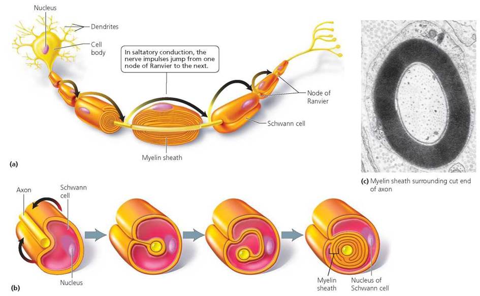
FIGURE 7.3. The myelin sheath, (a) An axon protected by a myelin sheath. The Schwann cells that form the myelin sheath are separated by nodes of Ranvier—areas of exposed axon that allow for saltatory conduction, (b) The myelin sheath forms from multiple wrappings of Schwann cell plasma membranes, (c) An electron micrograph of the cut end of a myelinated axon.
Long axons are myelinated, but short axons may not be. Why?
Saltatory conduction, which increases the speed of conduction, is possible only on myelinated axons. The increased speed is important when nerve impulses must travel long distances.
A single Schwann cell encloses only a small portion, about 1 mm long, of an axon. The gaps between adjacent Schwann cells, where the axon is exposed to the extracellular environment, are called nodes of Ranvier. Their presence is crucial to how rapidly a neuron transmits messages. With the myelin sheath in place, a nerve impulse "jumps" successively from one node of Ranvier to the next in a type of transmission called saltatory conduction (saltare, to jump), which is up to 100 times faster than signal conduction would be on an unmyelinated axon of the same diameter. Not surprisingly, the axons responsible for conducting signals over long distances are typically myelinated.
To get a sense of how this "jumping" mode of transmission increases the speed at which a message travels, think of the different ways a ball can be moved down the court during a basketball game. When only seconds are left in the game, dribbling the ball the length of the court would take too much time. Passing the ball through a series of players is faster. Likewise, an impulse passed from one node to the next, as occurs in myelinated nerves, moves faster than one traveling uniformly along the full length of the axon, as occurs in unmyelinated nerves.
The importance of the myelin sheath becomes dramatically clear in people with multiple sclerosis, a disease in which the myelin sheaths in the brain and spinal cord are progressively destroyed. The damaged regions of myelin become hardened scars called scleroses (hence the name of the disease) that interfere with the transmission of nerve impulses. Short-circuiting between normally unconnected conduction paths delays or blocks the signals going from one brain region to another. Depending on the part of the nervous system affected, the result can be paralysis or the loss of sensation, including loss of vision.
Nerve Impulses
The neuron membrane is specialized for communication. A nerve's message, which is called a nerve impulse or an action potential, is an electrochemical signal caused by sodium ions (Na+) and potassium ions (K+) crossing the neuron's membrane to enter and leave the cell.
Plasma Membrane of a Neuron
Like most living membranes, the plasma membrane of a neuron is selectively permeable; it allows some substances through but not others. The membrane contains many pores, called ion channels, that ions are able to pass through without using cellular energy. Each ion channel is designed to allow only certain ions to pass through. For example, sodium channels allow the passage of only sodium ions, and potassium channels allow the passage of only potassium ions. In this way, ion channels function as molecular sieves. Some channels are permanently open. Others are regulated by a "gate"—a protein that changes shape in response to changing conditions, either opening the channel, which allows ions to pass through, or closing it, which prevents ions from crossing the membrane.
The cell membrane also contains sodium-potassium pumps, which are special proteins in the cell membrane that actively transport sodium and potassium ions across the membrane. These pumps use cellular energy in the form of ATP to move the ions against their concentration gradients. Each pump ejects sodium ions (Na+) from within the cell while bringing in potassium ions (K+).
Resting Potential
It will be easier to understand the movement of ions during an action potential if we first consider a neuron that is not transmitting an action potential—that is, a neuron in its resting state. As we will see, however, resting is hardly the word to describe what is going on at this stage. The membrane of a resting neuron maintains a difference in the electrical charges near the two membrane surfaces (the surface facing inside the cell and the surface facing outside the cell), keeping the inside surface more negative than the outside one. This charge difference across the membrane, called the resting potential, results from the unequal distribution of ions across the membrane.
In a resting neuron, sodium and potassium ions are unequally distributed across the plasma membrane. There are more sodium ions outside the membrane than inside. Furthermore, there are more potassium ions inside than outside. Potassium ions tend to leak out because they are more concentrated inside the axon. (Recall from Chapter 3 that substances tend to move from an area of higher concentration to one of lower concentration.) To a lesser extent, sodium ions leak in. However, sodium-potassium pumps maintain the resting potential of a neuron by pumping out sodium ions while moving potassium ions back in. The cell also contains some negatively charged ions that are too large to pass through the membrane.
The result of this unequal distribution of ions is that the inner surface of a resting neuron's membrane is typically about 70 mV (millivolts) more negative than the outer surface. This voltage is the resting potential, and it is about 5% of the strength of a size AA flashlight battery.
Although the neuron's sodium-potassium pumps consume a lot of energy to maintain the resting potential, the energy is not wasted. The resting potential allows the neuron to respond more quickly than it could if the membrane were electrically neutral in its resting state. This situation is somewhat analogous to keeping your car's battery charged so that the car will start as soon as you turn the key.
Action Potential
Now we are ready to consider what happens when a neuron is stimulated—that is, when it receives some kind of excitatory signal. The action potential, or nerve impulse, resulting from such stimulation can be described briefly as a sudden reversal in the charge difference across the membrane, followed by the restoration of the original charge difference. Let's take a closer look at these two parts of a nerve impulse.
1. Sodium ions (Na+) enter the axon. An excitatory stimulus causes the gates on sodium channels to open. Sodium ions then enter the neuron, and their positive charge begins to reduce the negative charge within. Reduction of the charge difference across the membrane is called depolarization. The action potential begins when membrane depolarization reaches a certain value called the threshold. When the threshold is reached, the gates on more sodium channels open. Enough sodium ions enter through the open gates to create a net positive charge in that region (about +30 mV), as shown in Figure 7.4.
2. Potassium (K+) ions leave the axon. About halfway through the action potential, the gates on potassium channels open. Potassium ions now leave the cell. The exodus of potassium ions with their positive charge causes the interior of the neuron to become negative once again relative to the outside. The outward flow of potassium ions returns the membrane potential close to its resting value. Restoration of the charge difference across the membrane is called repolarization.
As noted, at the end of an action potential, the charge distribution across the membrane returns to the resting potential. However, there are slightly more sodium ions and slightly fewer potassium ions inside the cell than before. This alteration is corrected by the sodium-potassium pump, which restores the original distribution of sodium and potassium ions. The action of the sodium-potassium pump is slow. Therefore, it does not contribute directly to the events of the action potential.
To summarize, the action potential is a reversal of the charge difference across the membrane caused by the inward flow of sodium ions, followed immediately by restoration of the original charge difference caused by the outward flow of potassium ions (Figure 7.5). These changes occur sequentially along the axon, like a wave rippling away from the cell body.
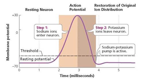
FIGURE 7.5. A graphic representation of an action potential. Voltage across the membrane can be measured by electrodes placed inside and outside the axon. The graph shows the changes in voltage that accompany an action potential.
The action potential is described as a wave of changes that travels down the neuron's plasma membrane because the events just described do not occur simultaneously along the entire length of the axon. Instead, as sodium ions enter the cell at one location along the membrane, and the charge inside the membrane becomes less negative in that region of the cell, the change in charge causes the opening of the sodium channel gates in an adjacent part of the membrane. Due to this sequential opening of gates, the change in charge travels down the length of the axon. Once started, action potentials do not diminish—just like the last domino in a falling row falls with the same energy as the first. Moreover, the intensity of the nerve impulse does not vary with the strength of the stimulus that triggered it. If an action potential occurs at all, it is always of the same intensity as any other action potential. This "all-or-nothing" aspect of nerve cell conduction is similar to the firing of a gun in that the force of the bullet is not changed by how hard you pull the trigger.
Immediately after an action potential occurs, the neuron cannot be stimulated again for a brief instant called the refractory period. During the refractory period, the sodium channels are closed and cannot be reopened. Consequently, a new action potential cannot yet be generated. Because of the refractory period, a prolonged stimulus that is above threshold can cause only a series of discrete nerve impulses, not a single, larger, sustained impulse. For this reason, increasing the strength of a stimulus will increase the frequency of impulses. For example, the frequency of nerve impulses increases with the heat of an object touched. However, the frequency can increase only to a point. The inability of the sodium gates to open during the refractory period is also the reason that the nerve impulses cannot reverse and go backward toward the cell body.
Stop and think
“Red tides” in the ocean are caused by proliferation of dinoflagellates. These single-celled marine algae contain a chemical called saxitoxin (STX), extremely small concentrations of which prevent sodium channels in mammalian neurons from opening. The clams, scallops, and mussels that consume the dinoflagellates are insensitive to the toxin, but the STX accumulates in their tissues. What effect would you expect STX to have on nerve transmission in humans who accidentally consume tainted shellfish?
Synaptic Transmission
When a nerve impulse reaches the end of an axon, in almost all cases the message must be relayed to the adjacent cell across a small gap that the impulse cannot jump across. To transmit the message to the adjacent cell requires a brief change of the medium of communication from an electrochemical signal to a chemical signal. Therefore, when an action potential reaches the end of the axon, a chemical is released from the axon's tip. That chemical, called a neurotransmitter, diffuses across the gap and conveys the message to the adjacent cell.
The junction between a neuron and another cell is called a synapse. The structure of a synapse between two neurons is shown in Figure 7.6. The gap between the cells is called the synaptic cleft. Recall that the axon branches near the end of its length. Each branch ends with a small bulblike swelling called a synaptic knob. The neuron sending the message is the presynaptic neuron (meaning "before the synapse"). The neuron receiving the message is the postsynaptic neuron ("after the synapse").
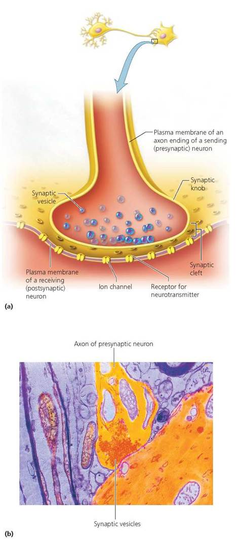
FIGURE 7.6. Structure of a synapse. (a) The synaptic knob at the end of the axon on the presynaptic neuron is separated from the dendrite or cell body of the postsynaptic neuron by a small gap called a synaptic cleft. Within the synaptic knob are small sacs, called synaptic vesicles, filled with neurotransmitter molecules, (b) An electron micrograph of a synapse.
Release of the Neurotransmitter and the Opening of Ion Channels
Let's consider the events that occur in the synapse as the message is sent from one neuron to the next (Figure 7.7).
1. The nerve impulse reaches the axon ending of the presynaptic neuron.
2. Synaptic knobs release packets of neurotransmitter.
Within the synaptic knobs of the presynaptic neuron, the neurotransmitter is contained in tiny sacs called synaptic vesicles. When a nerve impulse reaches the synaptic knob, the gates of calcium ion channels in the membrane there open. Calcium ions move into the knob, causing the membranes of the synaptic vesicles to fuse with the plasma membrane at the synaptic knob and to dump the enclosed neurotransmitter into the synaptic cleft.
3. Neurotransmitter diffuses across the synaptic cleft.
4. Neurotransmitter binds with receptors on the membrane of the postsynaptic neuron. A receptor is a protein that recognizes a particular neurotransmitter, much as a lock "recognizes" a key. The only cells a neurotransmitter can stimulate are cells that have receptors specific for that particular neurotransmitter. Thus, only certain neurons can be affected by a given neurotransmitter.
5. When a neurotransmitter binds to its receptor, an ion channel is opened. The binding of the neurotransmitter to a receptor causes the opening of an ion channel in the postsynaptic neuron. The response that is triggered as a result depends on the type of ion channel the receptor opens. It is the receptor that determines which ion channels will open and what the effect of a given neurotransmitter will be.
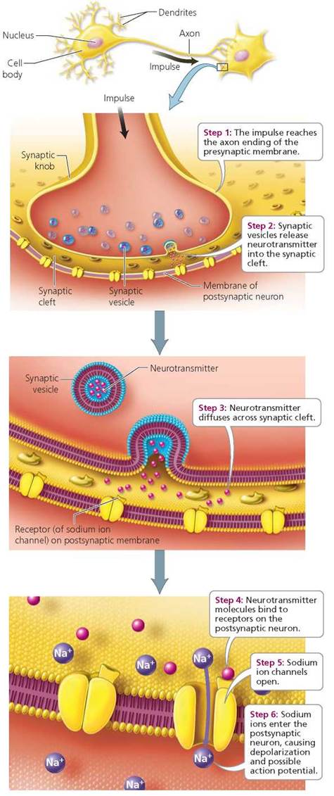
FIGURE 7.7. Transmission across an excitatory synapse
An excitatory synapse is one where the binding of the neurotransmitter to the receptor opens sodium channels, allowing sodium ions to enter and increasing the likelihood that an action potential will begin in the postsynaptic cell. In contrast, an inhibitory synapse is one where the binding of the neurotransmitter opens different ion channels, which decreases the likelihood that an action potential will be generated in the postsynaptic neuron. In this case, the cell's interior becomes more negatively charged than usual. As a result, the cell will require larger than usual amounts of an excitatory neurotransmitter in order to reach threshold.
Summation of Input from Excitatory and Inhibitory Synapses
A neuron may have as many as 10,000 synapses with other neurons at the same time (Figure 7.8). Some of these synapses will have excitatory effects on the postsynaptic membrane. Others will have inhibitory effects. The summation (combined effects) of excitatory and inhibitory effects on a neuron at any given moment determines whether an action potential is generated. This integration of input from large numbers of different kinds of synapses gives the nervous system fine control over neuronal responses, just as having both an accelerator and a brake gives you finer control over the movement of a car.
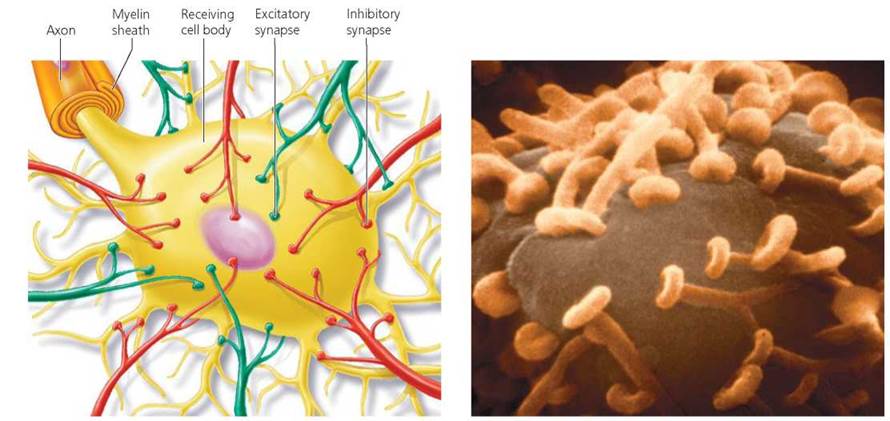
FIGURE 7.8. A neuron may have as many as 10,000 synapses at which it receives input from other neurons. Some synapses have an excitatory effect on the membrane of the postsynaptic neuron and increase the likelihood that the neuron will fire. Other synapses have an inhibitory effect and reduce the likelihood that the postsynaptic neuron will fire. The net effect of all the synapses determines whether an action potential is generated in the postsynaptic neuron. (The shape of the synaptic knobs shown in this electron micrograph is distorted as a result of the preparation process.)
Health Issue
Neurotransmitters and Disease
Neurotransmitters affect our movements, memory, and emotions. It should not be surprising, then, that changes in neurotransmitter level can cause disorders.
Alzheimer’s Disease and Acetylcholine
Alzheimer’s disease is progressive and results in loss of memory, particularly for recent events, followed by sometimes severe personality changes. In Alzheimer's disease, the parts of the brain important in memory and intellectual functioning (hippocampus and cerebral cortex; see Chapter 8) lose large numbers of neurons. Some of the neurons in these regions use the neurotransmitter acetylcholine, which may decrease in level by as much as 90% in a person with Alzheimer's and possibly explains the loss of memory and mental capacity. In addition, a brain affected by Alzheimer's disease is pocked with clusters of proteins, some between the neurons (amyloid plaques) and others within the neurons (neurofibrillary tangles). The amyloid plaques and neurofibrillary tangles are the prime suspects for the cause of the death of acetylcholine-producing neurons.
In hypothesizing that the loss of acetylcholine is responsible for some of the Alzheimer's symptoms, researchers and physicians have attempted to treat Alzheimer's disease with drugs meant to raise or at least maintain acetylcholine levels. Although such drugs (Aricept, Exelon, and Reminyl) do improve the memory and intellectual ability of some people with Alzheimer's, they do not help in all cases. Moreover, any improvement is rapidly lost when a person stops taking the drugs.
Depression, Serotonin, Dopamine, and Norepinephrine
We all describe ourselves as feeling depressed at times. However, more than 19 million Americans experience depression that lasts for weeks, months, or years and interferes with their ability to function in daily life. This condition is considered clinical depression, and it can affect anyone. It is thought to be related in some way to insufficient levels of the neurotransmitter serotonin as well as of dopamine and norepinephrine. Signs of depression can include a loss of interest and pleasure in the activities and hobbies that were previously pleasurable; anxiety; sleep problems; decreased energy; and feelings of sadness, hopelessness, worthlessness, and guilt. Depression takes the joy out of life and complicates certain medical conditions such as heart disease, cancer, diabetes, epilepsy, and osteoporosis.
Depression can be treated successfully. Unfortunately, few of the millions of people suffering from depression recognize the symptoms and seek help. Antidepressant drugs affect the functioning of the neurotransmitters responsible for the problem: norepinephrine and serotonin. Older medications affected both neurotransmitters simultaneously. Newer ones, including Prozac, Zoloft, and Paxil, specifically affect serotonin functioning, increasing the level of serotonin in the synapse by reducing its rate of removal. Other antidepressants (such as Welbutrin) increase norepinephrine and dopamine functioning by reducing their rates of removal from the synapse.
Parkinson’s Disease and Dopamine
Actor Michael J. Fox and former heavyweight champion Muhammad Ali have Parkinson's disease (Figure 7.A), a progressive disorder that results from the death of dopamine-producing neurons deep in the brain's movement control center. A person with Parkinson's disease moves slowly, usually with a shuffling gait and a hunched posture, and may suffer from involuntary muscle contractions that may cause tremors (involuntary rhythmic shaking) of the hands or head, because of muscles alternately contracting and relaxing; or they may cause muscle rigidity, because of muscles contracting continuously. This muscle rigidity may cause sudden “freezing” in the middle of a movement.
As the dopamine-producing neurons in the brain's movement control center die, dopamine levels begin to fall. Initially, the symptoms are subtle and are often written off as part of the aging process. By the time the Parkinson's diagnosis is apparent, 80% of the neurons in this small area of the brain may already have died.
Attempts to treat Parkinson's disease have focused on replacing dopamine or helping the brain get by with the remaining dopamine. Unfortunately, swallowing pills containing dopamine does not help, because dopamine is prevented from reaching the brain by the blood-brain barrier that you will read about in Chapter 8. Instead, patients are given other substances that can reach the brain. The most common and effective treatment combines two drugs: l-dopa, an amino acid that the brain converts to dopamine, and carbidopa, which prevents dopamine from forming outside of the brain and causing undesirable side effects. However, this treatment does not stop the steady loss of dopamine-producing neurons, so it loses effectiveness as the disease progresses. Some patients are treated with drugs that enhance their levels of dopamine by inhibiting the enzyme that breaks down dopamine.
Questions to Consider
• Few people would question the justifiability of providing drugs that elevate neurotransmitter function to someone who is depressed or suicidal in order to help the person live a normal life. However, researchers believe that the levels of key neurotransmitters also affect personality traits, such as shyness or impulsiveness. If so, we may someday be able to design our own personalities. Should minor personality problems be treated with drugs? Should a personality “flaw” like shyness or impulsiveness be treated?
• Schizophrenia, a mental illness characterized by hallucinations and disordered thoughts and emotions, is caused by an imbalance between the neurotransmitters dopamine and glutamate in one part of the brain (the midbrain). To verify that a drug is useful in treating schizophrenia, controlled experiments must be performed. In some experiments this means giving patients powerful antipsychotic drugs with unknown side effects. In other experiments this means taking patients off medication to see whether they would suffer a psychotic relapse. Participation in such experiments now requires written, informed consent. Every detail of the experiment and its potential risks must be presented in writing. Do you think that someone with schizophrenia will understand the consent form? Should researchers be held accountable if they did not know about certain risks? Should the research be published if the participants did not give informed consent?

FIGURE 7.A. Actor Michael J. Fox and former heavyweight champion Muhammad Ali have Parkinson’s disease, a progressive debilitating disease characterized by slowed movements, tremors, and muscle rigidity. The symptoms result from insufficient amounts of the neurotransmitter dopamine, caused by the death of dopamine-producing nerve cells in a movement control center of the brain. The Michael J. Fox Foundation for Parkinson’s Research (www.michaeljfox.org) funds research on the early diagnosis and treatment of Parkinson’s disease.
Removal of Neurotransmitter
After being released into a synapse, neurotransmitters are quickly removed so their effects are temporary. If they were not removed, they would continue to excite or inhibit the postsynaptic membrane indefinitely. Depending on the neurotransmitter, disposal is accomplished in one of two ways. First, enzymes can deactivate a neurotransmitter. For example, the enzyme acetylcholinesterase removes the neurotransmitter acetylcholine from synapses where it has been released. Second, the neurotransmitter may be actively pumped back into the presynaptic knob.
What would you do?
The organophosphate insecticides Malathion, Parathion, and Diazinon are poisons that kill insects by excessively boosting the activity at certain synapses in the insect's body. They work by inhibiting acetylcholinesterase. As a result, acetylcholine accumulates in the synapses and has a continuous effect. Because they have the same effect in the human body, these insecticides accidentally poison approximately 500,000 people around the world each year, primarily farm workers. As a consumer, are you willing to pay more for food that is not sprayed with pesticides to protect the lives of farm workers?
Roles of Different Neurotransmitters
As we have seen, neurotransmitters are the chemical means of communication within the nervous system. There are dozens of neurotransmitters, carrying messages among neurons and between neurons and muscles or glands. The activities of neurotransmitters produce our thoughts and feelings and enable us to interact appropriately with the world around us. Some neurotransmitters produce different effects on different types of cells.
Acetylcholine and norepinephrine are neurotransmitters that act in both the peripheral and the central nervous systems. Both have either excitatory or inhibitory effects, depending on where they are released. As we see in Chapter 8, most internal organs receive input from neurons that release acetylcholine and from neurons that release norepinephrine. Norepinephrine stimulates most organs but inhibits certain others. Whatever the effect of norepinephrine on any particular organ, acetylcholine will have the opposite effect.
Acetylcholine is also the neurotransmitter released at every neuromuscular junction (the junction of a motor neuron and a skeletal muscle cell), where it triggers contraction of voluntary (skeletal) muscles. We can see how important the nerve activation of muscle is whenever the interaction of nerve and muscle is interrupted. An example of such interruption is myasthenia gravis, an autoimmune disease in which the body's defense mechanisms attack the acetylcholine receptors at neuromuscular junctions. With any repeated movement, the amount of acetylcholine released with each nerve impulse decreases after the neurons have fired a few times in succession. The low number of acetylcholine receptors in people with myasthenia gravis makes such people extremely sensitive to even the slightest decline in acetylcholine availability. As a result, a person with myasthenia gravis has little muscle strength, and their repeated movements become feeble quite rapidly. Drugs that inhibit acetylcholinesterase are prescribed to prevent the breakdown of acetylcholine, elevating the level of acetylcholine in the neuromuscular junction.
About 50 neurotransmitters are used by the central nervous system for communication between the neurons in our brains. Why so many? One reason seems to be that different neurotransmitters are involved with different behavioral systems. Norepinephrine, for instance, is important in the regulation of mood, in the pleasure system of the brain, and in arousal. Norepinephrine is thought to produce an energizing "good" feeling. It is also thought to be essential in hunger, thirst, and the sex drive. Serotonin is thought to promote a generalized feeling of well-being. Dopamine helps regulate emotions. It is also used in pathways that control complex movements. A change in the level of a neurotransmitter affects the behaviors controlled by neurons that communicate using that neurotransmitter (see the Health Issue, Neurotransmitters and Disease).
Looking ahead
In this chapter, we considered the structure and function of neurons. In the next chapter, we will see how neurons are organized to form a functional nervous system.
Highlighting the Concepts
Cells of the Nervous System (pp. 116-117)
• The nervous system has two types of specialized cells: neurons (nerve cells) and neuroglial cells.
• Neuroglial cells outnumber neurons. They provide structural support for neurons, supply nerve growth factors, and form myelin sheaths around certain axons.
• There are three general categories of neurons. Sensory (or afferent) neurons conduct information from the sensory receptors toward the central nervous system. Motor (or efferent) neurons conduct information away from the central nervous system to an effector. Association neurons (interneurons) are positioned between sensory and motor neurons and are located in the central nervous system.
Structure of Neurons (pp. 117-119)
• Neurons are specialized for communicating with other cells. A typical neuron has a cell body containing the organelles that maintain the cell. Many branching fibers called dendrites conduct messages toward the cell body. A single long axon conducts impulses away from the cell body.
• An axon may be enclosed in an insulating layer called a myelin sheath. Neuroglia called Schwann cells form the myelin sheath by wrapping their plasma membranes repeatedly around the axon. The myelin sheath greatly increases the rate at which impulses are conducted along an axon; it also plays a role in the regeneration of cut axons in the peripheral nervous system.
Nerve Impulses (pp. 119-122)
• The message conducted by a neuron, called a nerve impulse or action potential, is caused by sodium ions (Na+) and potassium ions (K+) crossing the neuron's plasma membrane to enter and leave the cell.
• Ion channels in the plasma membrane are small pores through which ions move without the use of cellular energy. Ion channels are usually specific to one or a few types of ions.
• The sodium-potassium pump uses cellular energy in the form of ATP to pump sodium ions out of the cell and potassium ions into the cell against their concentration gradients.
• In the resting state, a neuron has an electrical potential difference, called the resting potential, across its plasma membrane. The resting potential is generated by an unequal distribution of charges across the membrane that makes the neuron more negative inside than outside; there are many negatively charged proteins inside the cell. Sodium ions are in greater concentration outside the neuron, and potassium ions are in greater concentration inside.
• The action potential begins when a region of the membrane suddenly becomes permeable to sodium ions. If enough sodium ions enter to reach a threshold, an action potential is generated. The gates on sodium channels open, and many sodium ions enter the cell, making its interior in that region of the membrane temporarily positive (depolarization). Potassium ions then leave the cell, making that region inside once again more negative than the outside (repolarization). The same events are repeated all along the axon in a wave of depolarization and repolarization called an action potential.
• At the end of an action potential, the sodium-potassium pump moves sodium ions out of the neuron and potassium ions into the neuron, restoring the original ion distribution.
• Once initiated, an action potential sweeps to the end of the axon without diminishing in strength.
• During the brief refractory period immediately following an action potential, the neuron cannot be stimulated.
Synaptic Transmission (pp. 122-126)
• The point where one neuron meets another is called a synapse. The neuron sending the message (the presynaptic neuron) and the neuron receiving the message (the postsynaptic neuron) are separated by a small gap called the synaptic cleft.
• The arrival of a nerve impulse at the axon's end causes calcium ions to enter the presynaptic cell there. These ions cause synaptic vesicles that store neurotransmitters to fuse with the plasma membrane of the presynaptic neuron and release their contents into the synaptic cleft. The neurotransmitter then diffuses across the gap and binds to receptors on the membrane of the postsynaptic neuron.
• If the synapse is excitatory, sodium ions enter the postsynaptic cell and increase the likelihood that it will generate a nerve impulse. If the synapse is inhibitory, the charge difference across the membrane of the receiving neuron is increased, reducing the likelihood that the postsynaptic neuron will generate a nerve impulse.
• Postsynaptic cells integrate excitatory and inhibitory input from many cells. If the threshold is reached, an action potential is generated in the postsynaptic cell.
• The neurotransmitter is quickly removed from the synapse either by enzymatic breakdown or by transport back into the presynaptic neuron.
• Acetylcholine, epinephrine, and norepinephrine are neurotransmitters used in both the peripheral and central nervous systems.
• Many neurotransmitters are found in the brain. Different ones are active in different behavioral systems. Disturbances in brain chemistry affect mood and behavior.
Reviewing the Concepts
1. What are the functions of neuroglial cells? p. 116
2. List the three types of neurons, and give their general functions. p. 117
3. Draw a typical neuron and label the following: cell body, nucleus, dendrites, and axon. p. 118
4. Explain how a myelin sheath is formed. What are the functions of the myelin sheath? pp. 118-119
5. Describe the distribution of sodium ions and potassium ions during a neuron's resting state. Explain how the movements of these ions affect the charge difference across the membrane. pp. 119-120
6. Why is the resting potential important? p. 120
7. What happens to sodium ions at the beginning of an action potential? p. 120
8. Describe the events that bring about the restoration of the charge difference across the membrane (repolarization). p. 120
9. How is the original ion distribution of sodium and potassium ions restored? p. 120
10. What is the refractory period? p. 120
11. Draw a synapse between two neurons. Label the following: presynaptic neuron, postsynaptic neuron, synaptic cleft, synaptic vesicles, neurotransmitter molecules, and receptors. р. 122
12. How do the events at an excitatory synapse differ from those at an inhibitory synapse? p. 123
13. How is the action of a neurotransmitter terminated? p. 125
14. Choose the incorrect statement:
a. Neurotransmitters diffuse across the myelin sheath.
b. In an inhibitory synapse, the neurotransmitter makes it less likely that an action potential will be generated in the postsynaptic (after the synapse) neuron.
с. Neurotransmitters are stored in synaptic vesicles.
d. Most interneurons are found in the central nervous system.
15. In botulism, a type of food poisoning, the poison produced by bacteria in the spoiled food prevents the person's synaptic vesicles from fusing with the neuron's membrane. You would expect that this effect would
a. cause excessive destruction of neurotransmitter in the synaptic cleft.
b. destroy myelin.
c. prevent the message of the presynaptic cell from reaching the postsynaptic cell.
d. cause neurotransmitter to clog the synaptic cleft.
16. In a resting neuron
a. potassium ions are more concentrated outside the membrane than inside.
b. the inside is more negative than the outside.
c. sodium ions are more concentrated inside than outside.
d. action potentials are being generated.
17. You are a neurophysiologist trying to identify the function of a particular nerve cell. You notice that this nerve cell fires immediately before a person's pinky finger bends. You correctly conclude that this axon
a. may be part of a sensory neuron carrying information from the finger toward the brain.
b. may be part of a motor neuron carrying information from the brain toward the pinky.
c. is without a doubt part of an interneuron.
d. is dead.
18. The synaptic cleft
a. is a chemical that allows two neurons to communicate with one another.
b. is a gap between two neurons.
c. is a gap between two Schwann cells forming the myelin sheath.
d. allows saltatory conduction.
19. The _____ is an insulating layer formed by Schwann cells that increases the rate of an action potential.
20. The _____ uses cellular energy to move sodium ions out of the axon and potassium ions into the axon.
Applying the Concepts
1. Indira has taken the sedative Valium, a drug that has a molecular shape similar to that of the neurotransmitter GABA (an inhibitory neurotransmitter). When Valium binds to GABA receptors, it causes the same effects as the binding of GABA itself. Based on this information, explain why Valium has a calming effect on the nervous system.
2. A nerve gas (diisopropyl fluorophosphate) that was once used in chemical warfare blocks the action of acetylcholinesterase, the enzyme that breaks down acetylcholine in the synapse. What effects would you expect this gas to have at the synapses that use acetylcholine?
3. Mohammed has a kidney condition that raises the level of potassium ions in the extracellular fluid that bathes cells. What effect would you expect this condition to have on his ability to generate nerve impulses (action potentials)?
4. Kerry is experiencing a weakness in her legs. A physician tells her that she has Guillain-Barre syndrome, which is a progressive but reversible condition in which the myelin sheath is lost from parts of the nervous system. Explain why the loss of myelin would cause weakness in Kerry's legs.
5. Ouabain is a drug that causes a neuron to lose its resting potential. What effect would ouabain have on the ability of a neuron to generate action potentials?
Becoming Information Literate
Attention deficit/hyperactivity disorder (ADHD) is caused by a neurotransmitter imbalance. Write a short article for the health section of a local newspaper describing the symptoms, causes, and treatment of ADHD. Use at least three reliable sources (books, journals, or websites). List each source you considered, and explain why you chose the three sources you used.