THE LIVING WORLD
Unit Four. The Evolution and Diversity of Life
16. Prokaryotes: The First Single-Celled Creatures
16.3. The Simplest Organisms
Judging from fossils in ancient rocks, prokaryotes have been plentiful on earth for over 2.5 billion years. From the diverse array of early living forms, a few became the ancestors of the great majority of organisms alive today. Several ancient forms including cyanobacteria have survived; others gave rise to other prokaryotes and to the second great group of prokaryotes, the Archaea; still others probably became extinct millions or even billions of years ago. The fossil record indicates that eukaryotic cells, being much larger than prokaryotes and exhibiting elaborate shapes in some cases, did not appear until about 1.5 billion years ago. Therefore, for at least 1 billion years prokaryotes were the only organisms that existed.
Today, prokaryotes are the simplest and most abundant form of life on earth. In a spoonful of farmland soil, 2.5 billion bacteria may be present. In 1 hectare (about 2.5 acres) of wheat land in England, the weight of bacteria in the soil is approximately equal to that of 100 sheep!
It is not surprising, then, that prokaryotes occupy a very important place in the web of life on earth. They play a key role in cycling minerals within the earth’s ecosystems. In fact, photosynthetic bacteria were, in large measure, responsible for the introduction of oxygen into the earth’s atmosphere. Bacteria are responsible for some of the most deadly animal thing we eat and on everything we touch.
The Structure of a Prokaryote
The essential character of prokaryotes can be conveyed in a simple sentence: Prokaryotes are small, simply organized, single cells that lack an organized nucleus. Therefore, bacteria and archaea are prokaryotes; their single circle of DNA is not confined by a nuclear membrane in a nucleus, as in the cells of eukaryotes. Too tiny to see with the naked eye, the cells of prokaryotes are simple in form. Figure 16.4 illustrates the various shapes of prokaryotes. Many exist as single cells, either rod-shaped (bacilli), spherical (cocci), or spirally coiled (spirilla), some with large flagella. Other kinds of prokaryotes aggregate into filaments, and a few even form stalked structures.
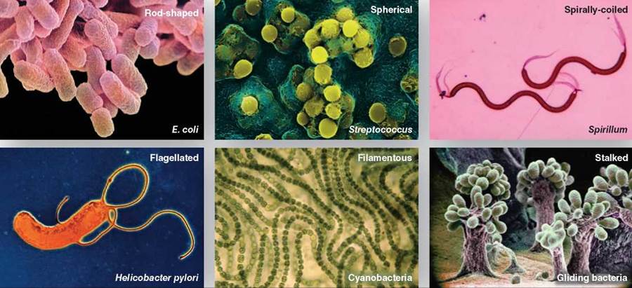
Figure 16.4. Prokaryotes come in many shapes.
The prokaryotic cell’s plasma membrane is encased within a cell wall. The cell wall of bacteria is made of pep- tidoglycan, a network of polysaccharide molecules linked together by peptide interbridgi Many species of bacteria have a cell wall composed of layers of peptidoglycan, represented by the purple rodlike structures in the diagram shown here.
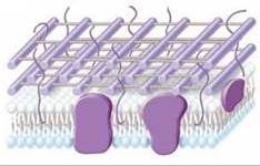
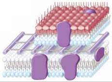
Other species have an outer membrane composed of large molecules of lipopolysaccharide (the red lipids in the diagram to the right) with chains of sugars attached to them covering a thinner peptidoglycan cell wall. Bacteria are commonly classified by the presence or absence of this membrane as gram-positive, with no outer membrane (as on the facing page), or gram-negative, possessing an outer membrane (as shown here). The name refers to the Danish microbiologist Hans Gram, who developed a staining process that stains the cell types differently. A purple dye is retained in the thicker peptidoglycan layer in the cell walls of gram-positive bacteria and they stain purple. In bacteria with an outer membrane, the peptidoglycan layer is thinner and does not retain the purple dye, which is washed away easily. A counterstain with a red dye is retained and so the cells stain red, not purple. The outer membranes of gram-negative bacteria make them resistant to antibiotics that attack the bacterial cell wall. That is why penicillin, which targets the protein cross-links of the bacterial cell wall, is effective only against gram-positive bacteria. Outside the cell wall and membrane, many bacteria have a gelatinous layer called a capsule.
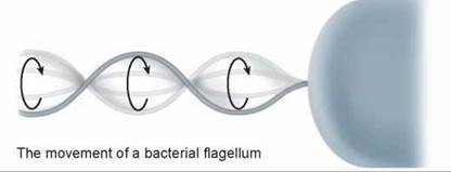
Many kinds of bacteria possess threadlike flagella, long strands of protein that may extend out several times the length of the cell body. Bacteria swim by twisting these flagella in a corkscrew motion. Some bacteria also possess multiple shorter flagella called pili (singular, pilus), which act as docking cables, helping the cell to attach to surfaces or other cells. When exposed to harsh conditions (dryness or high temperature), some bacteria form thick-walled endospores around their DNA and a small bit of cytoplasm. These endospores are highly resistant to environmental stress and may germinate to form new active bacteria even after centuries.
How Prokaryotes Reproduce
Prokaryotes reproduce using a process called binary fission, in which an individual cell simply increases in size and divides in two. Following replication of the prokaryotic DNA, the plasma membrane and cell wall grow inward and eventually divide the cell by forming a new wall from the outside.
Some bacteria can exchange genetic information by passing plasmids from one cell to another in a process called conjugation. A plasmid is a small, circular fragment of DNA that replicates outside the main bacterial chromosome. In bacterial conjugation, seen in figure 16.5, the pilus of the donor cell extends out and contacts a recipient cell 1, forming a passageway called a conjugation bridge between the two cells. The pilus draws the two cells close together. The plasmid in the donor cell begins to replicate its DNA 2, passing the replicated copy out across the bridge and into the recipient cell 3 where a complementary strand is synthesized 4. The recipient cell now contains some of the genetic material found in the donor cell 5. Genes that produce antibiotic resistance in bacteria are often transferred from one bacterial cell to another through conjugation. In addition to conjugation, bacteria can also obtain genetic information by taking up DNA from the environment (transformation; see figure 11.1) or from bacterial viruses (discussed later in this chapter; see figure 16.11).
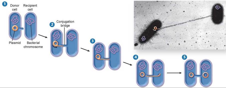
Figure 16.5. Bacterial conjugation.
Donor cells contain a plasmid that recipient cells lack. The plasmid replicates itself and transfers the copy across a conjugation bridge. The remaining strand of the plasmid serves as a template to build a replacement. When the single strand enters the recipient cell, it serves as a template to assemble a double-stranded plasmid. When the process is complete, both cells contain a complete copy of the plasmid.
Key Learning Outcome 16.3. Prokaryotes are the smallest and simplest organisms, a single cell with no internal compartments or organelles. They divide by binary fission.