THE LIVING WORLD
Unit Six. Animal Life
26. Maintaining the Internal Environment
26.4. The Mammalian Kidney
In humans, the kidneys are fist-sized organs located in the region of the lower back (figure 26.10a). Each kidney receives blood from a renal artery, and it is from this blood that urine is produced. Urine drains from each kidney through a ureter, which carries the urine to a urinary bladder. Urine passes out of the body through the urethra. Within the kidney, the mouth of the ureter flares open to form a funnel-like structure, the renal pelvis. The renal pelvis, in turn, has cup-shaped extensions that receive urine from the renal tissue. This tissue is divided into an outer renal cortex (containing blood vessels in figure 26.10b) and an inner renal medulla (containing the cup-shaped structures). Together, these structures perform filtration, reabsorption, secretion, and excretion.
The mammalian kidney is composed of roughly 1 million nephrons (figure 26.10c), each of which is composed of four regions:
1. Filter. The filtration device at the top of each nephron is called a Bowman’s capsule. Within each capsule an arteriole enters and splits into a fine network of vessels called a glomerulus (labeled 1 in the figure). The walls of these capillaries act as a filtration device. Blood pressure forces fluid through the capillary walls. These walls withhold proteins and other large molecules in the blood, while passing water, small molecules, ions, and urea, the primary waste product of metabolism.
2. Proximal tubule. The Bowman’s capsule empties into the proximal tubule, which reclaims most of the water (75%), as well as molecules useful to the body, such as glucose and a variety of ions.
3. Renal tube. The proximal tubule is connected to a long, narrow tube called a renal tubule (labeled 2 through 4), which is bent back on itself in its center. This long, hairpin loop, called the loop of Henle, is a reabsorption device. As the filtrate passes, the renal tubule extracts another 10% of water in the descending loop.
4. Collecting duct. The tube empties into a large collection tube called a collecting duct 5. The collecting duct operates as a water conservation device, reclaiming another 14% of water from the urine so that it is not lost from the body. Human urine is four times as concentrated as blood plasma—that is, the collecting ducts remove much of the water from the filtrate passing through the kidney. Your kidneys achieve this remarkable degree of water conservation by a simple but superbly designed mechanism: The duct bends back alongside the nephron tube, and the duct is permeable to urea. Urea passes out of the collecting duct by diffusion. This greatly increases the local salt (urea) concentration in the tissue surrounding the tube, causing water in urine to pass out of the tube by osmosis. The salty tissue sucks up water from the urine like blotting paper, passing it on to blood vessels that carry it out of the kidneys and back to the bloodstream.
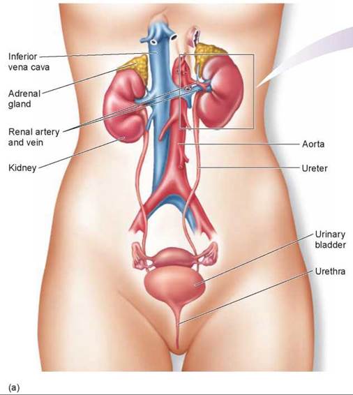
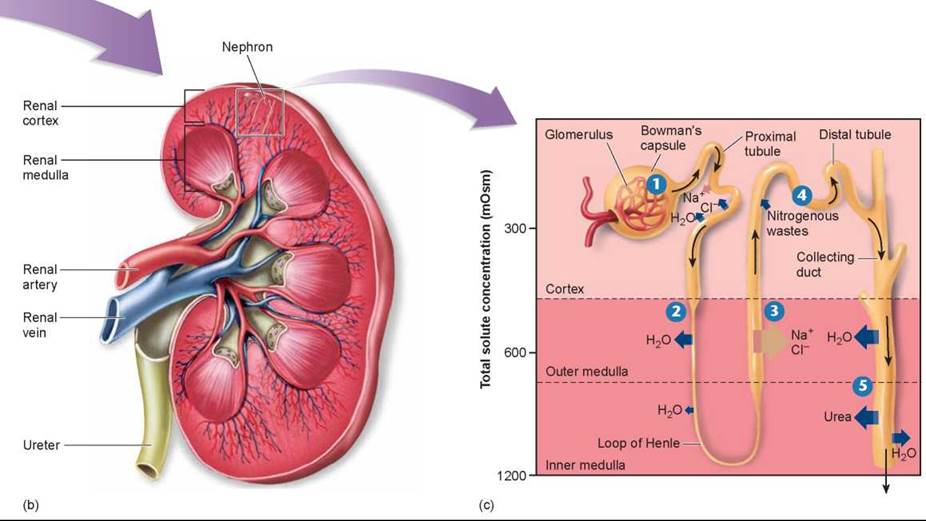
Figure 26.10. The mammalian urinary system contains two kidneys, each of which contain about a million nephrons that lie in the renal cortex and renal medulla.
(a) The urinary system consists of the kidneys, the ureters, which transport urine from the kidneys to the urinary bladder, and the urethra. (b) The kidney is a bean-shaped, reddish brown organ and contains about 1 million nephrons. (c) The glomerulus is enclosed within a filtration device called a Bowman's capsule. Blood pressure forces liquid from blood through the glomerulus and into the proximal tubule of the nephron, where glucose and small proteins are reabsorbed from the filtrate. The filtrate then passes through a loop arrangement consisting of the proximal tubule, the loop of Henle, and the collecting duct, all of which act to remove water from the filtrate. The water is then collected by blood vessels and transported out of the kidney to the systemic (body) circulation.
The formation of urine within the mammalian kidney involves the movement of several kinds of molecules between nephrons and the capillaries that surround them. Five steps are involved and indicated in figure 26.10c: pressure filtration 1, reabsorption of water 2, selective reabsorption of ions 3, tubular secretion 4, and further reabsorption of water 5.
Pressure Filtration. Driven by the blood pressure, small molecules are pushed across the thin walls of the glomerulus to the inside of the Bowman’s capsule 1. Blood cells and large molecules like proteins cannot pass through, and as a result the blood that enters the glomerulus is divided into two paths: nonfilterable blood components that are retained and leave the glomerulus in the bloodstream, and filterable components that pass across and leave the glomerulus in the urine. This filterable stream is called the glomerular filtrate. It contains water, nitrogenous wastes (principally urea), nutrients (principally glucose and amino acids), and a variety of ions.
Reabsorption of Water. Filtrate from the glomerulus passes down the proximal tubule into the descending arm of the loop of Henle. The walls of this descending arm are impermeable to either salts or urea but are freely permeable to water. Because the surrounding tissue has a high concentration of urea (for reasons we discuss later), water passes out of the descending arm by osmosis 2, leaving behind a more concentrated filtrate.
Selective Reabsorption. At the turn in the loop, the walls of the tubule become permeable to salts but much less permeable to water. As the concentrated filtrate passes up this ascending arm, these nutrients pass out into the surrounding tissue 3, where they are carried away by blood vessels. In the upper region of the ascending arm are active transport channels that pump out salt (NaCl). Left behind in the filtrate is the urea that initially passed through the glomerulus as nitrogenous waste. At this point in the tubule, the urea concentration is becoming very high.
Tubular Secretion. In the distal tubule, substances are also added to the urine by a process called tubular secretion 4. This active transport process secretes into the urine other nitrogenous wastes such as uric acid and ammonia, as well as excess hydrogen ions.
Further Reabsorption of Water. The tubule then empties into a collecting duct that passes back through the tissue of the kidney. Unlike the tubule, the lower portions of the collecting duct are permeable to urea, some of which diffuses out into the surrounding tissue (that is why the tissue surrounding the descending arm of the loop of Henle has a high urea concentration indicated by the darker pink color in the figure). The high urea concentration in the tissue causes even more water to pass outward from the filtrate by osmosis 5. The filtrate that is left in the collecting duct after salts, nutrients, and water have been removed is urine.
Key Learning Outcome 26.4. The mammalian kidney pushes waste molecules through a filter and then reclaims water and useful metabolites and ions from the filtrate before eliminating the residual urine.
Biology and Staying Healthy
How Hormones Control Kidney's Functions
As in all mammals and birds, the amount of water excreted in your urine varies according to the changing needs of your body. Acting through the mechanisms described in this chapter, your kidneys excrete a hypertonic urine when your body needs to conserve water.
If you drink a lot of water, your kidneys will excrete a hypotonic urine. As a result, the volume of your blood, your blood pressure, and the salt levels of your blood plasma are all maintained relatively constant by the kidneys, no matter how much water you drink. The kidneys also regulate the plasma Na+ and K+ concentrations and blood pH within narrow limits. These homeostatic functions of the kidneys are coordinated primarily by hormones, which are chemical signals produced in one part of the body that influence events in other parts. Hormones are the subject of chapter 30.
Antidiuretic hormone stimulates the reabsorption of water by the kidneys.
This action completes a negative feedback loop and helps to maintain homeostasis of blood volume and osmolality.
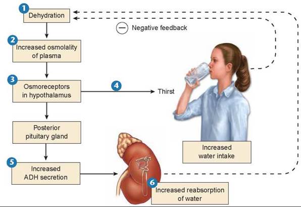
Antidiuretic Hormone. Antidiuretic hormone (ADH) is produced by a portion of your brain called the hypothalamus. The primary stimulus for ADH secretion into your bloodstream is an increase in the osmolality (concentration of salt) of the blood plasma. The salt in each milliliter of plasma increases when you are dehydrated or when you eat salty food. Consider the stimulus of dehydration shown in the figure here. Dehydration 1 causes an increase in the solute concentrations in the blood 2. Osmoreceptors in the hypothalamus 3 respond to the elevated blood osmolality by triggering a sensation of thirst 4 and by an increase in the secretion of ADH 5.
ADH causes the walls of the distal tubules and collecting ducts in your kidneys (see figure 26.10c) to become more permeable to water, thus increasing the amount of water they absorb from the urine as it passes through your kidneys 6. The increased absorption of water from the kidneys feeds back to the osmoreceptors (the dashed line), causing a reduction in the secretion of ADH. When the secretion of ADH is reduced, the walls become less permeable, and you excrete more water in your urine.
Under conditions of maximal ADH secretion, you excrete only 600 milliliters of highly concentrated urine per day. A person who lacks ADH has the disorder known as diabetes insipidus and constantly excretes a large volume of dilute urine. Such a person may become severely dehydrated and succumb to dangerously low blood pressure.
Aldosterone. Sodium ion is the major solute in your blood plasma. When the blood concentration of Na+ falls, the blood osmolality also falls. This drop in osmolality inhibits ADH secretion, causing more water to remain in the collecting duct for excretion in your urine. As a result, your blood volume and blood pressure decrease. A decrease in extracellular Na+ also causes more water to be drawn into your cells by osmosis, partially offsetting the drop in plasma osmolality but further decreasing your blood volume and blood pressure. If Na+ deprivation is severe, the blood volume may fall so low that there is insufficient blood pressure to sustain your life. For this reason, salt is necessary for life. Many animals have a "salt hunger” and actively seek salt. This is why a "salt lick” will attract deer.
A drop in blood Na+ concentration is normally compensated by the kidneys under the influence of another hormone, aldosterone, which is also secreted by your brain. Indeed, under conditions of maximal aldosterone secretion, Na+ may be completely absent from the urine.
The reabsorption of Na+ is followed by Cl" and by water, so aldosterone has the net effect of promoting the retention of both salt and water. It thereby helps to maintain your blood volume, osmolality, and pressure.