CONCEPTS IN BIOLOGY
PART II. CORNERSTONES: CHEMISTRY, CELLS, AND METABOLISM
4. Cell Structure and Function
Cadavers Source of Healing Power
Is there a risk?
Most people think that bones are lifeless; however, they are actually living, growing tissues. Bone is composed of many kinds of living cells surrounded by a bone matrix made of minerals and proteins. Certain bone cells are a source of red and white blood cells while others are engaged in a process known as bone remodeling—much like the process a sculptor uses when creating a piece of art from clay. Remodeling occurs throughout life and is the result of the absorption of bone followed by the formation of new bone. Cells known as osteoblasts are responsible for adding new bone matrix, whereas osteoclasts remove old matrix. Remodeling takes place as a result of bone loss due to stress, disease or breakage.
Following certain accidents or diseases, bone cells are unable to remodel the damage. However, researchers have discovered that healing can be promoted by using protein bone matrix from cadavers—dead bodies. This tissue is rich in growth factors that signal bone cells to multiply and promote repair. Are there drawbacks? There is only a limited supply of this matrix because it comes from human donors, and there is a risk of transmitting viruses to the recipient.
• What is the basic makeup of a cell?
• How do growth factors signal cells?
• In order to become a bone matrix organ donor, should a person undergo screening to check for hidden viruses?
ü Background Check
Concepts you should already know to get the most out of this chapter:
• The atomic and molecular nature of matter (chapter 2)
• Some molecules can be very large (chapter 3)
• There are millions of different kinds of molecules and that each kind of molecule has specific physical properties (chapter 2)
• Kinetic molecular theory (chapter 2)
4.1. The Development of the Cell Theory
The cell theory states that all living things are made of cells. The cell is the basic structural and functional unit of living things and is the smallest unit that displays the characteristics of life. However, the concept of a cell did not emerge all at once but, rather, was developed and modified over several centuries. It is still being modified today. The ideas of hundreds of people were important in the development of the cell theory, but certain key people can be identified.
Some History
The first person to use the term cell was Robert Hooke (1635-1703) of England. He used a simple kind of microscope to study thin slices of cork from the bark of a cork oak tree (figure 4.1). He saw many cubicles fitting neatly together, which reminded him of the barren rooms (cells) in a monastery. He used the term cell when he described his observations in 1665 in the publication Micrographia, the first picture book of science to come off the press, with 38 beautiful engravings. The book became a best-seller. The tiny cork boxes Hooke saw, and described in his book were, in fact, only the cell walls that surrounded the once living portions of these plant cells.
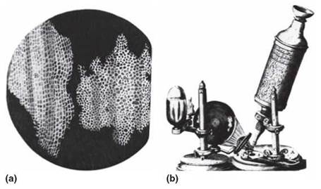
FIGURE 4.1. Hooke's Observations
(a) The concept of a cell has changed considerably over the past 300 years. Robert Hooke’s idea of a cell was based on his observation of slices of cork (cell walls of the bark of the cork oak tree). (b) Hooke constructed his own simple microscope to be able to make these observations.
We now know that the cell wall of a plant cell is produced on the outside of the cell and is composed of the complex carbohydrate called cellulose. It provides strength and protection to the living contents of the cell. Although the cell wall appears to be a rigid, solid layer of material, it is actually composed of many interwoven strands of cellulose molecules. Thus, most kinds of molecules pass easily through it.
Anton van Leeuwenhoek (1632-1723), a Dutch merchant who sold cloth, was one of the first individuals to carefully study magnified cells. He apparently saw a copy of Hooke’s Micrographia and began to make his own microscopes, so that he could study biological specimens. He was interested in magnifying glasses, because magnifiers were used to count the number of threads in cloth. He used a very simple kind of microscope that had only one lens. Basically, it was a very powerful magnifying glass (figure 4.2). What made his microscope better than others of the time was his ability to grind very high-quality lenses. He used his skill at lens grinding to make about 400 lenses during his lifetime. One of his lenses was able to magnify 270 times. Van Leeuwenhoek made thousands of observations of many kinds of microscopic objects. He also made very detailed sketches of the things he viewed with his simple microscopes and communicated his findings to Robert Hooke and the Royal Society of London. His work stimulated further investigation of magnification techniques and descriptions of cell structures.
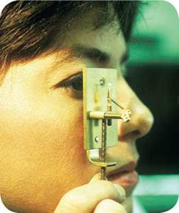
FIGURE 4.2. Anton van Leeuwenhoek's Microscope
Although van Leeuwenhoek’s microscope had only one lens, the lens quality was so good that he was able to see cells clearly. This replica of his microscope shows that it is a small, simple apparatus.
When van Leeuwenhoek discovered that he could see things moving in pond water using his microscope, his curiosity stimulated him to look at a variety of other things. He studied many things such as blood, semen, feces, and pepper, for example. He was the first to see individual cells and recognize them as living units, but he did not call them cells. The name he gave to the “little animals” he saw moving around in the pond water was animicules.
Although Hooke, van Leeuwenhoek, and others continued to make observations, nearly 200 years passed before it was generally recognized that all living things are made of cells and that these cells can reproduce themselves. In 1838, Mathias Jakob Schleiden of Germany stated that all plants are made up of smaller cellular units. In 1839, Theodor Schwann, another German, published the idea that all animals are composed of cells.
Soon after the term cell caught on, it was recognized that the cell wall of plant cells was essentially lifeless and that it was really the contents of the cell that had “life.” This living material was termed protoplasm, which means first-formed substance. Scientists used the term protoplasm to distinguish between the living portion of the cell and the nonliving cell wall. As better microscopes were developed, people began to distinguish two different regions of protoplasm. One region, called the nucleus, appeared as a central body within a more fluid material surrounding it. Today, we know the nucleus is the part of a cell that contains the genetic information. Cytoplasm was the name given to the fluid portion of the protoplasm surrounding the nucleus. Although the term protoplasm is seldom used today, the term cytoplasm is still common.
The development of special staining techniques, better light microscopes, and ultimately powerful electron microscopes revealed that the cytoplasm contains many structures, called organelles (little organs). Further research has shown that each kind of organelle has certain functions related to its structure.
Basic Cell Types
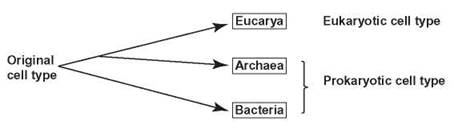
All living things are cells or composed of cells, and all cells share three basic traits: They all have an outer membrane, cytoplasm, and genetic material. However, about 400 years of research has revealed a variety of differences among cells. For example, we know that while all the cells in your body have been derived from one, single, fertilized egg cell, bone cells show structural differences in comparison to brain cells. They not only look different under the microscope, but perform very different metabolically. As scientists studied the cells of even more diverse organisms such as bacteria, plants, animals, fungi, algae, and protozoans, it became clear that there were even greater differences. Some of these differences were structural; others only became evident by doing chemical analysis. As a result of these investigations, biologists have categorized cells into two general types: eukaryotic and prokaryotic (noneukaryotic) cells (figure 4.3). The cells of plants, animals, fungi, protozoa, and algae are eukaryotic, and are placed in a category called Eucarya. All eukaryotic cells have their genetic material surrounded by a nuclear membrane forming the cellular nucleus. They also have a large number and variety of complex organelles, each specialized in the metabolic function it performs. In general, they are large in comparison to noneukaryotic cells. There are two categories of prokaryotic cells: Bacteria and Archaea. Neither of these cell types has a nuclear membrane; therefore they lack a cellular nucleus. In addition, they display unique chemical and metabolic characteristics but do not have the variety and number of organelles seen in eukaryotes. Bacteria and Archaea are classified into a group referred to as the Prokaryotes. From studying a vast amount of data, biologists have tried to understand the evolutionary relationship among these cell types. The previous hypothesized evolutionary relationship among these cell types was:
![]()
However, current data points to a different evolutionary pattern:
The fossil record shows evidence of prokaryotes 3.5 billion years ago. Eucarya show up in the fossil record about 1.8 billion years ago.
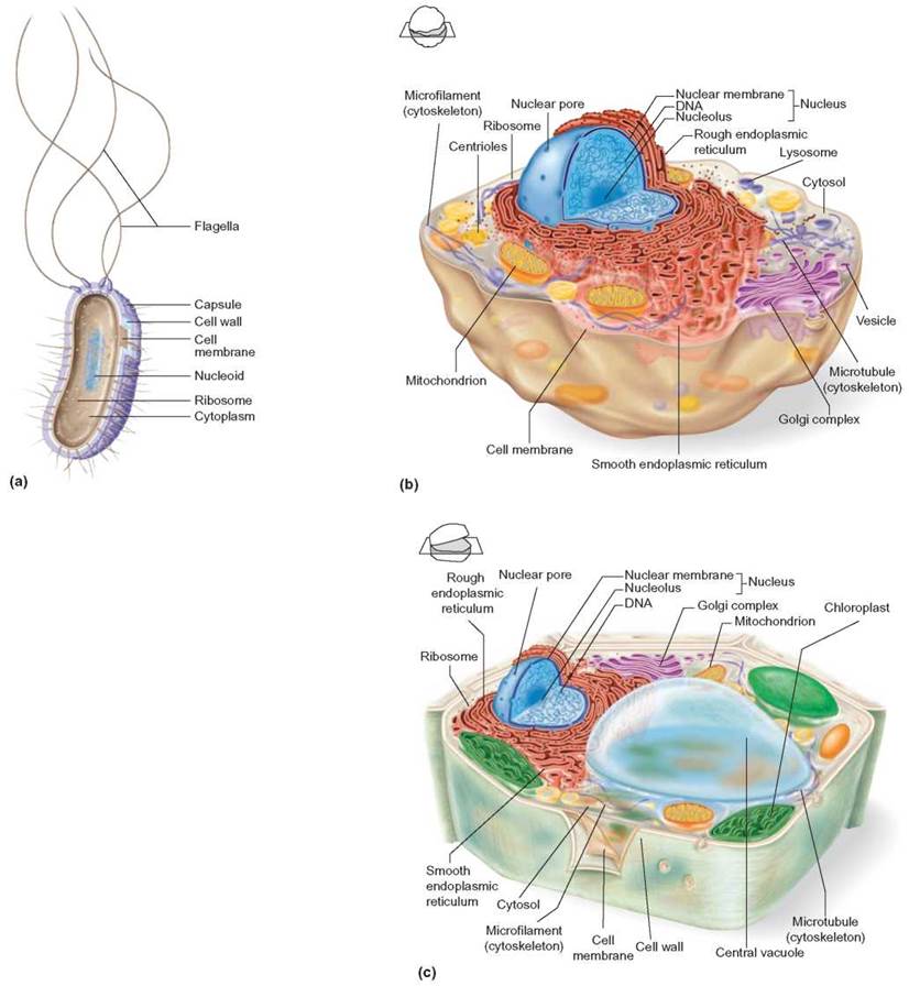
FIGURE 4.3. Major Cell Types
There are two major types of cells eukaryotic and noneukaryotic. Eukaryotic cells are 10 to 100 times larger than noneukaryotic cells such as this (a) bacterium. These drawings (not to scale) highlight the structural differences between them. The generalized eukaryotic cells are (b) an animal and (c) a plant cell.
4.1. CONCEPT REVIEW
1. Describe how the concept of the cell has changed over the past 200 years.
2. What features do all cell types have in common?