THE LIVING WORLD
Unit Six. Animal Life
27. How the Animal Body Defends Itself
27.5. T Cells: The Cellular Response
When macrophages process foreign antigens, they trigger the cellular immune response, illustrated in figure 27.9. As shown in step 1, macrophages secrete interleukin-1, which stimulates cell division and proliferation of T cells. Helper T cells become activated when they bind to the complex of MHC proteins and antigens presented to them by the macrophages. The helper T cells then secrete interleukin-2 2, which stimulates the proliferation of cytotoxic T cells 3, which recognize and destroy infected body cells. Cytotoxic T cells destroy infected cells only if those cells display the foreign antigen together with their MHC proteins 4.
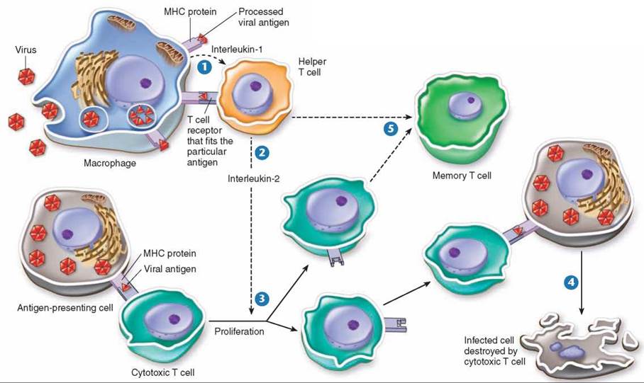
Figure 27.9. The T cell immune defense.
After a macrophage has processed an antigen, it releases interleukin-1, signaling helper T cells to bind to the antigen-MHC protein complex. This triggers the helper T cell to release interleukin-2, which stimulates the multiplication of cytotoxic T cells. In addition, proliferation of cytotoxic T cells is stimulated when a T cell with a receptor that fits the antigen displayed by an antigen-presenting cell binds to the antigen- MHC protein complex. Body cells that have been infected by the antigen are destroyed by the cytotoxic T cells. As the infection subsides, suppressor T cells "turn off" the immune response (not shown here).
The body makes millions of different versions of T cells. Each version bears a single, unique kind of receptor protein on its membrane, a receptor able to bind to a particular antigen- MHC protein complex on the surface of an antigen-presenting cell. Any cytotoxic T cell whose receptor fits the particular antigen-MHC protein complex present in the body begins to multiply rapidly, soon forming large numbers of T cells 3 capable of recognizing the complex containing the particular foreign antigen. Large numbers of infected cells can be quicklyeliminated, because the single T cell able to recognize the invading virus is amplified in number to form a large clone of identical T cells, all able to carry out the attack. Any of the body’s cells that bear traces of viral infection are destroyed. The method used by cytotoxic T cells to kill infected body cells is similar to that used by natural killer cells and complement— they puncture the plasma membrane of the infected cell. Following an infection, some activated T cells give rise to memory T cells 5 that remain in the body, ready to mount an attack quickly if the antigen is encountered again.
Cytotoxic T cells will also attack any foreign version of the MHC proteins. Thus even though the immune system did not evolve in vertebrates as a defense against tissue transplants, their immune systems will attack transplanted tissue and cause graft rejection. It is for this reason that relatives are often sought for kidney transplants—their MHC proteins are genetically closer to the recipient. The drug cyclosporine is often given to transplant patients because it inactivates cytotoxic T cells.
There is some evidence that cancer cells alter their “self’ markers in a way that can be detected by immune cells, creating so-called “cancer-specific antigens,” but the potential role of such modified cell surface markers in immunological surveillance against cancer is not well understood.
Key Learning Outcome 27.5. The cellular immune response is carried out by T cells, which mount an immediate attack on infected cells, killing any that present unusual surface antigens.