THE LIVING WORLD
Unit Six. Animal Life
The spinal cord is a cable of neurons extending from the brain down through the backbone, which is the view in figure 28.15. The cross section through the spinal cord in figure 28.16 shows a darker gray area in the center that consists of neuron cell bodies, which form a column down the length of the cord. This column is surrounded by a sheath of axons and dendrites, which make the outer edges of the cord white because they are coated with myelin. The spinal cord is surrounded and protected by a series of bones called the vertebrae. Spinal nerves pass out to the body from between the vertebrae. Messages between the body and the brain run up and down the spinal cord, like an information highway.
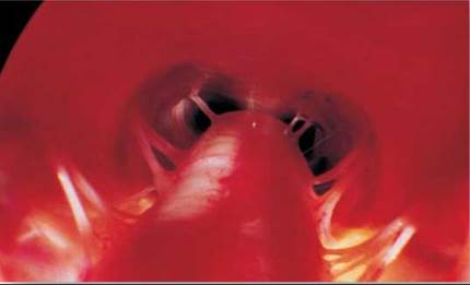
Figure 28.15. A view down the human spinal cord.
Pairs of spinal nerves can be seen extending out from the spinal cord. Along these nerves, the brain and spinal cord communicate with the body.
In each segment of the spine, motor nerves extend out of the spinal cord to the muscles. Motor nerves from the spine control most of the muscles below the head. This is why injuries to the spinal cord often paralyze the lower part of the body. A muscle is paralyzed and cannot move if its motor neurons are damaged.
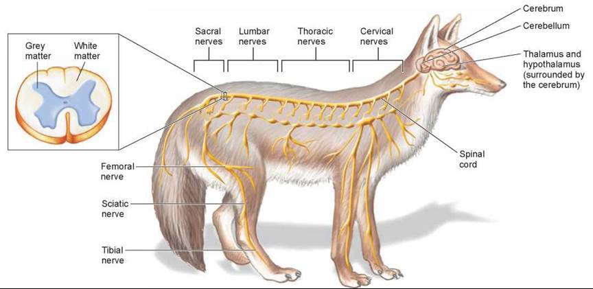
Figure 28.16. The vertebrate nervous system.
The brain is colored tan and the spinal cord and nerves are colored yellow.
Spinal Cord Regeneration
In the past, scientists have tried to repair severed spinal cords by installing nerves from another part of the body to bridge the gap and act as guides for the spinal cord to regenerate. But most of these experiments have failed because the nerve bridges did not go from white matter to gray matter. Also, there is a factor that inhibits nerve growth in the spinal cord. After discovering that fibroblast growth factor stimulates nerve growth, neurobiologists tried gluing on the nerves, from white to gray matter, with fibrin that had been mixed with the fibroblast growth factor.
Three months later, rats with the nerve bridges began to show movement in their lower bodies. In further analyses of the experimental animals, dye tests indicated that the spinal cord nerves had regrown from both sides of the gap. Many scientists are encouraged by the potential to use a similar treatment in human medicine. However, most spinal cord injuries in humans do not involve a completely severed spinal cord; often, nerves are crushed. Also, although the rats with nerve bridges did regain some locomotory ability, tests indicated that they were barely able to walk or stand.
Key Learning Outcome 28.7. The spinal cord, protected in vertebrates by a backbone, extends motor nerves to the muscles below the head.